. 2011 Dec 20;108(51):20766-71.
doi: 10.1073/pnas.1115141108. Epub 2011 Dec 5.

Paraquat neurotoxicity is mediated by the dopamine transporter and organic cation transporter-3

Affiliations

Paraquat neurotoxicity is mediated by the dopamine transporter and organic cation transporter-3

Phillip M Rappold et al. Proc Natl Acad Sci U S A. .

Abstract

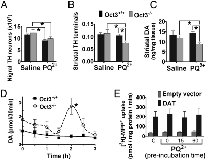
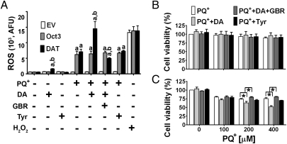
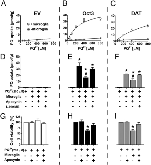
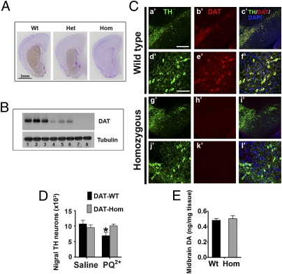
The herbicide paraquat (PQ) has increasingly been reported in epidemiological studies to enhance the risk of developing Parkinson's disease (PD). Furthermore, case-control studies report that individuals with genetic variants in the dopamine transporter (DAT, SLC6A) have a higher PD risk when exposed to PQ. However, it remains a topic of debate whether PQ can enter dopamine (DA) neurons through DAT. We report here a mechanism by which PQ is transported by DAT: In its native divalent cation state, PQ(2+) is not a substrate for DAT; however, when converted to the monovalent cation PQ(+) by either a reducing agent or NADPH oxidase on microglia, it becomes a substrate for DAT and is accumulated in DA neurons, where it induces oxidative stress and cytotoxicity. Impaired DAT function in cultured cells and mutant mice significantly attenuated neurotoxicity induced by PQ(+). In addition to DAT, PQ(+) is also a substrate for the organic cation transporter 3 (Oct3, Slc22a3), which is abundantly expressed in non-DA cells in the nigrostriatal regions. In mice with Oct3 deficiency, enhanced striatal damage was detected after PQ treatment. This increased sensitivity likely results from reduced buffering capacity by non-DA cells, leading to more PQ(+) being available for uptake by DA neurons. This study provides a mechanism by which DAT and Oct3 modulate nigrostriatal damage induced by PQ(2+)/PQ(+) redox cycling.


Conflict of interest statement

The authors declare no conflict of interest.

Figures

Fig. 1.
Fig. 1.
PQ2+ injection increases striatal neurotoxicity and DA overflow in mice with Oct3 deficiency. (A–C: neurotoxicity study) Oct3−/− mice and Oct3+/+ littermates (10–12 wk old) were injected with PQ2+ (10 mg/kg, i.p., every second day for a total of 10 injections) or saline. Seven days after the last injection, mice were processed for stereological cell counting (A), striatal tyrosine hydroxylase immunoreactivity (B, optical density), or HPLC measurement of DA content (C). n = 5 animals per group for A and B and n = 5–9 per group for C. *P < 0.05, analyzed by two-way ANOVA followed by the Newman–Keuls post hoc test. (D: in vivo microdialysis study) Oct3−/− and Oct3+/+ littermates (10–12 wk old) were stereotactically implanted with microdialysis probes into the right striatum. After 2 h of equilibration, dialysates were collected every 30 min for 1 h before PQ2+ injection (15 mg/kg, i.p.) for baseline measurements (pooled for 0 time point) and for an additional 3 h after the injection, followed by HPLC analyses for DA. n = 7 animals per group. Area under the curve was generated using GraphPad Prism followed by a two-tailed t test. *P < 0.05 compared with the Oct3+/+ group. (E: transport study) Uptake of tritiated MPP+ was assessed in EM4 cells (modified human embryonic kidney cells; SI Materials and Methods) with stable expression of DAT or empty vector control. Uptake reaction mediated by DAT was assessed in cells preincubated with 500 μM PQ2+ up to 60 min and compared with the control group (“C”) without PQ2+ preincubation. n = 3–5 independent experiments in quadruplicate.
Fig. 2.
Fig. 2.
PQ+ is transported by both Oct3 and DAT to induce cytotoxicity. Stable EM4 cells expressing empty vector control (A), Oct3 (B), or DAT (C) were cultured in 24-well plates for 24 h. Cells were then washed and incubated with varying concentrations of PQ2+ with or without 0.5 mM sodium dithionite (SDT) in degassed Krebs Ringer Hepes (KRH) buffer to convert PQ2+ to PQ+. After 20 min, cells were washed and then collected for HPLC analysis. To determine the effects of PQ+ on the formation of reactive oxygen species (ROS) (D–F), these cell types were treated for 24 h with the indicated concentrations of PQ2+ with or without 0.5 mM SDT. After 20 min, cells were washed and grown in regular medium. After 24 h, cells were incubated with 5 μM dihydroethidium and analyzed for ROS levels using flow cytometry (AFU: arbitrary fluorescence unit). H2O2 (30 μM) was incubated with cells for 20 min as a positive control for ROS production. To assess whether the observed ROS production would lead to cytotoxicity, cell viability was performed (G–I). Cells were treated with PQ2+ and SDT as shown in D–F, washed, and grown in cell culture medium for another 48 h before a 3-(4,5-dimethylthiazol-2-yl)-2,5-diphenyltetrazolium bromide (MTT) assay was performed. n = 4 independent experiments in quadruplicate, analyzed by two-way ANOVA followed by the Newman–Keuls post hoc test. *P < 0.05 compared with the respective control groups without SDT.
Fig. 3.
Fig. 3.
DA enhances ROS production and cytotoxicity induced by PQ+. EM4 cells with empty vector, Oct3, or DAT expression were treated with 200 μM PQ2+ plus 0.5 mM SDT for 20 min. Then the cells were washed and incubated in culture medium with or without 50 μM DA, 50 μM tyramine, or 1 μM GBR12909 (a DAT inhibitor). H2O2 (30 μM for 1 h) was used as a positive control for ROS production. After 48 h, cells were assessed for ROS levels using flow cytometry (A). In a parallel set of experiments, cells with empty vector (B) and DAT (C) treated with varying concentrations of PQ2+ as described in A were assessed for cell viability using a MTT assay. n = 4 independent experiments in quadruplicate, analyzed by one-way ANOVA followed by the Newman–Keuls post hoc test. aP < 0.05 compared with respective EV; bP < 0.05 compared with the respective Oct3 groups; *P < 0.05.
Fig. 4.
Fig. 4.
Microglia promote the conversion of PQ2+ to PQ+ through NADPH oxidase. Stable EM4 cells expressing Oct3, DAT, or empty vector control were grown on glass coverslips for 24 h. These coverslips were then transferred to six-well plates on which a monolayer of human microglial immortalized cells (CHME5) had been plated for 24 h. Different concentrations of PQ2+ were added to these cocultures. After 24 h of PQ2+ addition, the uptake of PQ+ into EM4 cells was evaluated by removing the coverslips from the cocultures and collecting cells for HPLC measurement (A–C). To determine whether increased uptake of PQ+ was mediated by NADPH oxidase or nitric oxide synthase (NOS), apocynin and l-NAME, respectively, were added to the cocultures (D–F). Inhibition of NADPH oxidase by apocynin, but not NOS by l-NAME, significantly reduced PQ+ transport into Oct3 or DAT cells. n = 4 independent experiments, analyzed by one-way ANOVA followed by the Newman–Keuls post hoc test. *P < 0.05 compared with the PQ2+ alone group; #P < 0.05 compared with PQ2+ plus microglia group as well as PQ2+ plus microglia plus the l-NAME group. The reduced uptake of PQ+ in the presence of apocynin resulted in less cytotoxicity (G–I) as assessed by using an MTT assay 48 h later. n = 4 independent experiments, analyzed by one-way ANOVA followed by the Newman–Keuls post hoc test. *P < 0.05 compared with all other groups.
Fig. 5.
Fig. 5.
Mutant DAT hypomorphic mice are resistant to PQ neurotoxicity. (A) Coronal striatal sections from wild type (Wt), heterozygous (Het), and homozygous (Hom) mutant DAT mice were immunostained with a DAT antibody (brown, diaminobenzidine chromogen) and counterstained with Nissl to reveal the structure of other brain regions. (B) For a more quantitative comparison, Western blotting was performed. Lanes 1–3 (Wt), lanes 4–6 (Het), and lanes 7–8 (Hom) are from eight animals. (C) Immunofluorescence of TH and DAT in coronal midbrain sections containing the substantia nigra and ventral tegmental area. [Scale bars: 400 μm (a′–c′, g′–i′), 100 μm (d′–f′, j′–l′).] No apparent differences in morphology of midbrain DA neurons was noted between mutant and wild-type mice. Stereological cell counting confirmed comparable population of nigral DA neurons (D) and midbrain DA levels (E) in these mutant mice. (D) To assess the effects of PQ toxicity in these animals, DAT wild-type mice and their homozygous mutant littermates (∼10 wk old) were injected with PQ2+ (10 mg/kg, i.p., every second day for a total of 10 injections) or saline. Seven days after the last injection, mice were processed for stereological cell counting. n = 4–5 animals per group, analyzed by two-way ANOVA followed by the Newman–Keuls post hoc test. *P < 0.05 compared with the control saline-treated group.

References

    1. Dauer W, Przedborski S. Parkinson's disease: Mechanisms and models. Neuron. 2003;39:889–909. - PubMed
    1. Ritz BR, et al. Dopamine transporter genetic variants and pesticides in Parkinson's disease. Environ Health Perspect. 2009;117:964–969. - PMC - PubMed
    1. Tanner CM, et al. Rotenone, paraquat and Parkinson's disease. Environ Health Perspect. 2011;119:866–872. - PMC - PubMed
    1. Day BJ, Patel M, Calavetta L, Chang LY, Stamler JS. A mechanism of paraquat toxicity involving nitric oxide synthase. Proc Natl Acad Sci USA. 1999;96:12760–12765. - PMC - PubMed
    1. Langston JW, Ballard P, Tetrud JW, Irwin I. Chronic Parkinsonism in humans due to a product of meperidine-analog synthesis. Science. 1983;219:979–980. - PubMed

Publication types

LinkOut - more resources