Review
. 2012 Jan;4(1):65-74.
doi: 10.1039/c1ib00124h. Epub 2011 Dec 5.

What yeast and cardiomyocytes share: ultradian oscillatory redox mechanisms of cellular coherence and survival

Affiliations
Review

What yeast and cardiomyocytes share: ultradian oscillatory redox mechanisms of cellular coherence and survival

David Lloyd et al. Integr Biol (Camb). 2012 Jan.

Abstract

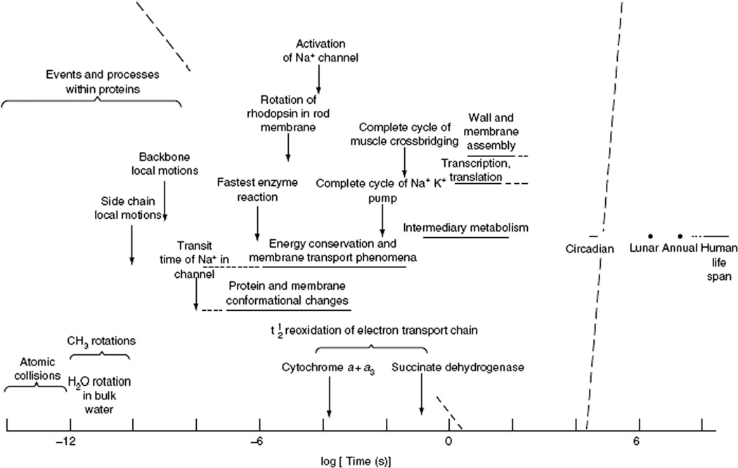
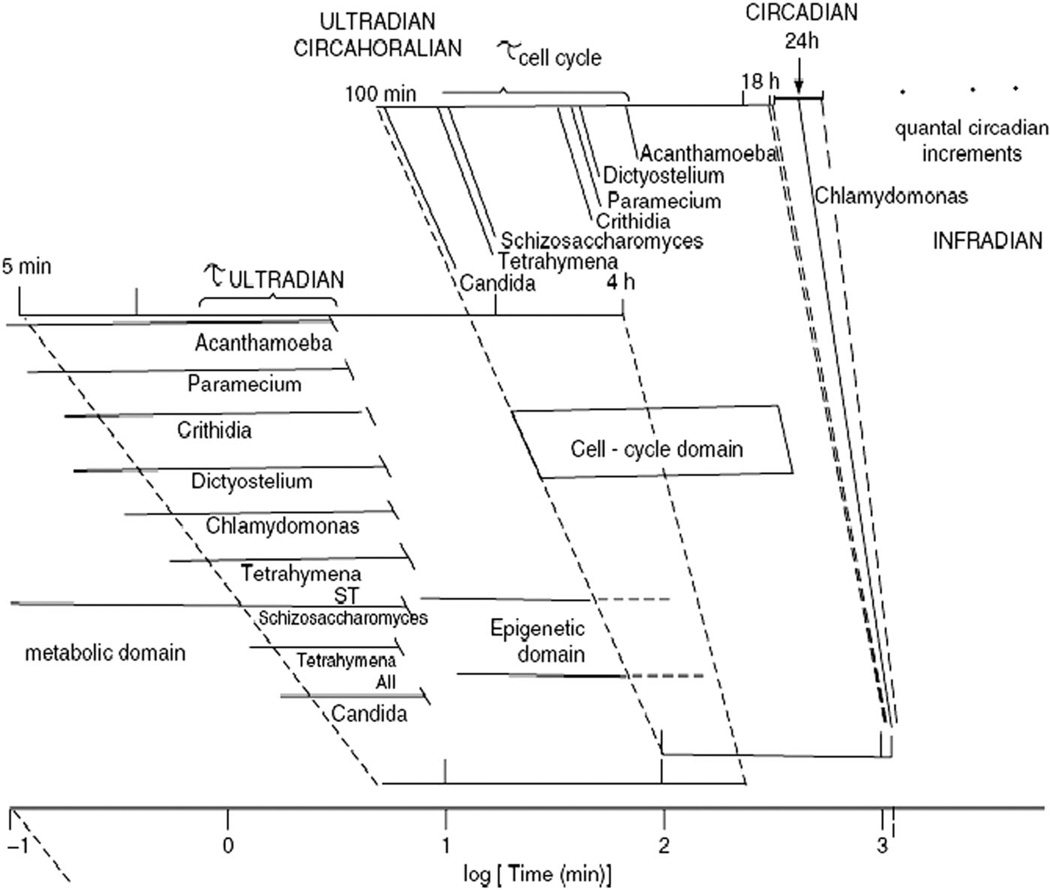
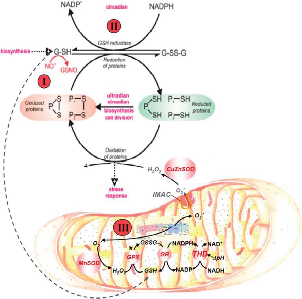
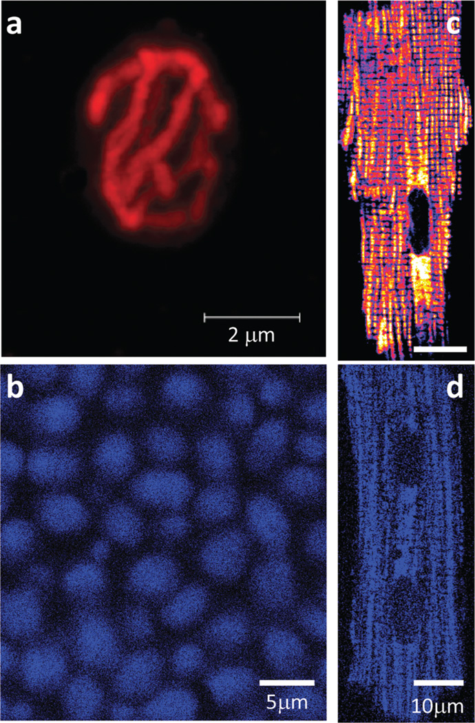
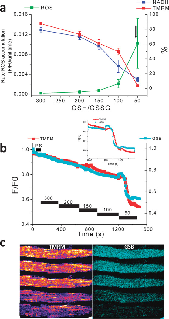
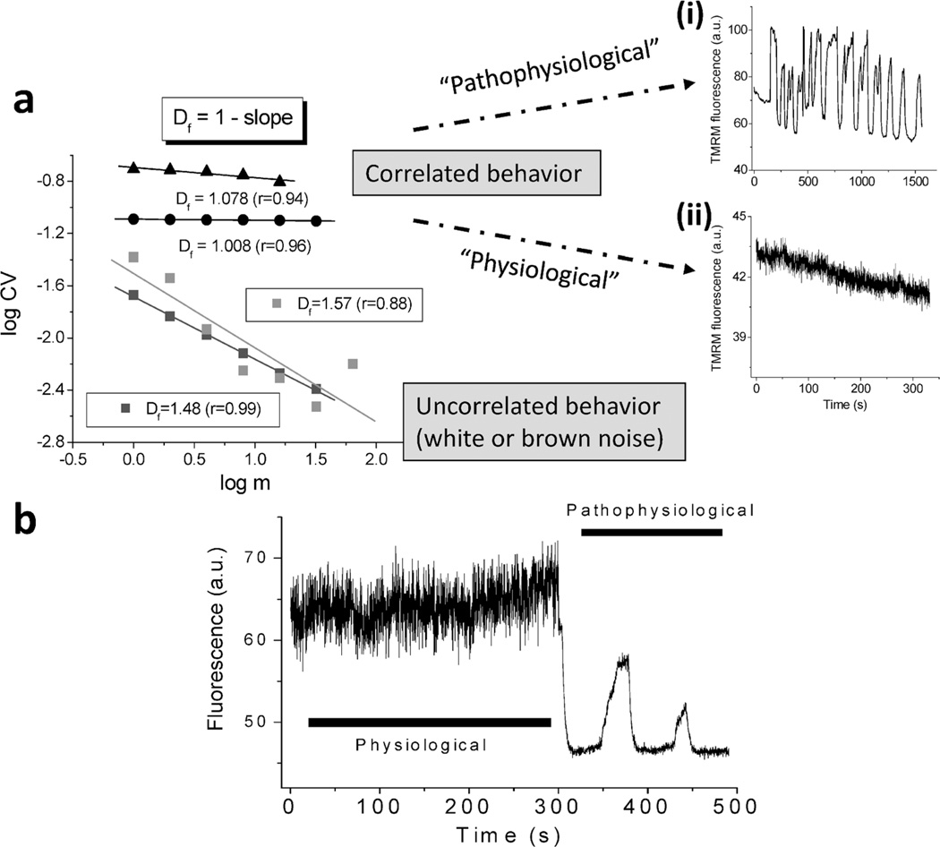
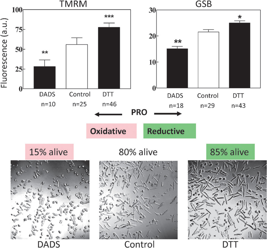
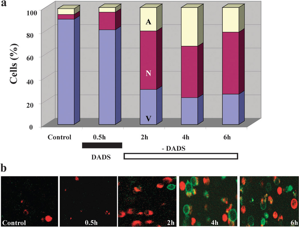
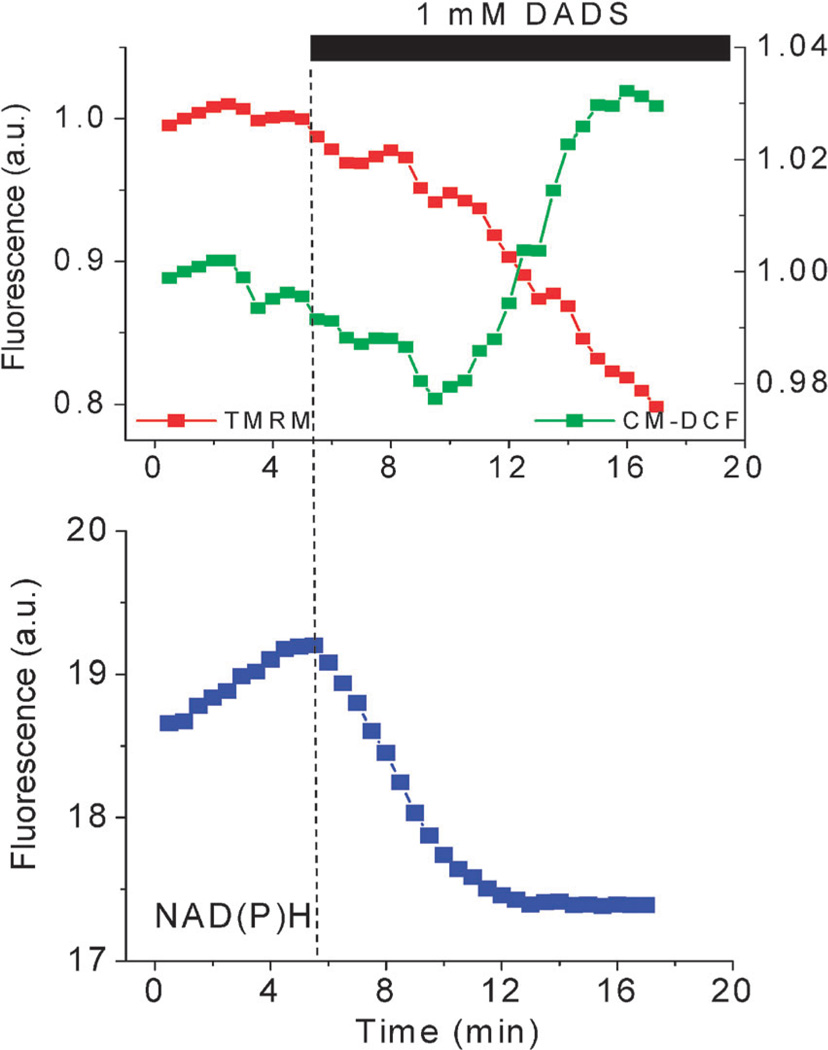
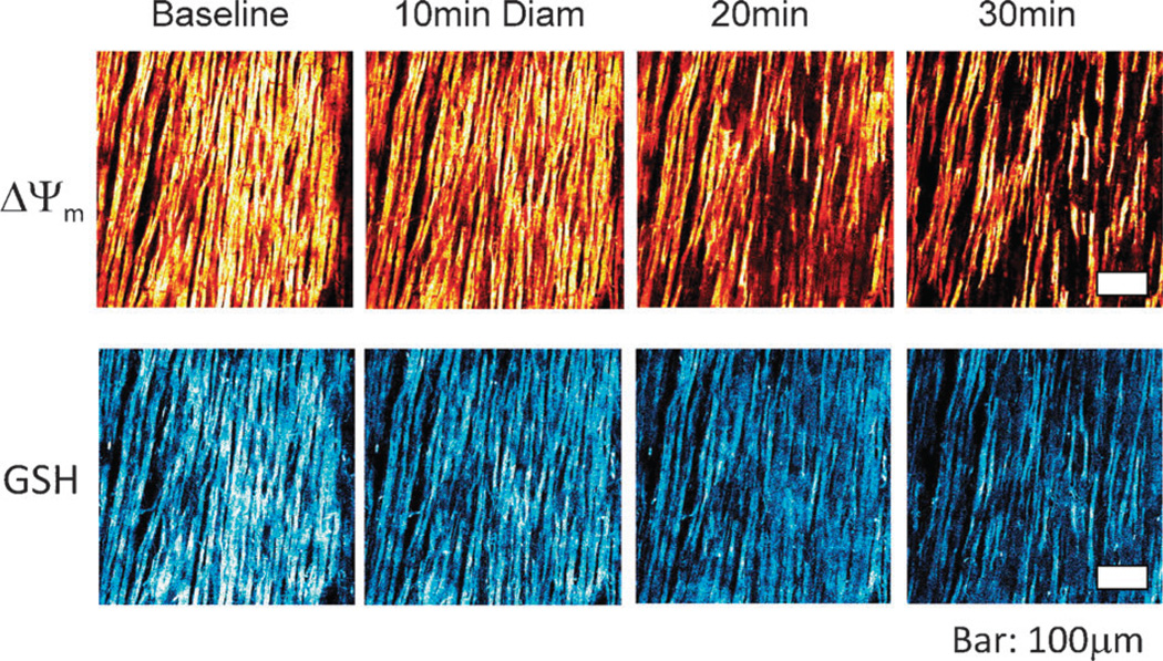
The coherent and robust, yet sensitively adaptable, nature of organisms is an astonishing phenomenon that involves massive parallel processing and concerted network performance at the molecular level. Unravelling the dynamic complexities of the living state underlines the essential operation of ultradian oscillations, rhythms and clocks for the establishment and maintenance of functional order simultaneously on fast and slower timescales. Non-invasive monitoring of respiration, mitochondrial inner membrane potentials, and redox states (especially those of NAD(P)H, flavin, and the monochlorobimane complex of glutathione), even after more than 50 years research, continue to provide both new insights and biomedical applications. Experiments with yeast and in cardiac cells reveal astonishing parallels and similarities in their dynamic biochemical organization.


Figures

Fig. 1
Fig. 1
The temporal structure of biological organization as characterized by the relaxation times of biological processes. The different time scales exhibited by living systems are shown by the typical relaxation times (after perturbation) in decades on a logarithmic scale. This way of representation accounts for the broad temporal scale spanned by the occurrence of biological processes, i.e. from picoseconds [10−12 s] to many years. The biological processes indicated represent many of the main molecular and cellular functional properties as well as those related to conformational changes in molecules (e.g. following ligand interactions), metabolism, energy transduction, solute transport, action potentials in neurons and cardiomyocytes, macromolecule polymerization and cell growth and division. (Reproduced from Lloyd and Rossi (2008) in Ultradian Rhythms from Molecules to Mind).
Fig. 2
Fig. 2
Periodic phenomena across temporal scales. The period of cyclic processes in the ultradian (<24 h), circadian (24 h) and infradian (>24 h) time domains are shown in a logarithmic scale. Several clocks (temperature-compensated biological mechanisms) can be found in the ultradian temporal domain, in addition to the ubiquitous circadian clock. A prominent ultradian clock is the circahoralian (about 1 h period) that is indicated as occurring in many lower eukaryotes and is always of shorter duration that the cell division cycle, as shown in the figure. (Reproduced from Lloyd and Rossi (2008) in Ultradian Rhythms from Molecules to Mind).
Fig. 3
Fig. 3
Redox cycling of intracellular thiols at the core of yeast and heart rhythmicity. The scheme shows that generation of rhythms entails the cycling of cytoplasmic and mitochondrial proteins between their oxidized and reduced states mainly driven by ROS and the redox potential of the thiols pool. Mitochondria are the main source of ROS produced by the respiratory chain; oxidative stress results from an imbalance between ROS production and ROS scavenging. The glutathione and thioredoxin redox potentials, and the absolute concentrations of reduced (GSH) and oxidized (GSSG) glutathione, modulate ROS emission in mitochondria. Mitochondrial GSH and thioredoxin (Trx2) regeneration in the matrix are essential for keeping the ROS balance. In yeast, numerous processes (magenta I–III) have proven to be oscillatory and we propose that ensembles of oscillators are coupled via this primordial mechanism. In heart, the redox cycling involves at least glutathione, Trx2, and NAD(P)H couples in the mitochondrial matrix, as modulators of ROS generation. Perturbation analysis of the yeast ultradian system utilising NO+ donors, 5-nitro-2-furaldehyde(i), d,l-butathionine (S,R)-sulphoximine(ii) or protonophores(iii), confirms the central role of this redox system. The numbers on the figure represent the site of perturbation. (Reproduced from Aon MA, Roussel MR, Cortassa S, B. O’Rourke, Murray DB, et al. (2008) The Scale-Free Dynamics of Eukaryotic Cells. PLoS ONE 3(11): e3624. doi: 10.1371/journal.pone.0003624).
Fig. 4
Fig. 4
Mitochondria in yeast and cardiomyocytes. (a,b) Confocal microscopy of a Saccharomyces cerevisiae cell from an aerobically-grown culture loaded in (a) with tetramethyl rhodamine ethyl ester (TMRE). (b) autofluorescence (NAD(P)H) during perfusion of untreated tethered organisms with 5mM glucose in aerated Na phosphate-buffered saline. (c,d) Two photon laser scanning fluorescence microscopy of a freshly isolated cardiomyocyte loaded with (c) TMRE or after (d) autofluorescence. Notice the reticulate or lattice organization of the mitochondrial network in yeast and heart, respectively.
Fig. 5
Fig. 5
ROS production, and ΔΨm, NADH, and GSH status as a function of GSH:GSSG ratio in saponin-permeabilized cardiomyocytes. Myocytes were resuspended and loaded with TMRM (100 nM) and CM-H2DCFDA (2 µM) for at least 20 min. After loading, the excess dye was washed out, and the cells were briefly superfused with a permeabilizing solution and then continuously perfused with an intracellular solution containing different GSH/GSSG ratios as indicated. ΔΨm, oxidation of the ROS probe, and NADH redox state were simultaneously monitored using two-photon fluorescence excitation. (a) rate of oxidation of the ROS probe (F/F0/unit time), NADH (in % of initial fluorescence before permeabilization), and tetramethyl rhodamine methyl ester, TMRM (ΔΨm in % of initial fluorescence before permeabilization) obtained at different GSH/GSSG ratios (3 mM GSH concentration). The arrow in (a) denotes the point at which ΔΨm irreversibly collapsed. (b) Mitochondrial GSH redox status and ΔΨm level in response to decreasing the GSH/GSSG ratio in saponin-permeabilized cardiomyocytes; timing of the depletion of reduced glutathione levels and ΔΨm collapse. Inset, note that the rapid oxidation of the mitochondrial GSB signal occurs ~30 s before the irreversible collapse of ΔΨm. (c) Montage of a permeabilized cardiomyocyte loaded with the ΔΨm and GSH sensors and sampled at the different GSH/GSSG ratios. Notice the oxidation of the GSH pool and mitochondrial membrane collapse (bottom row). (Modified from Aon, Cortassa, Maack, O’Rourke (2007) J. Biol. Chem. 28, 21889–21900).
Fig. 6
Fig. 6
Multi-oscillatory behavior in a self-organized (synchronous) continuous culture of S. cerevisiae. Relative membrane-inlet mass spectrometry (MIMS) signals of the m/z = 32 component corresponding to dissolved O2 versus time. Time is given in hours after the start of fermentor continuous operation. Periods of ~13 h, ~40 min and ~4 min can be detected (see the sub-panel). The biological bases for all three oscillatory outputs of the yeast culture have been confirmed by exclusion of the possible influences of variations of aeration or stirring, pulsed medium addition, cycles of NaOH addition and pH variation, or cycles of temperature control. (Reproduced from Aon MA, Roussel MR, Cortassa S, O’Rourke B, Murray DB, et al. (2008) The Scale-Free Dynamics of Eukaryotic Cells. PLoS ONE 3(11): e3624. doi: 10.1371/journal.pone.0003624).
Fig. 7
Fig. 7
Physiological and pathophysiological behavior of the network of coupled mitochondrial oscillators in cardiac cells. (a) Temporally correlated behavior of the mitochondrial network in cardiac cells. The statistical analysis of the TMRM signal showed that the mitochondrial network of the heart cell functions as a highly correlated network of oscillators. Shown in left panel are the results obtained by Relative Dispersional Analysis (RDA) as a log-log plot of the CV (=SD/mean) of the fluorescence distribution obtained at increasing values of the aggregation parameter, m. This gives a fractal dimension, Df, close to 1.0, either for myocytes showing large (“pathophysiological”) oscillations in ΔΨm (top right panel A) or those under “physiological” conditions (bottom right panel). A completely random process gives Df ~ 1.5. (b) Before the mitochondrial network reaches criticality, the ΔΨm oscillates at high frequencies and small amplitudes. After criticality, the network behavior evolves into “pathophysiological” behavior characterized by low-frequency, high amplitude oscillations.
Fig. 8
Fig. 8
Effect on cell viability and ΔΨm (TMRM) and GSH (GSB) pool of a pro-reductive or a pro-oxidative shift in redox status. (a, b) Cardiomyocytes in the absence (control) or in the presence of 2 mM dithiothreitol (DTT) or 1 mM diallyldisulphide (DADS) were preincubated for 2 h and examined by two-photon microscopy. The pro-oxidative shift in the intracellular thiol pool significantly decreases both the levels of intracellular GSH and ΔΨm with respect to the control (a); in contrast, both variables are increased in the presence of DTT (b). Eighty five percent of cardiomyocytes became non-viable in the presence of DADS (c, left panel) whereas DTT protected them to even slightly, but significantly, higher (85%) values (c, right panel) than the control (80%; c, mid panel) (n = 400; 2 experiments).
Fig. 9
Fig. 9
Kinetics of cell death induction in Candida albicans by a short treatment with DADS. (a) Culture-grown C. albicans cells were exposed to 0.5 mM DADS for 30 min at room temperature, while untreated cells remained as control. Cell death was monitored after DADS removal by sampling at the times specified, in which the cells were washed and subjected to protoplasting. Cell death by apoptosis and necrosis was monitored with Alexa Fluor 488 annexin V and propidium iodide on coverslips coated with polylysine, in a thermostated chamber at 30 °C, on the stage of a Nikon E600FN upright microscope. (b) Images of green and red fluorescence were recorded by two photon microscopy. Representative snapshots of the kinetics of appearance of necrotic (red) or apoptotic (green) cells are shown. Key to symbols: A, apoptotic; N, necrotic; V, viable; DADS, diallyl disulphide. (Reproduced from Lemar, Aon, Cortassa, O’Rourke, Muller and Lloyd (2007) Yeast 24, 695–706).
Fig. 10
Fig. 10
DADS-triggered oxidative stress, ΔΨm depolarization, and cell death. Freshly isolated guinea pig cardiomyocytes were loaded with TMRM and the ROS probe CM-H2DCFDA at 37 °C and imaged by two-photon laser scanning fluorescence microscopy in a perfusion chamber. After exposure to the thiol-oxidizing agent DADS (1 mM), the oxidation of the ROS probe steadily increased after oxidation of the NAD(P)H pool triggering ΔΨm depolarization and sudden cell death.
Fig. 11
Fig. 11
Imaging of ΔΨm (top) and GSB (bottom) in intact guinea pig hearts using 2-photon microscopy after exposure to diamide. Hearts were perfused under normoxic conditions in the presence of 200 µM diamide. Mitochondria remained polarized until approximately 20 min of exposure, at which point Ψm, began to heterogeneously depolarize (top row), and GSB (the adduct marker of GSH, bottom row) became markedly oxidized in parallel with the onset of arrhythmias. Bar is equal to 100 µm. (Modified from Brown, Aon, Frasier, Sloan, Maloney, Anderson, O’Rourke (2010) J. Mol. Cell. Cardiol. 48, 673–679).

Similar articles

Cited by

References

    1. Lloyd D, Murray DB. Trends Biochem. Sci. 2005;30(7):373–377. - PubMed
    1. Lloyd D, Aon MA, Cortassa S. Scientific World Journal. 2001;1:133–145. - PMC - PubMed
    1. Yates FE. Methods Enzymol. 1992;210:636–675. - PubMed
    1. Lloyd D, Murray DB. FEBS Lett. 2006;580(12):2830–2835. - PubMed
    1. Lloyd D, Murray DB. Bio Essays. 2007;29(5):465–473. - PubMed

Publication types

Substances