. 2011 Dec 19;208(13):2675-89.
doi: 10.1084/jem.20102037. Epub 2011 Dec 5.

A genome-wide RNAi screen in mouse embryonic stem cells identifies Mp1 as a key mediator of differentiation

Affiliations

A genome-wide RNAi screen in mouse embryonic stem cells identifies Mp1 as a key mediator of differentiation

Bart A Westerman et al. J Exp Med. .

Abstract

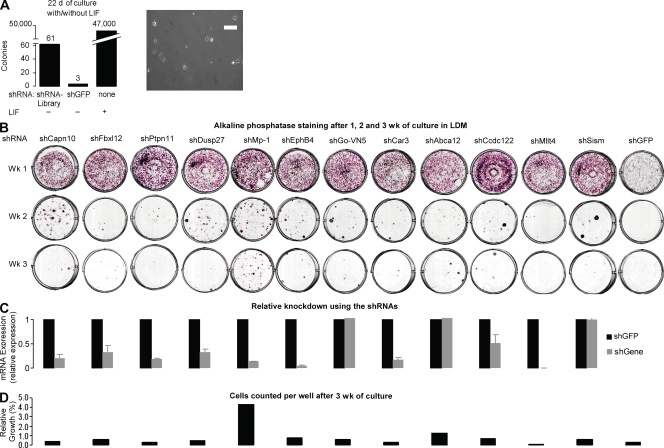
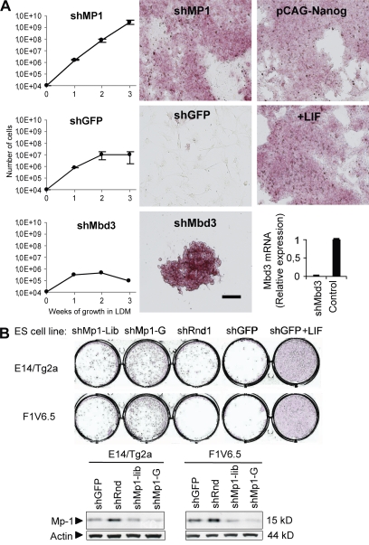
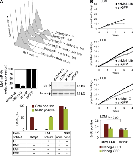
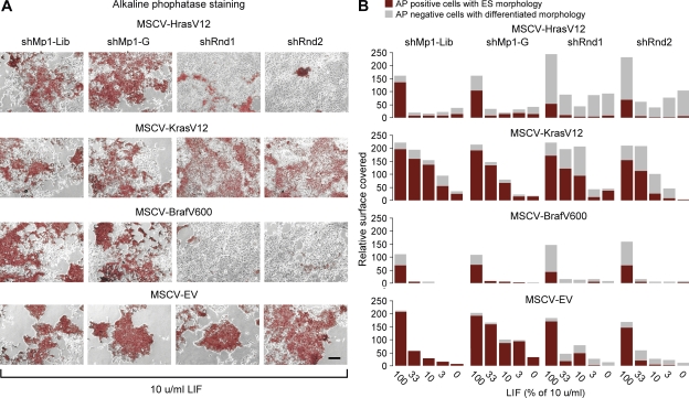
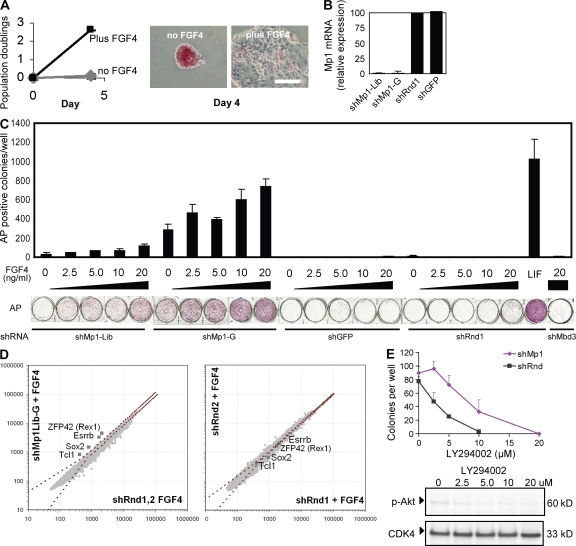
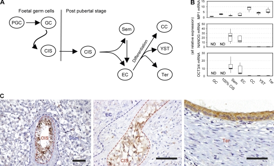
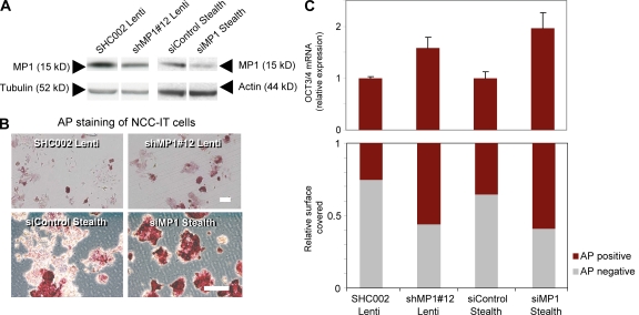
Despite intense investigation of intrinsic and extrinsic factors that regulate pluripotency, the process of initial fate commitment of embryonic stem (ES) cells is still poorly understood. We used a genome-wide short hairpin RNA screen in mouse ES cells to identify genes that are essential for initiation of differentiation. Knockdown of the scaffolding protein Mek binding protein 1 (Mp1, also known as Lamtor3 or Map2k1ip1) stimulated self-renewal of ES cells, blocked differentiation, and promoted proliferation. Fibroblast growth factor 4 (FGF4) signaling is required for initial fate commitment of ES cells. Knockdown of Mp1 inhibited FGF4-induced differentiation but did not alter FGF4-driven proliferation. This uncoupling of differentiation and proliferation was also observed when oncogenic Ras isoforms were overexpressed in ES cells. Knockdown of Mp1 redirected FGF4 signaling from differentiation toward pluripotency and up-regulated the pluripotency-related genes Esrrb, Rex1, Tcl1, and Sox2. We also found that human germ cell tumors (GCTs) express low amounts of Mp1 in the invasive embryonic carcinoma and seminoma histologies and higher amounts of Mp1 in the noninvasive carcinoma in situ precursor and differentiated components. Knockdown of Mp1 in invasive GCT cells resulted in resistance to differentiation, thereby showing a functional role for Mp1 both in normal differentiation of ES cells and in germ cell cancer.


Figures

Figure 1.
Figure 1.
shRNA screen for genes influencing mouse ES cell differentiation. Mouse ES cells were retrovirally transduced with a shRNA library targeting 16,000 genes (or shGFP specific). Cells were grown in LDM for 1 wk, and this was repeated two times. Colonies remaining after 3 wk were pooled, and 300 recovered shRNAs were sequenced. (A) Bar graphs show the absolute number (above bars) of recovered ES colonies after 3 wk of culture. The seeding density of the ES cells 1 d after plating is shown on the right. (B–D) The identified shRNAs were reintroduced into mouse ES cells, which were grown for one, two, or three replatings. (B) At the indicated time points, cells were stained with AP. (C) Efficiency of target gene knockdown (relative to shGFP) was measured by qPCR. (D) Cells in each well were counted and compared with +LIF conditions (100%) after 3 wk of culture. Error bars, standard deviation. Data represent two independent experiments. Bar, 100 µm.
Figure 2.
Figure 2.
Knockdown of Mp1 in E14T ES cells resembles Nanog overexpression or growth in the presence of LIF. (A) Growth curves (left) of ES cells infected with shMp1, the negative control shGFP, or the positive control shMbd3 when grown in LDM for 3 wk. After 3 wk, colonies were stained for AP. Additional positive controls were transfection with pCAG-Nanog or addition of LIF. Knockdown of Mbd3, as measured with qPCR, is shown below the Mbd3 growth curve. (B) AP staining shows that knockdown of Mp1 in two other ES cell lines, E14/Tg2a and F1V6.5, inhibits differentiation when these cells were grown in LDM for 2 wk. Knockdown was performed using the shRNA identified from the library (shMp1-Lib) and an additional shRNA indicated by shMp1-G. Negative controls were shGFP and a shRNA containing a random sequence (shRnd1). Western blot analysis shows the knockdown levels of Mp1 in these cell lines (right). Data represent at least two independent experiments. Error bars, standard deviation. Bar, 100 µm.
Figure 3.
Figure 3.
Knockdown of Mp1 inhibits differentiation and stimulates proliferation in ES cells. (A) FACS plot of E14T-Nanog-GFP reporter ES cells containing a randomly targeted Nanog promoter fragment that drives GFP expression. Reporter activity was assayed after growing the cells in LDM for 4 d after knockdown of Mp1 or after using a control shRNA containing a random sequence (shRnd1). Knockdown was performed using the shRNA identified from the library (shMp1-Lib) and an additional shRNA indicated by shMp1-G. Positive control cells were grown in the presence of LIF for 4 d. Knockdown of Mp1 was confirmed by qPCR (bottom) and Western blotting (middle right). Knockdown of Mp1 in the absence of Bmp4 maintains OCT3/4 expression (positive fraction is shown in red) while suppressing Nestin expression (positive fraction is shown in green). Positive controls were: undifferentiated ES cells and subventricular zone neural stem cells. (B) Knockdown of Mp1 gives a proliferation advantage in the absence (top) and presence (middle) of LIF in the total population of seeded cells, but also in Nanog-GFP–positive ES cells that were cultured 4 d without LIF (bottom). Error bars, standard deviation. Data represent two independent experiments.
Figure 4.
Figure 4.
Ras/Raf-induced differentiation can be inhibited by Mp1 knockdown. (A) AP stainings show the effect of overexpression of constitutively active forms of Ras/Raf on ES cells combined with or without knockdown of Mp1. (B) The red bars in the histogram show the relative surface covered by AP-positive cells with an ES cell morphology and the gray bars show the surface covered by AP-negative cells with a differentiated morphology. When constitutively active Hras-V12 was overexpressed, this led to a strong induction of differentiation. Knockdown of Mp1 was, for the most part, able to inhibit this differentiation capacity of HrasV12. When constitutively active Kras-V12 was overexpressed, the inhibition of differentiation was less pronounced, and this weaker effect was inhibited by Mp1 knockdown. Overexpression of the constitutively active form of Braf, BrafV600, led to strong differentiation, which was comparable to HrasV12. This strong differentiation effect could be inhibited largely by Mp1 knockdown. When control cells were transduced with an empty vector, differentiation was observed only in the absence of LIF. Data represent more than two independent experiments. Bar, 100 µm.
Figure 5.
Figure 5.
Mp1 knockdown uncouples proliferation and differentiation of ES cells in the absence of LIF. (A) Growth curve and graphs showing that addition of FGF4 to FGF4-deficient ES cells leads to both proliferation and differentiation. (B) qPCR showing Mp1 knockdown in FGF4-deficient ES cells. (C) FGF4-deficient ES cells were infected with Mp1 shRNAs or control shRNAs and cells were subsequently treated with increasing doses of FGF4 to induce differentiation in the absence of LIF. Knockdown of the positive control gene Mbd3 gave only a very minor increase in AP-positive colonies. (D) Microarray analysis shows that FGF4-deficient ES cells that have knockdown of Mp1 combined with FGF4 treatment have enhanced expression of the pluripotency genes Znf42 (Rex1), Esrrb, Sox2, and Tcl1 when compared with cells that were infected with control shRNAs and were treated simultaneously with FGF4. The dashed red stripes show standard deviations of duplicate experiments. (E) Titration of the PI3K inhibitor LY294002 showed enhanced resistance to this drug by cells with Mp1 knockdown. Increasing the LY294002 dose led to inhibition of phosphorylated Akt. CDK4 was used as a loading control. Error bars, standard deviation. Data represent two independent experiments. Bar, 100 µm.
Figure 6.
Figure 6.
GCTs of invasive EC and Sem show low expression of MP1. (A) Schematic outline of GCT progression. Primordial germ cells (PGCs) can progress into CIS which can give rise to Sem and/or EC, of which the latter can progress to Ter, CC, and YST. (B) Bar plot showing the relative RNA expression levels of MP1, OCT3/4, and NANOG in different stages of human GCTs as measured by qPCR. Horizontal lines represent the median value, boxes the upper and lower quartiles, and lines the maximal and minimal measured value. PGCs (n = 5) have lower expression of MP1 than CIS (n = 6; P < 0.05). CC (n = 3) and Ter (n = 6) have higher expression of MP1 than EC (n = 7) or Sem (n = 10; P < 0.05), similar to YS (n = 6), which has higher expression of MP1 than Sem (n = 12; P < 0.05). (C) Immunohistochemistry shows that preinvasive CIS are positive for MP1, whereas both Sem and EC in the same specimen have low to undetectable levels of MP1. MP1 is expressed in differentiated components of Ter tumors in ∼15% of the patients. Panel A was adapted from R. Lothe, with permission. Bars 100 µm.
Figure 7.
Figure 7.
Knockdown of MP1 in NCC-IT cells prevents differentiation. Knockdown of MP1 in NCC-IT cells (A) prevents differentiation as a result of SU-5402 treatment at 30 µM, as shown by AP staining (B), enhanced OCT3/4 mRNA expression, and enhanced numbers of cells AP-positive cells (C) with an undifferentiated morphology versus AP negative/weak cells with a differentiated morphology. Error bars, standard deviation. Bars, 100 µm. Data represent two independent experiments.

Similar articles

Cited by

References

    1. Andrews P.W. 2002. From teratocarcinomas to embryonic stem cells. Philos. Trans. R. Soc. Lond. B Biol. Sci. 357:405–417 10.1098/rstb.2002.1058 - DOI - PMC - PubMed
    1. Beddington R.S., Robertson E.J. 1989. An assessment of the developmental potential of embryonic stem cells in the midgestation mouse embryo. Development. 105:733–737 - PubMed
    1. Boeuf H., Hauss C., Graeve F.D., Baran N., Kedinger C. 1997. Leukemia inhibitory factor-dependent transcriptional activation in embryonic stem cells. J. Cell Biol. 138:1207–1217 10.1083/jcb.138.6.1207 - DOI - PMC - PubMed
    1. Boyer L.A., Lee T.I., Cole M.F., Johnstone S.E., Levine S.S., Zucker J.P., Guenther M.G., Kumar R.M., Murray H.L., Jenner R.G., et al. 2005. Core transcriptional regulatory circuitry in human embryonic stem cells. Cell. 122:947–956 10.1016/j.cell.2005.08.020 - DOI - PMC - PubMed
    1. Brons I.G., Smithers L.E., Trotter M.W., Rugg-Gunn P., Sun B., Chuva de Sousa Lopes S.M., Howlett S.K., Clarkson A., Ahrlund-Richter L., Pedersen R.A., Vallier L. 2007. Derivation of pluripotent epiblast stem cells from mammalian embryos. Nature. 448:191–195 10.1038/nature05950 - DOI - PubMed

Publication types

MeSH terms

Substances

Associated data