. 2012 Apr;31(4):621-9.
doi: 10.1007/s10067-011-1899-7. Epub 2011 Dec 7.

MRI identifies plantar plate pathology in the forefoot of patients with rheumatoid arthritis

Affiliations

MRI identifies plantar plate pathology in the forefoot of patients with rheumatoid arthritis

Heidi J Siddle et al. Clin Rheumatol. 2012 Apr.

Abstract

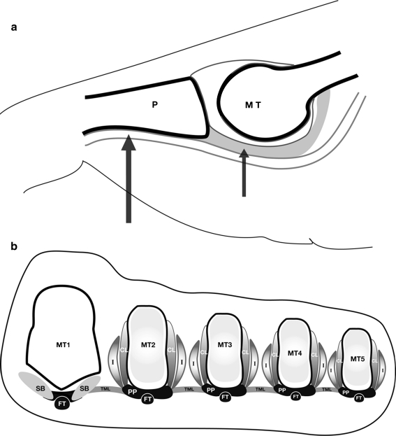
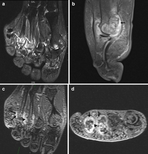
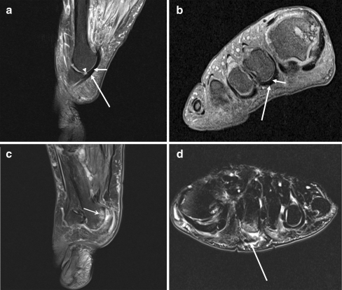
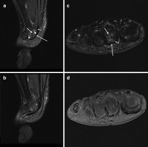
Previous cadaveric studies have suggested that forefoot deformities at the metatarsophalangeal (MTP) joints in patients with rheumatoid arthritis (RA) might result from the failure of the ligamentous system and displacement of the plantar plates. This study aimed to examine the relationship between plantar plate pathology and the rheumatoid arthritis magnetic resonance imaging score (RAMRIS) of the lesser (second to fifth) MTP joints in patients with RA using high-resolution 3 T magnetic resonance imaging (MRI). In 24 patients with RA, the forefoot was imaged using 3 T MRI. Proton density fat-suppressed, T2-weighted fat-suppressed and T1-weighted post gadolinium sequences were acquired through 96 lesser MTP joints. Images were scored for synovitis, bone marrow oedema and bone erosion using the RAMRIS system and the plantar plates were assessed for pathology. Seventeen females and 7 males with a mean age of 55.5 years (range 37-71) and disease duration of 10.6 years (range 0.6-36) took part in the study. Plantar plate pathology was most frequently demonstrated on MRI at the fifth MTP joint. An association was demonstrated between plantar plate pathology and RAMRIS-reported synovitis, bone marrow oedema and bone erosion at the fourth and fifth MTP joints. In patients with RA, 3 T MRI demonstrates that plantar plate pathology at the lesser MTP joints is associated with features of disease severity. Plantar plate pathology is more common at the fourth and fifth MTP joints in subjects with RA in contrast to the predilection for the second MTP reported previously in subjects without RA.


Figures

Fig. 1
Fig. 1
a Sagittal illustration through the lesser MTP joint, the plantar plate (short arrow) inserts into the proximal phalanx (P) and the flexor tendon (long arrow) lies adjacent to the plantar plate. b Short axis illustration through the forefoot, proximally the plantar plate (PP) attaches to the metatarsal shaft (MT) with the two collateral ligaments (CL) providing the most substantial attachment of the plantar plate to the metatarsal. The dorsal interosseous tendon (I) with the accessory CL insert into the dorsal aspect of the plantar plate. The flexor tendon sheath (FT) lies immediately plantar to the plantar plate and the deep transverse metatarsal ligament (TML) runs between the metatarsals attaching to the plantar plates
Fig. 2
Fig. 2
Images through the MTP joints of a patient with RA showing synovitis, bone marrow oedema and bone erosion. a Long axis T2-weighted fat-suppressed image showing extensive bone marrow oedema (white arrows). b Sagittal post contrast T1-weighted image showing synovitis (black arrow). The plantar plate is absent and synovitis extends from the joint to the flexor tendon. c Long axis and d short axis reconstructions from T1-weighted post contrast 3D VIBE images, again showing synovitis (black arrows) and erosion of the metatarsal heads (black arrowheads)
Fig. 3
Fig. 3
a Sagittal intermediate-weighted and b short axis T1-weighted fat suppressed images through the second MTP joint of a patient with RA showing an intact plantar plate (arrow), deep to the flexor tendon (long arrow). Small amount of high signal is seen at the insertion of the plantar plate into the proximal phalanx centrally (arrowhead), a common finding. (Cystic change is noted in first metatarsal head). c Sagittal intermediate-weighted and d short axis T2-weighted fat-suppressed images through the second MTP joint of a patient with RA. There is dislocation of the joint and the plantar plate is not visualised. There is bone marrow oedema (open arrow). On short axis section, fluid can be seen tracking from the joint around the flexor tendon sheath (arrow)
Fig. 4
Fig. 4
a and b (adjacent slices) Sagittal intermediate-weighted, c short axis T2-weighted fat-suppressed and d short axis T1 fat suppressed images through the second MTP joint demonstrating a tear (between arrowheads) of the plantar plate (arrow). The flexor tendon (long arrow) is adjacent to the bone distally without interposed plantar plate. There is extensive bone marrow oedema (open arrow)

Similar articles

Cited by

References

    1. Vidigal E, Jacoby RK, Dixon AS, Ratliff AH, Kirkup J. The foot in chronic rheumatoid arthritis. Ann Rheum Dis. 1975;34(4):292–297. doi: 10.1136/ard.34.4.292. - DOI - PMC - PubMed
    1. Grondal L, Tengstrand B, Nordmark B, Wretenberg P, Stark A. The foot: still the most important reason for walking incapacity in rheumatoid arthritis: distribution of symptomatic joints in 1,000 RA patients. Acta Orthop. 2008;79(2):257–261. doi: 10.1080/17453670710015067. - DOI - PubMed
    1. Johnston RB, 3rd, Smith J, Daniels T, Johnston RB, 3rd, Smith J, Daniels T. The plantar plate of the lesser toes: an anatomical study in human cadavers. Foot Ankle Int. 1994;15(5):276–282. - PubMed
    1. Stainsby GD. Pathological anatomy and dynamic effect of the displaced plantar plate and the importance of the integrity of the plantar plate-deep transverse metatarsal ligament tie-bar. Ann R Coll Surg Engl. 1997;79(1):58–68. - PMC - PubMed
    1. Yao L, Do HM, Cracchiolo A, Farahani K, Yao L, Do HM, Cracchiolo A, Farahani K. Plantar plate of the foot: findings on conventional arthrography and MR imaging. AJR Am J Roentgenol. 1994;163(3):641–644. - PubMed

Publication types