. 2012 Mar;64(3):808-20.
doi: 10.1002/art.34317.

Requirement of myeloid cell-specific Fas expression for prevention of systemic autoimmunity in mice

Affiliations

Requirement of myeloid cell-specific Fas expression for prevention of systemic autoimmunity in mice

Carla M Cuda et al. Arthritis Rheum. 2012 Mar.

Abstract

Objective: The death receptor Fas is a critical mediator of the extrinsic apoptotic pathway, and its role in mediating lymphoproliferation has been extensively examined. The present study was undertaken to investigate the impact of myeloid cell-specific loss of Fas.

Methods: Mice with Fas flanked by loxP sites (Fas(flox/flox) ) were crossed with mice expressing Cre under control of the murine lysozyme M gene promoter (Cre(LysM) ), which functions in mature lysozyme-expressing cells of the myelomonocytic lineage. The genotype for Cre(LysM) Fas(flox/flox) mice was verified by polymerase chain reaction and flow cytometric analysis. Flow cytometric analysis was also used to characterize myeloid, dendritic, and lymphoid cell distribution and activation in bone marrow, blood, and spleen. Luminex-based assays and enzyme-linked immunosorbent assays were used to measure serum cytokine/chemokine and immunoglobulin levels. Renal damage or dysfunction was examined by immunohistochemical and immunofluorescence analysis.

Results: Cre(LysM) Fas(flox/flox) mice exhibited a systemic lupus erythematosus (SLE)-like disease that included leukocytosis, splenomegaly, hypergammaglobulinemia, antinuclear autoantibody and proinflammatory cytokine production, and glomerulonephritis. Loss of Fas in myeloid cells increased levels of both Gr-1(low) and Gr-1(intermediate) blood monocytes and splenic macrophages and, in a paracrine manner, incited activation of conventional dendritic cells and lymphocytes in Cre(LysM) Fas(flox/flox) mice.

Conclusion: Taken together, these results suggest that loss of Fas in myeloid cells is sufficient to induce inflammatory phenotypes in mice, reminiscent of an SLE-like disease. Thus, Fas in myeloid cells may be considered a suppressor of systemic autoimmunity.


Figures

Figure 1
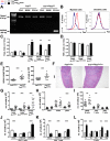
Figure 1. Myeloid cell-specific Fas-deficiency disrupts bone marrow myeloid lineage homeostasis
A. Fas message. RT-PCR on bone marrow-derived macrophages from young Fasflox/flox (WT, n=2) and CreLysMFasflox/flox (n=4) mice derived from bone marrow single-cell suspensions plated in the presence of growth factors. Bands: Fasfloxed-allele and Fasdeleted-allele. B. Fas protein. Bone marrow cells from young Fasflox/flox (n=2) and CreLysMFasflox/flox (n=4) mice stained for flow cytometric analysis. Histograms of %CD11b+ cells expressing Fas. C. Myeloid cell distribution. Bone marrow cells from aged Fasflox/flox (n=17 or 8) and CreLysMFasflox/flox (n=15 or 7) mice stained for flow cytometric analysis. CD11b+F4/80-Gr-1high granulocyte, CD11b+F4/80+Gr-1intermediate macrophage, and CD11b+F4/80+Gr-1low macrophage numbers. Unpaired, two-tailed Student's t-test: **-p<0.01.
Figure 2
Figure 2. Myeloid cell-specific Fas-deficiency disrupts circulating monocyte and lymphocyte homeostasis
A. Fas message. RT-PCR on CD11b+-sorted blood leukocytes from young Fasflox/flox (WT, n=2) and CreLysMFasflox/flox (n=4) mice. Bands: Fasfloxed-allele and Fasdeleted-allele. B. Fas protein. Blood leukocytes from young Fasflox/flox (n=2) and CreLysMFasflox/flox (n=4) mice stained for flow cytometric analysis. Histograms of %CD11b+ cells expressing Fas. C. Total leukocytes from aged mice. D-G. Blood leukocytes from aged Fasflox/flox (n=8-17) and CreLysMFasflox/flox (n=7-15) mice stained for flow cytometric analysis. Counts based on 60 uL blood volume. D. Myeloid cell distribution. CD11b+Gr-1high granulocyte, CD11b+Gr-1intermediateCD62L+ classical monocyte, and CD11b+Gr-1lowCD62L- nonclassical monocyte numbers. E. Lymphocyte distribution. B220+, CD4+, and CD8+ cell numbers. F-G. T-cell activation. Naïve-CD44-CD62L+, memory-CD44+CD62L+, and effector-CD44+CD62L- CD4+ (F) and CD8+ (G) T-cell numbers. Unpaired, two-tailed Student's t-test: *-p<0.05, **-p<0.01, ***-p<0.001.
Figure 3
Figure 3. Myeloid cell-specific Fas-deficiency disrupts splenic macrophage, dendritic cell, and lymphocyte homeostasis
A. Fas message. RT-PCR on CD4+-, B220+-, and CD11b+-sorted splenocytes from young Fasflox/flox (WT, n=2) and CreLysMFasflox/flox (n=4) mice. Bands: Fasfloxed-allele and Fasdeleted-allele. B. Fas protein. Splenocytes from young Fasflox/flox (n=2) and CreLysMFasflox/flox (n=4) mice stained for flow cytometric analysis. Histograms of %CD11b+F4/80+ and %CD11c+ cells expressing Fas. C-D. Apoptosis measurement. Splenocytes from young Fasflox/flox and CreLysMFasflox/flox (n=3) mice incubated with FasL (C) or TNFα (D) and stained for flow cytometric analysis. C-D %live cells converted to fold change over unstimulated condition. E. Spleen weight and splenocyte numbers from aged mice. F. Spleen pathology. H&E-stained sections from aged Fasflox/flox and CreLysMFasflox/flox mice. G-L. Splenocytes from aged Fasflox/flox (n=8-17) and CreLysMFasflox/flox (n=7-15) mice stained for flow cytometric analysis. G. Myeloid cell distribution. CD11b+F4/80-Gr-1high granulocyte, CD11b+F4/80+Gr-1intermediate and CD11b+F4/80+Gr-1low macrophage numbers. H. Dendritic cell distribution. Lymphoid (lDCs-CD11c+B220-CD8+), myeloid (mDCs-CD11c+B220-CD8-), and plasmacytoid (pDCs-CD11cintermediateB220+PDCA-1+) dendritic cell numbers. I. Lymphocyte distribution. B220+, CD4+, and CD8+ cell numbers. J-K. T-cell activation. Naïve-CD44-CD62L+, memory-CD44+CD62L+, and effector-CD44+CD62L- CD4+ (J) and CD8+ (K) T-cell numbers. L. B-cell distribution. Transitional (T1-B220+AA4.1+CD23-, T2-B220+AA4.1+CD23+), follicular (FO-CD19+CD21/35+CD23+), marginal zone (MZ-CD19+CD21/35+CD23low), and plasmablast (PB-CD19+B220lowCD138+CD21/35-CD23-) B-cell numbers. Unpaired, two-tailed Student's t-test: **-p<0.01, ***-p<0.001.
Figure 4
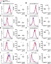
Figure 4. Altered splenic macrophage, dendritic cell, and CD4+ T-cell activation due to myeloid cell-specific Fas-deficiency
Splenocytes from aged Fasflox/flox (WT, n=8, red-line) and CreLysMFasflox/flox (n=7, blue-line) mice stained for flow cytometric analysis. A-F. Splenic macrophage activation. Represented by geometrical mean fluorescence intensity (MFI) of FcR (A), CD40 (C), and CD86 (E) expression on CD11b+F4-80+Gr-1low macrophages. MFI of FcR (B), CD40 (D), and CD86 (F) expression on CD11b+F4-80+Gr-1intermediate macrophages. G-I. Splenic dendritic cell activation. MFI of CD40 (G) and CD86 (H) expression on myeloid dendritic cells (mDCs-CD11c+B220-CD8-). MFI of MHC II expression (I) on plasmacytoid dendritic cells (pDCs-CD11cintermediateB220+PDCA-1+). J. Splenic T-cell activation. MFI of CD69 expression on CD4+ T-cells. Unpaired, two-tailed Student's t-test: *-p<0.05, **-p<0.01, ***-p<0.001.
Figure 5
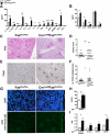
Figure 5. Myeloid cell-specific Fas-deficiency induces and SLE-like disease. A-B. Serum cytokine/chemokine and immunoglobulin levels
Serum from aged Fasflox/flox (WT, n=8) and CreLysMFasflox/flox (n=9) mice harvested for evaluation of cytokines/chemokines (A) and immunoglobulin subtypes (B). C-F. Kidney pathology. PAS- (C-D) and CD45- (E-F) stained sections from aged Fasflox/flox (n=8) and CreLysMFasflox/flox (n=9) mice. Pictures taken at 100x. Sections scored by a pathologist blinded to the study. G. Kidney IgG deposition. Sections from aged Fasflox/flox (n=8) and CreLysMFasflox/flox (n=9) mice stained with DAPI and FITC-conjugated anti-mouse IgG. Pictures taken at 200x. H-I. Serum autoantibody titers. Serum from aged Fasflox/flox (n=8) and CreLysMFasflox/flox (n=9) mice harvested for evaluation of both IgM (H) and IgG (I) autoantibody titers by ELISA. Unpaired, two-tailed Student's t-test: *-p<0.05, **-p<0.01, ***-p<0.001.
Figure 6
Figure 6. Myeloid cell-specific Fas-deficiency alters activation of MAPKs in peritoneal macrophages and increases susceptibility to in vivo TLR4 ligation
A, C-D. Young Fasflox/flox and CreLysMFasflox/flox (n=6) mice intraperitoneally injected with thioglycollate, peritoneal cells harvested after 3 days, and subjected to flow cytometric analysis. A. Peritoneal cell distribution. Total peritoneal cell, CD11b+F4/80high macrophage, and CD11b+F4/80-Gr-1high granulocyte numbers. B. LPS-induced endotoxic shock. Young Fasflox/flox (WT, n=10) and CreLysMFasflox/flox (n=9) mice intraperitoneally injected with LPS. Survival monitored for 7 days. Mantel-cox one-tailed Chi-square test. C-D. Constitutive activation status of peritoneal cells. Surface FcR, TLR4, CD86, TLR2, MHC II (C) (n=6) and intracellular phosphorylated p38, p65, JNK, Akt, and ERK (D) (n=3). C displayed as fold-change of values from CreLysMFasflox/flox mice divided by values from Fasflox/flox mice. Representative of two individual experiments. Unpaired, two-tailed Student's t-test: *-p<0.05.

Comment in

References

    1. Strasser A, Jost PJ, Nagata S. The many roles of FAS receptor signaling in the immune system. Immunity. 2009;30(2):180–92. - PMC - PubMed
    1. Cohen PL, Eisenberg RA. Lpr and gld: single gene models of systemic autoimmunity and lymphoproliferative disease. Annu Rev Immunol. 1991;9:243–69. - PubMed
    1. Stranges PB, Watson J, Cooper CJ, Choisy-Rossi CM, Stonebraker AC, Beighton RA, et al. Elimination of antigen-presenting cells and autoreactive T cells by Fas contributes to prevention of autoimmunity. Immunity. 2007;26(5):629–41. - PMC - PubMed
    1. Hao Z, Hampel B, Yagita H, Rajewsky K. T Cell-specific Ablation of Fas Leads to Fas Ligand-mediated Lymphocyte Depletion and Inflammatory Pulmonary Fibrosis. J Exp Med. 2004;199(10):1355–65. - PMC - PubMed
    1. Hao Z, Duncan GS, Seagal J, Su YW, Hong C, Haight J, et al. Fas receptor expression in germinal-center B cells is essential for T and B lymphocyte homeostasis. Immunity. 2008;29(4):615–27. - PMC - PubMed

Publication types

MeSH terms