. 2011 Dec;54(6):1901-12.
doi: 10.1002/hep.24557.

Expression of paramyxovirus V proteins promotes replication and spread of hepatitis C virus in cultures of primary human fetal liver cells

Affiliations
Free PMC article

Expression of paramyxovirus V proteins promotes replication and spread of hepatitis C virus in cultures of primary human fetal liver cells

Linda Andrus et al. Hepatology. 2011 Dec.
Free PMC article

Abstract

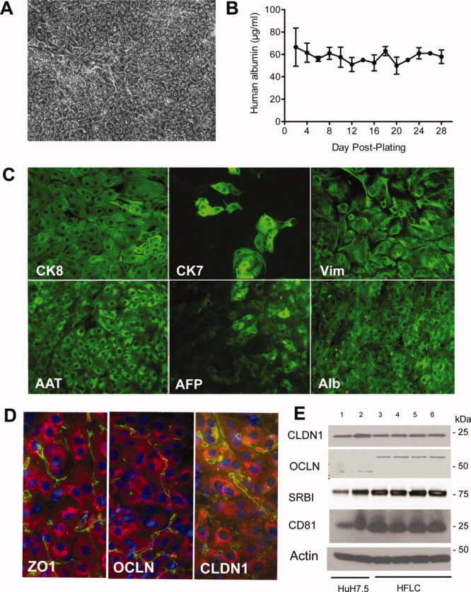
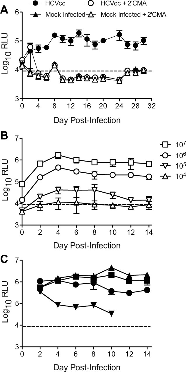
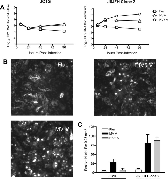
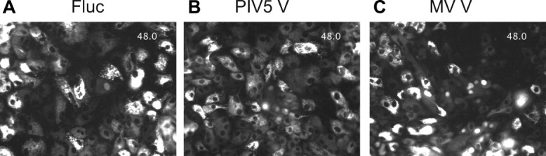
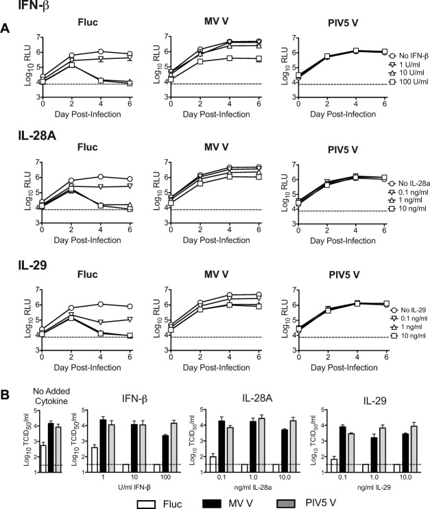
Here we demonstrate that primary cultures of human fetal liver cells (HFLC) reliably support infection with laboratory strains of hepatitis C virus (HCV), although levels of virus replication vary significantly between different donor cell preparations and frequently decline in a manner suggestive of active viral clearance. To investigate possible contributions of the interferon (IFN) system to control HCV infection in HFLC, we exploited the well-characterized ability of paramyxovirus (PMV) V proteins to counteract both IFN induction and antiviral signaling. The V proteins of measles virus (MV) and parainfluenza virus 5 (PIV5) were introduced into HFLC using lentiviral vectors encoding a fluorescent reporter for visualization of HCV-infected cells. V protein-transduced HFLC supported enhanced (10 to 100-fold) levels of HCV infection relative to untransduced or control vector-transduced HFLC. Infection was assessed by measurement of virus-driven luciferase, by assays for infectious HCV and viral RNA, and by direct visualization of HCV-infected hepatocytes. Live cell imaging between 48 and 119 hours postinfection demonstrated little or no spread of infection in the absence of PMV V protein expression. In contrast, V protein-transduced HFLC showed numerous HCV infection events. V protein expression efficiently antagonized the HCV-inhibitory effects of added IFNs in HFLC. In addition, induction of the type III IFN, IL29, following acute HCV infection was inhibited in V protein-transduced cultures.

Conclusion: These studies suggest that the cellular IFN response plays a significant role in limiting the spread of HCV infection in primary hepatocyte cultures. Strategies aimed at dampening this response may be key to further development of robust HCV culture systems, enabling studies of virus pathogenicity and the mechanisms by which HCV spreads in its natural host cell population.


Figures

Fig. 1
Fig. 1
Cultured HFLC are long-lived, express markers of differentiated hepatocytes, and are positive for the expression of HCV entry factors. (A) Phase contrast microscopy of HFLC 2 weeks postplating. (B) Albumin secretion by HFLC during 1 month of culture (mean and SD of three cultures). (C) Antibody staining for cytokeratin 8 (CK8), cytokeratin 7 (CK7), vimentin (Vim), alpha-1 antitrypsin (AAT), alpha fetoprotein (AFP), and albumin (Alb) in cultured HFLC 1 week postplating. Bound antibody was detected with AlexaFluor (AF)-488-conjugated antibody to immunoglobulins. (D) AF-488 (green) detection of bound antibodies to the tight junction protein zona occludens 1 (ZO1) and the HCV entry factors occludin (OCLN) and claudin 1 (CLDN1) in cultured HFLC 1 week postplating. Hepatocyte staining for AAT (red) was detected with AF-594-conjugated antibody to goat IgG. Nuclei are counterstained with DAPI (blue). (E) Western blot for HCV entry factors in lysates of Huh-7.5 cells and HFLC 1 week postplating: Lane 1, 15 μg Huh-7.5 lysate; Lane 2, 30 μg Huh-7.5; Lane 3, 30 μg HFLC. We also examined entry factor expression in HFLC transduced with each of the three lentiviral vectors described below. Lanes 4-6 correspond to 30 μg lysate from HFLC transduced with pseudoparticles encoding: Lane 4, firefly luciferase; Lane 5, parainfluenza virus 5 V protein; Lane 6, measles virus V protein. Lysates were prepared 6 days posttransduction. SRBI, scavenger receptor BI. Note that HFLC express the ≍60 kDa form of OCLN that is found in adult human liver, and absent or weakly expressed in Huh-7.5 cells.
Fig. 2
Fig. 2
Cultured HFLC are susceptible to infection with HCVcc. (A) Gaussia luciferase secretion by HFLC (ABR-15168, 23 weeks gestation) following infection with the reporter virus JC1G in the presence or absence of the polymerase inhibitor 2′CMA (2.2 μm). Mock-infected cells were incubated with culture medium alone. Polymerase inhibitor treatment was discontinued after day 6 of culture. Secreted luciferase was measured at each medium change (mean and SD of three cultures). RLU, relative light units. The dashed line indicates mean plus 2 SD of RLU detected in culture medium alone. (B) Doses of HCVcc required for establishment of detectable infection. HFLC (AECOM-052810, gestational age not available) were incubated with the indicated tissue culture infectious doses (TCID50) of JC1G for 5 hours, washed, and refed with HDM. Secreted luciferase was measured at each medium change (mean and SD of three cultures). (C) JC1G infection of four different HFLC preparations with ≍105 TCID50 of a single virus stock that was prepared in medium containing 10% v/v FBS.
Fig. 3
Fig. 3
PMV V protein expression enhances productive infection with JC1G in cultured HFLC. (A) One day postplating, HFLC (ABR-5591, 17 weeks gestation) were transduced with lentiviral vectors encoding firefly luciferase (Fluc), parainfluenza virus 5 V protein (PIV5 V), or measles virus V protein (MV V), or left nontransduced (NTD). Five days later cells were infected with 1 × 10⁶ tissue culture infectious doses (TCID50) JC1G reporter virus. Washed cells were cultured in HDM and the medium was removed and replaced every 2 days. Harvested supernatants were assayed for luciferase (left panel) and for levels of infectious virus by titration on Huh-7.5 cells (right panel). RLU, relative light units. Values show mean and SD of three cultures. The dashed line indicates the lower limit of detection for each assay. (B) Levels of infectious virus recovered from cell supernatants 2 weeks post-JC1G infection of four different HFLC preparations. Graphs show mean and SD of 2-4 cultures. Log-transformed TCID50 values within each experiment were analyzed by 1-way analysis of variance (ANOVA) with Bonferroni multiple comparison posttest. Values that differ significantly from both nontransduced (NTD) and Fluc-transduced cultures are indicated: *P < 0.05; **P < 0.01; ***P < 0.001. Fold differences relative to nontransduced cultures are summarized below the graphs.
Fig. 4
Fig. 4
J6JFH Clone 2 replicates to higher levels in HFLC than JC1G, and replication is enhanced by V protein expression. Four days posttransduction with lentiviral vectors, HFLC (AECOM-063010; gestational age not available) were incubated with 1 × 10⁷ TCID50 JC1G or J6JFH Clone 2 for 6 hours, washed three times, and cultured in HDM. (A) Levels of cell-associated HCV RNA detected during the first 4 days postinfection (mean and SD of 2 assays on 2 cultures). (B) Representative images for HFLC infected with J6JFH Clone 2. (C) Numbers of RFP-positive nuclei per microscope field on day 7 postinfection (mean and SD of six fields from two cultures).
Fig. 5
Fig. 5
Kinetics of J6JFH Clone 2 replication in cultured HFLC. (A) Five days posttransduction, HFLC (AECOM-031010, gestational age unavailable) were incubated with 1 × 10⁷ TCID50 J6JFH Clone 2, washed three times, and refed with HDM with or without of 2.2 μm 2′CMA. Cells were washed again and refed 24 hours postinfection. Treatment with 2′CMA was discontinued on day 7. Infectious virus in culture supernatants was determined by titration on Huh-7.5 cells. Values are mean and SD of four cultures. (B) Kinetics of appearance of RFP-positive nuclei during the first 3 days of culture following infection with J6JFH Clone 2 (ABR-8338; 20 weeks gestation). Values are mean and SD of nuclei counts from four fields of two cultures.
Fig. 6
Fig. 6
Live-cell imaging of J6JFH Clone 2 infection in PMV V protein- or Fluc-transduced HFLC. Five days posttransduction with lentiviral vectors, HFLC (ABR-3676; 19 weeks gestation) were infected with 1 × 10⁷ TCID50 J6JFH Clone 2. Washed cells were cultured in HDM for 48 hours prior to initiation of imaging. For each culture condition, 10 fields containing at least one cell with nuclear translocation of RFP were selected to monitor possible spread of infection. The figure shows the first frame of representative fields of cells transduced with (A). Firefly luciferase (Fluc). (B) Parainfluenza virus 5 V protein (PIV5 V) or (C) measles virus V protein (MV V). Full videos for each field are shown in Fig. 6A_Fluc.avi, Fig. 6B_PIV5 V.avi, and Fig. 6C_MV V.avi and are viewable in the online version of the manuscript. The time stamp indicates elapsed time in hours beginning at 48 hours postinfection.
Fig. 7
Fig. 7
V protein expression counteracts the HCV-inhibitory effects of added type I and type III interferons in HFLC. Four days posttransduction with lentiviral vectors, HFLC (ABR-1108; 19 weeks gestation) were incubated with 1 × 10⁶ TCID50 per well JC1G reporter virus for 5 hours. Cells were washed three times then refed with HDM containing serial 10-fold dilutions of IFN-β, IL-28A, or IL-29. The culture medium was aspirated and replaced every 2 days. Cytokine treatment was discontinued after the second day of culture. (A) JC1G reporter virus-derived luciferase activity detected in culture supernatants during the first 6 days of culture (mean and SD of two cultures). RLU, relative light units. (B) Levels of infectious HCV detected in cell culture supernatants on day 14 of culture (mean and SD of two assays on two cultures). TCID50, tissue culture infectious doses; Fluc, firefly luciferase; PIV5 V, parainfluenza virus 5 V protein; MV V, measles virus V protein.
Fig. 8
Fig. 8
V protein expression inhibits IL-29 protein induction following HCV infection. Four days posttransduction with lentiviral vectors, cultures of gradient-enriched HFLC (ABR-8338; 20 weeks gestation) were incubated for 5 hours with (A) 1 × 10⁶ TCID50 per well J6JFH Clone 2, or (B) with an equivalent dilution of supernatant from mock-electroporated Huh-7.5 cells. Cells were washed three times then refed with HDM. The culture medium was removed and replaced daily and supernatants were assayed for IL-29 by ELISA (mean and SD of two cultures). Comparable results were obtained in two other experiments. NTD, no transduction; Fluc, firefly luciferase; PIV5 V, parainfluenza virus 5 V protein; MV V, measles virus V protein.

References

    1. Altar HJ, Seeff LB. Recovery, persistence and sequelae in hepatitis C virus infection: a perspective on the long-term outcome. Semin Liv Dis. 2000;20:17–25. - PubMed
    1. Li XD, Sun L, Seth RB, Pineda G, Chen ZJ. Hepatitis C virus protease NS3/4A cleaves mitochondrial antiviral signaling protein off the mitochondria to evade innate immunity. Proc Natl Acad Sci U S A. 2005;102:17717–17722. - PMC - PubMed
    1. Otsuka M, Kato N, Moriyama M, Taniguchi H, Wang Y, Dharel N, et al. Interaction between the HCV NS3 protein and the host TBK1 protein leads to inhibition of cellular antiviral responses. HEPATOLOGY. 2005;41:1004–1012. - PubMed
    1. Lin W, Kim SS, Yeung E, Kamegaya Y, Blackard JT, Kim KA, et al. Hepatitis C virus core protein blocks interferon signaling by interaction with STAT1 SH2 domain. J Virol. 2006;80:9226–9235. - PMC - PubMed
    1. Gale M, Foy EM. Evasion of intracellular host defense by hepatitis C virus. Nature. 2005;436:939–945. - PubMed

Publication types