. 2012 Apr 12;119(15):3539-49.
doi: 10.1182/blood-2011-03-345215. Epub 2011 Dec 5.

Critical requirement for Stat5 in a mouse model of polycythemia vera

Affiliations

Critical requirement for Stat5 in a mouse model of polycythemia vera

Dongqing Yan et al. Blood. .

Abstract

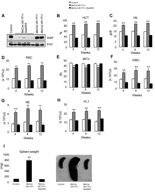
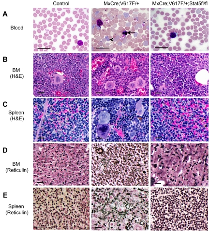
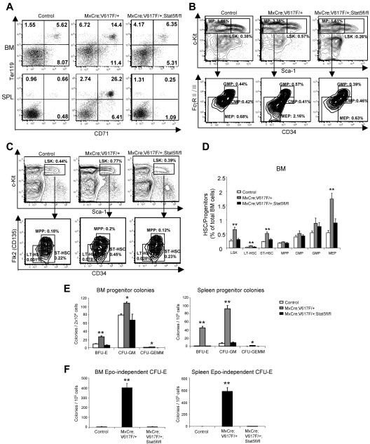
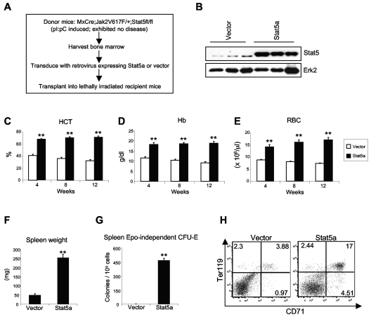
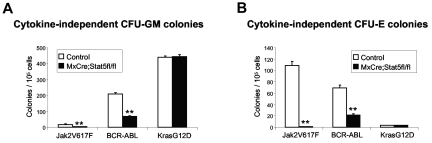
The JAK2V617F mutation has been identified in most cases of Ph-negative myeloproliferative neoplasms (MPNs) including polycythemia vera (PV), essential thrombocythemia (ET), and primary myelofibrosis (PMF). Expression of JAK2V617F results in constitutive activation of multiple signaling molecules/pathways. However, the key signaling downstream of JAK2V617F required for transformation and induction of MPNs remains elusive. Using a mouse genetic strategy, we show here that Stat5 is absolutely required for the pathogenesis of PV induced by Jak2V617F. Whereas expression of Jak2V617F in mice resulted in all the features of human PV, including an increase in red blood cells, hemoglobin, hematocrit, white blood cells, platelets, and splenomegaly, deletion of Stat5 in the Jak2V617F knockin mice normalized all the blood parameters and the spleen size. Furthermore, deletion of Stat5 completely abrogated erythropoietin (Epo)-independent erythroid colony formation evoked by Jak2V617F, a hallmark feature of PV. Re-expression of Stat5 in Stat5-deficient Jak2V617F knockin mice completely rescued the defects in transformation of hematopoietic progenitors and the PV phenotype. Together, these results indicate a critical function for Stat5 in the pathogenesis of PV. These findings also provide strong support for the development of Stat5 inhibitors as targeted therapies for the treatment of PV and other JAK2V617F-positive MPNs.


Figures

Figure 1
Figure 1
Deletion of Stat5 inhibits the development of MPN in Jak2V617F knockin mice. (A) Immunoblot analysis for total Stat5 protein show efficient deletion of Stat5 in the BM of MxCre;Jak2V617F/+;Stat5fl/fl mice after induction with pI:pC. Peripheral blood HCT (B), Hb (C), RBC (D), MCV (E), WBC (F), NE (G), and PLT (H) counts were assessed at 4, 8, and 12 weeks after pI:pC induction in control, MxCre;Jak2V617F/+, and MxCre;Jak2V617F/+;Stat5fl/fl mice (n = 15 for control; n = 17 for MxCre;Jak2V617F/+; n = 28 for MxCre;Jak2V617F/+;Stat5fl/fl mice at all time points). All the peripheral blood parameters in MxCre;Jak2V617F/+;Stat5fl/fl mice were within normal range and comparable with those in controls. Spleen weight/size (I) was significantly reduced in MxCre;Jak2V617F/+;Stat5fl/fl mice compared with MxCre; Jak2V617F/+ mice, and was identical to those of controls (12 weeks after induction with pI:pC; n = 15). Asterisks indicate significant differences by 1-way ANOVA (*P < .05; **P < .005). Data are shown as mean ± SEM
Figure 2
Figure 2
Histopathologic analysis. (A) Blood smears show decreased RBCs, leukocytes, and PLTs in induced MxCre;Jak2V617F/+;Stat5fl/fl mice compared with MxCre; Jak2V617F/+ mice. Arrows point to reticulocyte and nucleated immature RBC. (B) BM sections from induced MxCre;Jak2V617F/+ mice display hypercellularity with trilineage hyperplasia. In contrast, BM sections from induced MxCre;Jak2V617F/+;Stat5fl/fl mice are indistinguishable from the control BM. (C) Spleen sections from induced MxCre;Jak2V617F/+ mice exhibited a marked expansion of red pulp with clusters of megakaryocytes and immature erythroid progenitors. Noticeably, spleens from induced MxCre;Jak2V617F/+;Stat5fl/fl mice were normal and identical to control spleens. Reticulin staining on the BM (D) and spleen (E) sections showed fibrosis in the BM and red pulp of the spleens of older Jak2V617F knockin mice (24 weeks after induction), whereas Stat5-deficiency prevented the development of fibrosis in the BM and spleens of MxCre;Jak2V617F/+;Stat5fl/fl mice. Scale bars represent 20 μm in all images.
Figure 3
Figure 3
Effects of Stat5-deficiency on hematopoietic stem cells and progenitors expressing Jak2V617F. (A) Flow cytometric analysis revealed a marked reduction in CD71+Ter119+ erythroid precursors in the BM and spleens of induced MxCre;Jak2V617F/+;Stat5fl/fl mice compared with induced MxCre; Jak2V617F/+ mice; the CD71+Ter119+ population in the BM and spleens of MxCre;Jak2V617F/+;Stat5fl/fl mice was comparable with that of control mice. Representative dot plots from 7 independent experiments are shown. (B) Flow cytometric analysis of the LSK compartment (LinSca1+c-kit+) and subsets of myeloid progenitors (MP) including CMP (LinSca1c-kit+CD34+FcγRII/IIIlo), GMP (LinSca1c-kit+CD34+FcγRII/IIIhigh), and MEP (LinSca1c-kit+CD34FcγRII/III) in the BM from control (n = 4), MxCre;Jak2V617F/+ mice (n = 4) and MxCre;Jak2V617F/+; Stat5fl/fl (n = 5) mice. (C) HSC-enriched LSK compartments from the BM of control, MxCre;Jak2V617F/+ and MxCre;Jak2V617F/+; Stat5fl/fl mice were further analyzed for LT-HSC (LSKCD34Flk2), ST-HSC (LSKCD34+Flk2), and MPP (LSKCD34+Flk2+). Representative contour plots from 3 independent experiments are shown. (D) Percentages of LSK, LT-HSC. ST-HSC, MPP, CMP, GMP, and MEP are shown in histogram as mean ± SEM Data are presented as percentage of total cells. Asterisks show significant differences by 1-way ANOVA (**P < .005). Note that a significant decrease in LSK, LT-HSC, ST-HSC, and MEP compartments in the BM from MxCre;Jak2V617F/+;Stat5fl/fl mice compared with MxCre;Jak2V617F/+ mice and those populations were comparable with that observed in controls. (E) Analysis of hematopoietic progenitor colonies. BM (2 × 10⁴) and spleen (1 × 10⁵) cells from control (n = 3), MxCre;Jak2V617F/+ mice (n = 5), and MxCre;Jak2V617F/+;Stat5fl/fl mice (n = 5) were seeded in complete methylcellulose medium (Methocult M3434). BFU-E, CFU-GM, and CFU-GEMM colonies were counted on day 7. (F) Epo-independent CFU-E colonies. BM (1 × 10⁵) or spleen (1 × 10⁵) cells were plated in methylcellulose medium without any cytokine (Methocult M3234). CFU-E colonies were counted after 2 days. For BM CFU-E colonies, n = 4 for control and n = 6 for both MxCre;Jak2V617F/+ and MxCre;Jak2V617F/+;Stat5fl/fl mice; for spleen CFU-E colonies, n = 6 for control and n = 8 for both MxCre;Jak2V617F/+ and MxCre;Jak2V617F/+; Stat5fl/fl mice. Data are presented as mean ± SEM. Asterisks show significant differences by 1-way ANOVA (*P < .05; **P < .005).
Figure 4
Figure 4
Retroviral expression of Stat5a complements the deficiency of Stat5 in inducing PV by Jak2V617F. (A) Experimental design for rescue of Jak2V617F-induced PV by retroviral transduction/transplantation of Stat5a in the BM of Stat5-deficient Jak2V617F-expressing (MxCre;Jak2V617F/+;Stat5fl/fl) mice. (B) Immunoblot analysis of Stat5 protein levels in leukocytes 4 weeks after transplantation. Erk2 was used as a loading control. Peripheral blood HCT (C), Hb (D), and RBCs (E) were significantly increased in recipients receiving Stat5a-transduced BM (n = 4). Blood counts were measured at 4, 8, and 12 weeks after transplantation. Data are represented as mean ± SEM. Spleen weight (F) and Epo-independent CFU-E colonies (G) were also markedly elevated in mice receiving Stat5a-transduced BM compared with those receiving vector-transduced BM. Data are presented as mean ± SEM. Asterisks show significant differences by t test (**P < .005). (H) Flow cytometric analysis demonstrates a marked increase in CD71+Ter119+ erythroid precursors in the spleens of recipient mice transplanted with Stat5a-transduced BM compared with those with vector-transduced BM. Shown are representative dot plots from 3 independent experiments.
Figure 5
Figure 5
Stat5 is not required for transformation mediated by KrasG12D. BM from pI:pC-treated control and MxCre; Stat5fl/fl mice were infected with retroviruses expressing Jak2V617F, BCR-ABL, or KrasG12D. Transduced BM cells (1 × 10⁵) were then plated in methylcellulose medium without any cytokine (Methocult M3234). Cytokine-independent CFU-GM colonies (A) were counted after 7 days and CFU-E colonies (B) were counted after 2 days (n = 4). Asterisks show significant differences by t test (*P < .05; **P < .005). Data are shown as mean ± SEM.
Figure 6
Figure 6
Erk, Akt and Stat3 are dispensable for hematopoietic transformation induced by Jak2V617F. (A) Left panel: inhibition of Erk1/2 phosphorylation in the BM of Jak2V617F knockin mice by MEK inhibitor U0126. Right panel: BM cells from Jak2V617F knockin mice were plated in the methylcellulose medium in the absence of cytokine with indicated concentrations of U0126. CFU-E colonies were counted after 2 days. (B) Left panel: inhibition of Akt phosphorylation in the BM of Jak2V617F knockin mice by Akt inhibitor wortmannin. Right panel: BM cells from Jak2V617F knockin mice were plated in the methylcellulose medium in the absence of cytokine with indicated concentrations of wortmannin. CFU-E colonies were counted after 2 days. (C) Effect of dominant negative Stat3 (DN-Stat3) on phosphorylation/activation of Stat3 in the BM of Jak2V617F knockin mice and erythroid transformation mediated by Jak2V617F. BM from induced MxCre;V617F/+ mice were transduced with retrovirus expressing either DN-Stat3 or vector. After selection with puromycin (2 μg/mL) for 2 days, BM cells (1 × 10⁵) were plated in methylcellulose medium without any cytokine (Methocult M3234). CFU-E colonies were counted after 2 days. Notably, expression of DN-Stat3 in the BM of Jak2V617F knockin mice almost completely inhibited phosphorylation of Stat3 (left panel); however, erythroid transformation induced by Jak2V617F was only partially inhibited (right panel). Asterisks show significant differences (*P < .05; **P < .005). Data are shown as mean ± SEM.
Figure 7
Figure 7
The effects of Stat5 deficiency on signaling mediated by Jak2V617F in primary hematopoietic cells. BM from control, MxCre;Jak2V617F/+, and MxCre;Jak2V617F/+;Stat5fl/fl mice were factor-deprived for 15 hours. Cell lysates were prepared by direct boiling in 2× sample buffer and subjected to immunoblotting with phospho-specific antibodies against Stat5 or p70S6K, and total antibodies against Stat5, p70S6K, Bcl-xL, Cyclin D2, and Pim1. Erk2 was used as loading control.

Comment in

References

    1. James C, Ugo V, Le Couedic JP, et al. A unique clonal JAK2 mutation leading to constitutive signaling causes polycythemia vera. Nature. 2005;434(7037):1144–1148. - PubMed
    1. Levine RL, Wadleigh M, Cools J, et al. Activating mutation in the tyrosine kinase JAK2 in polycythemia vera, essential thrombocythemia, and myeloid metaplasia with myelofibrosis. Cancer Cell. 2005;7(4):387–397. - PubMed
    1. Baxter EJ, Scott LM, Campbell PJ, et al. Acquired mutation of the tyrosine kinase JAK2 in human myeloproliferative disorders. Lancet. 2005;365(9464):1054–1061. - PubMed
    1. Kralovics R, Passamonti F, Buser AS, et al. A gain-of-function mutation of JAK2 in myeloproliferative disorders. N Engl J Med. 2005;352(17):1779–1790. - PubMed
    1. Zhao R, Xing S, Li Z, et al. Identification of an acquired JAK2 mutation in polycythemia vera. J Biol Chem. 2005;280(24):22788–22792. - PMC - PubMed

Publication types