. 2011 Dec;85(6):1025-34.
doi: 10.4269/ajtmh.2011.11-0102.

Characterization of novel Leishmania infantum recombinant proteins encoded by genes from five families with distinct capacities for serodiagnosis of canine and human visceral leishmaniasis

Affiliations

Characterization of novel Leishmania infantum recombinant proteins encoded by genes from five families with distinct capacities for serodiagnosis of canine and human visceral leishmaniasis

Geraldo G S Oliveira et al. Am J Trop Med Hyg. 2011 Dec.

Abstract

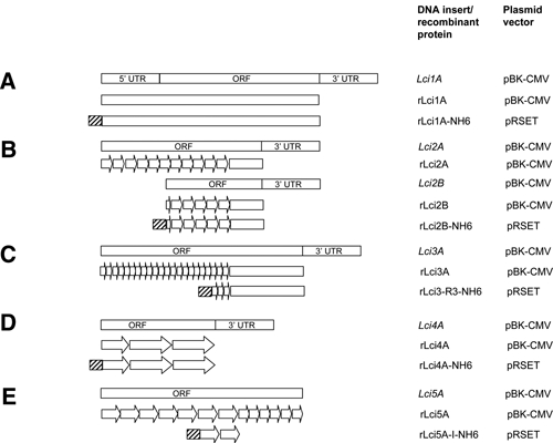
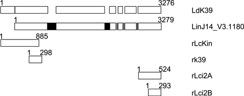
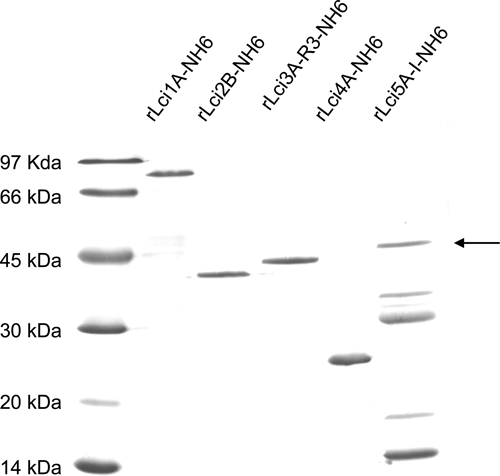
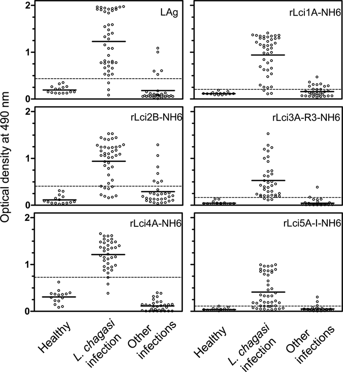
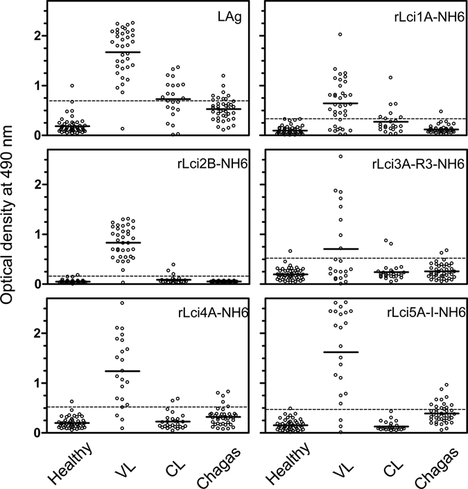
To expand the available panel of recombinant proteins that can be useful for identifying Leishmania-infected dogs and for diagnosing human visceral leishmaniasis (VL), we selected recombinant antigens from L. infantum, cDNA, and genomic libraries by using pools of serum samples from infected dogs and humans. The selected DNA fragments encoded homologs of a cytoplasmic heat-shock protein 70, a kinesin, a polyubiquitin, and two novel hypothetical proteins. Histidine-tagged recombinant proteins were produced after subcloning these DNA fragments and evaluated by using an enzyme-linked immunosorbent assays with panels of canine and human serum samples. The enzyme-linked immunosorbent assays with different recombinant proteins had different sensitivities (67.4-93.0% and 36.4-97.2%) and specificities (76.1-100% and 90.4-97.3%) when tested with serum samples from Leishmania-infected dogs and human patients with VL. Overall, no single recombinant antigen was sufficient to serodiagnosis all canine or human VL cases.


Figures

Figure 1.
Figure 1.
Schematic representation of various cDNAs and corresponding deduced recombinant proteins evaluated in this study. Maps were derived from sequences produced after direct sequencing of the Lci2A and Lci2B, Lci3A and Lci4A cDNAs and/or from the coding genomic sequences of the Lci1A and Lci5A, available at the GeneDb (www.genedb.org). A, Lci1A cDNA, including the 5′ untranslated region (UTR) segment, which is included in the recombinant protein rLci1A-NH6. B, Lci2A cDNA, highlighting the 11 39-amino acid repetitive segments and the segment common to Lci2B cDNA and which is present in recombinant protein rLci2B-NH6. C, Lci3A cDNA highlighting the 21 repetitive 14-amino acid motif segments and recombinant protein rLci3A-R3-NH6, which contain the last three repetitive segments. D, Lci4A cDNA sequence emphasizing the last two repeats and part of the third repeat and recombinant protein rLci4A-NH6. E, Lci5A-coding sequence and the recombinant protein rLci5A-I –NH6. Arrows and open boxes, which have not been drawn to represent protein segments in scale, represent repetitive amino acid motifs and non-repetitive polypeptide segments. Hatched represent His-tag provided by the vector. ORF = open reading frame.
Figure 2.
Figure 2.
Schematic representation of the full-length sequences or sequence fragments from selected kinesins from the Leishmania donovani complex. Sequences from LdK39 (kinesin of L. donovani18), the protein encoded by LinJ14_V3.1180 (kinesin of L. infantum, GeneDB), rLcKin and rk39 (L. chagasi kinesin fragments described by Evans and others7), and the rLci2A and rLci2B kinesin fragments reported here are represented. Open boxes represent overlapping regions, and closed boxes indicate sequences in protein encoded by LinJ14_V3.1180 that are absent in LdK39. The numbers indicate the position of the amino acids at the start and end of the polypeptides.
Figure 3.
Figure 3.
Repeats found in the recombinant proteins encoded by the Lci2 and Lci3 gene fragments. A, The 39-amino acid repetitive motifs of rLci2A and rLci2B, the consensus sequences of rLci2B, and the consensus sequences rK39 are presented showing the variability of the amino acid sequences. B, The different 14-amino acid repeats found in rLci3A (the number of copies of each repeat present in the sequence is indicated in parenthesis), the consensus sequences of the rLci3A repeats, and the three repeats present in the rLci3A-R3 recombinant protein are shown.
Figure 4.
Figure 4.
Recombinant proteins evaluated in this study. Affinity-purified, His-tagged, recombinant fragments (rLci1A-NH6, rLci2B-NH6, rLci3A-R3-NH6, rLci4A-NH6, and rLci5A-I-NH6) were analyzed by using run in denaturing 15% SDS-PAGE stained with Coomassie Blue. Arrow indicates non-degraded rLci5A-I-NH6 protein.
Figure 5.
Figure 5.
Reactivity of a panel of canine serum with Leishmania recombinant antigens assessed by an enzyme-linked immunosorbent assay. Serum samples from 46 dogs with visceral leishmaniasis (L. chagasi-infected), 31 dogs with other infections (4 with babesiosis, 20 with erhlichiosis, and 7 with demodicosis) and 15 healthy control animals were assayed with the various recombinant antigens produced in Escherichia coli (rLci1A-NH6, rLci2B-NH6, rLci3A-R3-NH6, rLci4A-NH6, and rLci5A-I-NH6) or with total L. chagasi lysate as antigen (LAg). Each symbol corresponds to the result obtained with an individual serum. Solid horizontal lines indicate mean optical densities. Dashed horizontal lines indicate cutoff values, which were calculated as described in the Materials and Methods.
Figure 6.
Figure 6.
Reactivity of a panel of human sera with Leishmania recombinant antigens assessed by an enzyme-linked immunosorbent assay. Serum samples 36 patients with visceral leishmaniasis (VL), 26 patients with cutaneous leishmaniasis (CL), 40 patients with Chagas' disease (Chagas) and 50 healthy controls were assayed with the same set of antigens evaluated in Figure 5. Each symbol corresponds to the result obtained with an individual serum. Solid horizontal lines indicate mean optical densities. Dashed horizontal lines indicate cutoff values, which were calculated as described in the Materials and Methods.

Similar articles

Cited by

References

    1. Desjeux P. Leishmaniasis: current situation and new perspectives. Comp Immunol Microbiol Infect Dis. 2004;27:305–318. - PubMed
    1. Ministry of Health, Brazil . Manual de Vigilância e Controle da Leishmaniose Visceral. Brasilia. Brazil: Ministry of Health; 2003.
    1. Desjeux P. The increase in risk factors for leishmaniasis worldwide. Trans R Soc Trop Med Hyg. 2001;95:239–243. - PubMed
    1. Mauricio IL, Howard MK, Stothard JR, Miles MA. Genomic diversity in the Leishmania donovani complex. Parasitology. 1999;119:237–246. - PubMed
    1. Lukes J, Mauricio IL, Schonian G, Dujardin JC, Soteriadou K, Dedet JP, Kuhls K, Tintaya KW, Jirku M, Chocholova E, Haralambous C, Pratlong F, Obornik M, Horak A, Ayala FJ, Miles MA. Evolutionary and geographical history of the Leishmania donovani complex with a revision of current taxonomy. Proc Natl Acad Sci USA. 2007;104:9375–9380. - PMC - PubMed

Publication types

MeSH terms