

HIV target cells in Schistosoma haematobium-infected female genital mucosa
- PMID: 22144444
- PMCID: PMC3225152
- DOI: 10.4269/ajtmh.2011.11-0135
HIV target cells in Schistosoma haematobium-infected female genital mucosa
Abstract
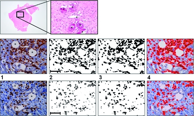
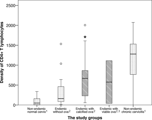
The parasite Schistosoma haematobium frequently causes genital lesions in women and could increase the risk of human immunodeficiency virus (HIV) transmission. This study quantifies the HIV target cells in schistosome-infected female genital mucosa. Cervicovaginal biopsies with and without schistosomiasis were immunostained for quantification of CD4(+) T lymphocytes (CD3, CD8), macrophages (CD68), and dendritic Langerhans cells (S100 protein). We found significantly higher densities of genital mucosal CD4(+) T lymphocytes and macrophages surrounding schistosome ova compared with cervicovaginal mucosa without ova (P = 0.034 and P = 0.018, respectively). We found no increased density of Langerhans cells (P = 0.25). This study indicates that S. haematobium may significantly increase the density of HIV target cells (CD4(+) T lymphocytes and macrophages) in the female genitals, creating a beneficial setting for HIV transmission. Further studies are needed to confirm these findings and to evaluate the effect of anti-schistosomal treatment on female genital schistosomiasis.
Figures
Similar articles
-
Schistosoma haematobium infection and CD4+ T-cell levels: a cross-sectional study of young South African women.PLoS One. 2015 Mar 13;10(3):e0119326. doi: 10.1371/journal.pone.0119326. eCollection 2015. PLoS One. 2015. PMID: 25768005 Free PMC article.
-
Increased vascularity in cervicovaginal mucosa with Schistosoma haematobium infection.PLoS Negl Trop Dis. 2011 Jun;5(6):e1170. doi: 10.1371/journal.pntd.0001170. Epub 2011 Jun 7. PLoS Negl Trop Dis. 2011. PMID: 21666790 Free PMC article.
-
Female genital schistosomiasis as a risk-factor for the transmission of HIV.Int J STD AIDS. 1994 Sep-Oct;5(5):368-72. doi: 10.1177/095646249400500517. Int J STD AIDS. 1994. PMID: 7819359 Review.
-
Cervicovaginal Immune Activation in Zambian Women With Female Genital Schistosomiasis.Front Immunol. 2021 Mar 2;12:620657. doi: 10.3389/fimmu.2021.620657. eCollection 2021. Front Immunol. 2021. PMID: 33737927 Free PMC article. Clinical Trial.
-
Early HIV-1 target cells in human vaginal and ectocervical mucosa.Am J Reprod Immunol. 2011 Mar;65(3):261-7. doi: 10.1111/j.1600-0897.2010.00939.x. Epub 2010 Dec 1. Am J Reprod Immunol. 2011. PMID: 21118402 Free PMC article. Review.
Cited by
-
A new mouse model for female genital schistosomiasis.PLoS Negl Trop Dis. 2014 May 1;8(5):e2825. doi: 10.1371/journal.pntd.0002825. eCollection 2014 May. PLoS Negl Trop Dis. 2014. PMID: 24786606 Free PMC article.
-
Decrease in Seminal HIV-1 RNA Load After Praziquantel Treatment of Urogenital Schistosomiasis Coinfection in HIV-Positive Men-An Observational Study.Open Forum Infect Dis. 2017 Sep 15;4(4):ofx199. doi: 10.1093/ofid/ofx199. eCollection 2017 Fall. Open Forum Infect Dis. 2017. PMID: 29181419 Free PMC article.
-
Eosinophil granule proteins ECP and EPX as markers for a potential early-stage inflammatory lesion in female genital schistosomiasis (FGS).PLoS Negl Trop Dis. 2014 Jul 17;8(7):e2974. doi: 10.1371/journal.pntd.0002974. eCollection 2014 Jul. PLoS Negl Trop Dis. 2014. PMID: 25033206 Free PMC article.
-
Schistosoma haematobium infection and CD4+ T-cell levels: a cross-sectional study of young South African women.PLoS One. 2015 Mar 13;10(3):e0119326. doi: 10.1371/journal.pone.0119326. eCollection 2015. PLoS One. 2015. PMID: 25768005 Free PMC article.
-
Cost-effectiveness of a community-based intervention for reducing the transmission of Schistosoma haematobium and HIV in Africa.Proc Natl Acad Sci U S A. 2013 May 7;110(19):7952-7. doi: 10.1073/pnas.1221396110. Epub 2013 Apr 15. Proc Natl Acad Sci U S A. 2013. PMID: 23589884 Free PMC article.
References
-
- Chenine AL, Shai-Kobiler E, Steele LN, Ong H, Augostini P, Song R, Lee SJ, Autissier P, Ruprecht RM, Secor WE. Acute Schistosoma mansoni infection increases susceptibility to systemic SHIV clade C infection in rhesus macaques after mucosal virus exposure. PLoS Negl Trop Dis. 2008;2:e265. - PMC - PubMed
-
- Feldmeier H, Krantz I, Poggensee G. Female genital schistosomiasis as a risk-factor for the transmission of HIV. AIDS. 1994;5:368–372. - PubMed
-
- Kjetland EF, Ndhlovu PD, Gomo E, Mduluza T, Midzi N, Gwanzura L, Mason PR, Sandvik L, Friis H, Gundersen SG. Association between genital schistosomiasis and HIV in rural Zimbabwean women. AIDS. 2006;20:593–600. - PubMed
-
- WHO . Report of an informal working group on urogenital schistosomiasis and HIV transmission, 1–2 October 2009. Geneva; Switzerland: 2009.
Publication types
MeSH terms
LinkOut - more resources
Full Text Sources
Medical
Research Materials
