. 2012 Feb;80(2):753-67.
doi: 10.1128/IAI.06101-11. Epub 2011 Dec 5.

Distinct compartmentalization of NF-κB activity in crypt and crypt-denuded lamina propria precedes and accompanies hyperplasia and/or colitis following bacterial infection

Affiliations

Distinct compartmentalization of NF-κB activity in crypt and crypt-denuded lamina propria precedes and accompanies hyperplasia and/or colitis following bacterial infection

Parthasarathy Chandrakesan et al. Infect Immun. 2012 Feb.

Abstract

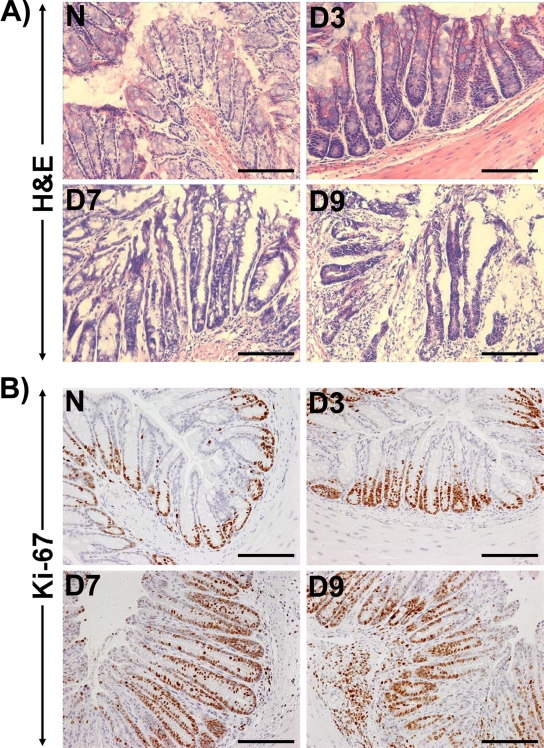
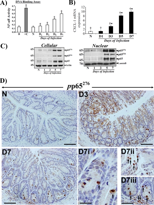
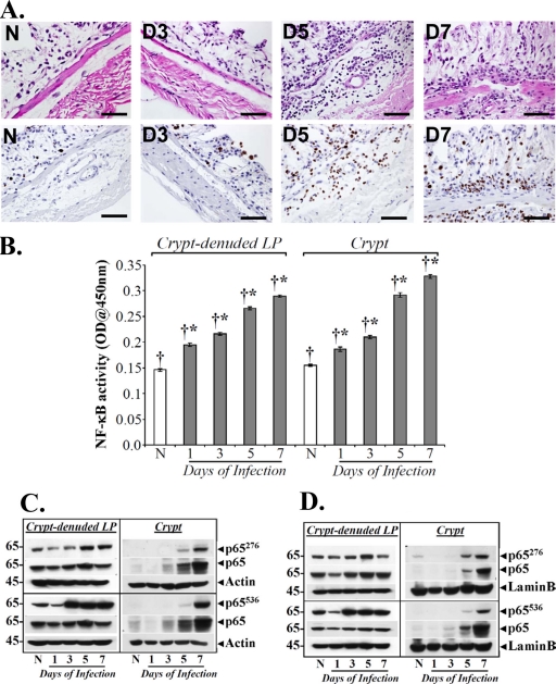
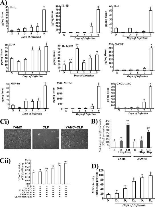
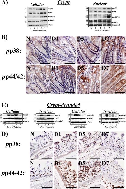
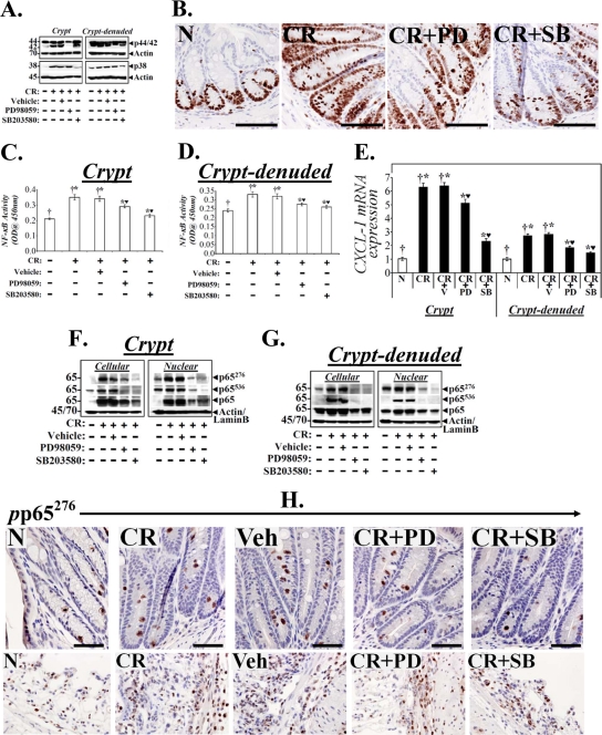
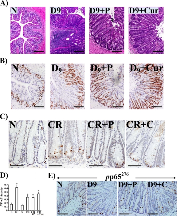
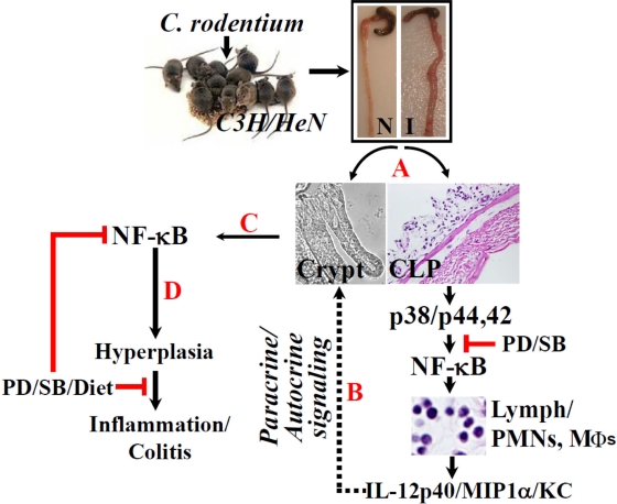
Citrobacter rodentium induces transmissible murine colonic hyperplasia (TMCH) and variable degrees of inflammation and necrosis depending upon the genetic background. Utilizing C. rodentium-induced TMCH in C3H/HeNHsd inbred mice, we observed significant crypt hyperplasia on days 3 and 7 preceding active colitis. NF-κB activity in the crypt-denuded lamina propria (CLP) increased within 24 h postinfection, followed by its activation in the crypts at day 3, which peaked by day 7. Increases in interleukin-α1 (IL-1α), IL-12(p40), and macrophage inflammatory protein 1α (MIP-1α) paralleled NF-κB activation, while increases in IL-1α/β, IL-6/IL-12(p40)/granulocyte colony-stimulating factor (G-CSF)/keratinocyte-derived chemokine (KC)/monocyte chemotactic protein 1 (MCP-1), and MIP-1α followed NF-κB activation leading to significant recruitment of neutrophils to the colonic mucosa and increased colonic myeloperoxidase (MPO) activity. Phosphorylation of the crypt cellular and nuclear p65 subunit at serines 276 and 536 led to functional NF-κB activation that facilitated expression of its downstream target, CXCL-1/KC, during TMCH. Distinct compartmentalization of phosphorylated extracellular signal-regulated kinase 1 and 2 ([ERK1/2] Thr(180)/Tyr(182)) and p38 (Thr(202)/Tyr(204)) in the CLP preceded increases in the crypts. Inhibition of ERK1/2 and p38 suppressed NF-κB activity in both crypts and the CLP. Dietary administration of 6% pectin or 4% curcumin in C. rodentium-infected mice also inhibited NF-κB activity and blocked CD3, F4/80, IL-1α/β, G-CSF/MCP-1/KC, and MPO activity in the CLP while not affecting NF-κB activity in the crypts. Thus, distinct compartmentalization of NF-κB activity in the crypts and the CLP regulates crypt hyperplasia and/or colitis, and dietary intervention may be a novel strategy to modulate NF-κB-dependent protective immunity to facilitate crypt regeneration following C. rodentium-induced pathogenesis.


Figures

Fig 1
Fig 1
Hyperplasia precedes inflammation in an inbred mouse strain genetically susceptible to inflammation and/or colitis. (A) H&E staining in the paraffin-embedded sections prepared from the distal colons of uninfected healthy (N, for normal) and C. rodentium (CR)-infected (days 3 to 9 [D3 to D9]) C3H inbred mice. Scale bar, 75 μm. (B) Crypt hyperplasia as measured by immunohistochemical labeling of Ki-67 as a marker of proliferation in the paraffin-embedded sections prepared from the distal colons of uninfected and C. rodentium-infected (days 3 to 9) C3H mice. Scale bar, 75 μm (n = 3 independent experiments).
Fig 2
Fig 2
Crypt hyperplasia in C3H mice in response to C. rodentium infection correlates with increases in NF-κB activity and subunit expression. (A) Nuclear extracts were prepared from the isolated crypts of uninfected healthy (N) mice and of mice at days 1 to 7 postinfection and assayed for NF-κB activity via a TransAM NF-κB-p65 Chemi Transcriptional Factor assay kit from Active Motif (n = 3; *, P < 0.05 versus control). (B) Real-time RT-PCR. Expression of CXCL-1/KC mRNA isolated from the crypts of the animals described for panel A was measured as a readout for NF-κB activity via real-time RT-PCR. †♥, P < 0.05 versus control (†) (n = 3 independent experiments). (C) Phosphorylation status of the p65 subunit in vivo. Relative levels of cellular and nuclear p65 subunit phosphorylated at Ser-276 and -536 (p65276/p65536) along with the total p65 subunit were determined in the colonic crypt extracts prepared from uninfected healthy (N) mice and from mice at days 1 to 7 postinfection by Western blotting (n = 3 independent experiments). (D) Immunohistochemical staining of the p65 subunit phosphorylated at Ser-276 in vivo. Paraffin-embedded sections prepared from mouse distal colons of uninfected healthy (N) mice and of mice at days 3 to 7 postinfection were stained with antibody specific for NF-κB p65 phosphorylated at Ser-276 (pp65276) and were analyzed with light microscopy. Magnifications, ×200 (D3 to D7) and ×400 (D7i to D7iii showing crypt and stromal staining). n = 3 independent experiments.
Fig 3
Fig 3
Separation of crypts and CLP from the distal colons of C3H mice. (A) H&E staining (upper panel) of the CLP prepared from the distal colons of uninfected healthy (N) and C. rodentium-infected (days 1 to 7) C3H mice. Scale bar, 100 μm; n = 3. The lower panel shows immunohistochemical labeling of Ki-67 as a marker of proliferation in the paraffin-embedded sections prepared from the distal colons of CLP of uninfected healthy (N) and C. rodentium-infected (days 1 to 7) C3H mice. Scale bar, 100 μm. (B) NF-κB activities measured in the CLP and crypts via a DNA binding assay. NF-κB p65 activities in the nuclear extracts of crypts and CLP from uninfected healthy (N) C3H mice and mice at days 1 to 7 postinfection were examined by utilizing a TransAM NF-κB p65 Chemi Transcriptional Factor assay kit from Active Motif. †*, P < 0.05 versus control (*); n = 3. OD, optical density (C and D) Kinetics of p65 phosphorylation at Ser-276 and -536 in the two compartments in vivo. Relative levels of phosphorylated (p65276/p65536) and total p65 subunit in the cellular (C) and nuclear (D) extracts prepared from isolated crypts and CLP of uninfected healthy (N) C3H mice and mice at days 1 to 7 postinfection were determined by Western blotting.
Fig 4
Fig 4
Distinct cytokine/chemokine expression and stromal-epithelial interaction in the regulation of NF-κB activity in vivo. (A) Expression of proinflammatory cytokines and chemokines in the distal colonic homogenates of uninfected healthy (N) and C. rodentium-infected (days 1 to 9) mice were measured using a Bio-Plex Cytokine assay kit as described by the manufacturer. Samples were analyzed on a Bio-Rad 96-well plate reader using a Bio-Plex array system and Bio-Plex Manager software. Each bar represents mean ± standard deviation. *, P < 0.05 versus control; †*, P < 0.05 versus the control (†) (n = 3). (B) Luciferase reporter assay to measure relative activation of NF-κB in response to C. rodentium infection. Both YAMC and JAWSII cell lines were transfected with pGL4.32(luc2P/NF-κB-RE/Hygro) vector for 36 h. The transfected cells were treated with or without C. rodentium for 3 h, washed to remove bacteria, and processed for measuring the relative levels of luciferase. The NF-κB activity changes are presented as percent change in luciferase units (n = 3 independent experiments). (C) Growth of YAMC and CLP cells in vitro (i) Both YAMC and CLP cells were propagated in appropriate medium at 33 and 37°C, respectively. For coculture studies, YAMC cells were layered onto CLP at a ratio of 1:1, and cocultures were incubated at 37°C for 24 h. Untreated or C. rodentium-infected cells were used to measure NF-κB activity via a TransAM NF-κB-p65 Chemi Transcriptional Factor assay from Active Motif (ii). †*, P < 0.05 versus control (†); †♥, P < 0.05 versus control (†), *♥○, P < 0.05 versus C. rodentium-infected CLP (CLP+CR) or C. rodentium-infected YAMC (YAMC+CR) (†* or †♥, respectively) (n = 3 independent experiments). (D) Measurement of MPO as a readout for assaying the extent of neutrophil recruitment to the colonic mucosa. MPO activity was measured in the colonic homogenates of uninfected healthy (N) and C. rodentium-infected (days 1 to 9) mice using a Fluoro MPO detection kit (Cell Technology, Inc., CA) according to the manufacturer's protocol. Each bar represents mean ± standard deviation (*, P < 0.05; n = 3 independent experiments).
Fig 5
Fig 5
Biochemical and immunohistochemical measurement of changes in the relative levels of p38-MAPK and p44/42-ERK during TMCH. Cellular and nuclear extracts (A and C) prepared from the crypts and CLP from the distal colons of uninfected healthy (N) mice of mice at days 1 to 7 postinfection were analyzed for the relative abundance of p38 and p44/42-ERK proteins by Western blot analysis (n = 3 independent experiments). (B and D) Immunohistochemical staining for pp38 and pp44/42 in the paraffin-embedded sections prepared from either whole distal colons (B) or CLP (D) of uninfected healthy (N) and C. rodentium-infected (days 1 to 7) C3H mice. Scale bar, 25 μm; n = 3 independent experiments.
Fig 6
Fig 6
Effect of p44/42-ERK1/2 and p38 inhibition on cell proliferation and NF-κB activity in vivo. Uninfected or C. rodentium-infected-infected C3H mice were injected once a day for 10 days with either vehicle or inhibitors specific for ERK1/2 (PD) or p38 (SB). At 2 h after the last injection, colonic crypts and CLP were isolated. (A) Representative Western blots showing total ERK1/2 and p38 in the two compartments. (B) Representative photomicrographs of paraffin-embedded sections stained with antibody to Ki-67: N, uninfected healthy mice; CR, C. rodentium-infected mice; CR+PD or CR+SB, C. rodentium-infected mice treated with specific ERK1/2 or p38 inhibitor. Scale bar = 50 μm (n = 3 independent experiments). (C and D) NF-κB activities measured via DNA binding assay in the crypts and CLP. Each bar represents mean ± standard deviation. †*, P < 0.05 versus control (†); *♥, P < 0.05 versus CR (†*) (n = 3). (E) Real-time RT-PCR. CXCL-1 expression in the crypts and CLP were measured via real-time RT-PCR. †*, P < 0.05 versus control (†); *♥, P < 0.05 versus C. rodentium-infected, vehicle-treated mice (CR+V; †*) (n = 3 independent experiments). (F and G) Crypts and CLP cellular and nuclear extracts prepared from the distal colons of the above group of animals were analyzed by Western blotting with antibodies specific for total p65 or the p65 subunit phosphorylated at Ser 276 (p65276) or Ser 536 (p65536). Actin or lamin B was the loading control, respectively (n = 3 independent experiments). (H) Representative photomicrographs of paraffin-embedded sections stained with antibody specific for phosphorylated p65276 (pp65276): N, uninfected healthy; CR, C. rodentium-infected; Veh, C. rodentium-infected and vehicle-treated; CR+PD or CR+SB, C. rodentium-infected and treated with specific ERK1/2 or p38 inhibitor, respectively. Scale bar = 30 μm (n = 3 independent experiments).
Fig 7
Fig 7
Anti-inflammatory and proproliferatory properties of pectin and curcumin diets. Paraffin-embedded sections prepared from the distal colons of uninfected healthy (N) mice, C. rodentium-infected mice at day 9 postinfection (D9), and C. rodentium-infected mice at day 9 treated with 6% pectin or 4% curcumin diets (D9+P or D9+Cur, respectively) were stained with H&E (A), stained for Ki-67 (B), or examined for apoptosis via TUNEL assay (C). Scale bar, 50 μm; n = 3. In panel B, please note significant increases in Ki-67 staining demonstrating proproliferatory activities of both pectin and curcumin. In panel C, please note significant inhibition of apoptosis in treated samples. (D) Both the pectin and curcumin diets promote NF-κB activity in the crypts. Colonic crypt NF-κB p65 activity in the nuclear extracts prepared from uninfected healthy (N) mice, C. rodentium-infected mice, and C. rodentium-infected mice treated with 6% pectin (P) or 4% curcumin (Cur) was measured by utilizing a TransAM NF-κB p65 Chemi Transcriptional Factor assay kit from Active Motif. *, P < 0.05 versus control (n = 3). (E) Staining for p65 phosphorylated at Ser-276 (pp65276) in the diet-treated samples. Panels show immunohistochemical staining of p65 phosphorylated at Ser-276 (pp65276) in the paraffin-embedded sections prepared from the distal colons of uninfected healthy (N) mice, C. rodentium-infected mice at day 9, or C. rodentium-infected mice treated with a 6% pectin (D9+P) or 4% curcumin (D9+C) diet, respectively (scale bar, 75 μm; n = 3).
Fig 8
Fig 8
Proposed mechanism of NF-κB's role in hyperplasia and/or inflammation in response to C. rodentium infection. Following isolation of crypts and CLP from uninfected (N) or C. rodentium-infected (I) distal colons of C3H mice (A), p38 and p44/42 MAPKs may be activated in the CLP, leading to NF-κB-induced recruitment of immune and inflammatory cells in association with release of proinflammatory cytokines/chemokines. This may facilitate NF-κB activation in the crypts (dotted lines; steps B and C), resulting in crypt hyperplasia. The combined paracrine/autocrine effort may eventually prime the mucosa toward inflammation and/or colitis (D). Both p38 (SB203580) and p44/42 (PD98059) inhibitors along with dietary interventions may significantly affect the infection outcome by modulating NF-κB activity in the two compartments.

Similar articles

Cited by

References

    1. Abraham C, Medzhitov R. 2011. Interactions between the host innate immune system and microbes in inflammatory bowel disease. Gastroenterology 140:1729–1737 - PMC - PubMed
    1. Barthold SW, Osbaldiston GW, Jonas AM. 1977. Dietary, bacterial, and host genetic interactions in the pathogenesis of transmissible murine colonic hyperplasia. Lab. Anim. Sci. 27:938–945 - PubMed
    1. Berg DJ, et al. 1996. Enterocolitis and colon cancer in interleukin-10-deficient mice are associated with aberrant cytokine production and CD4(1) TH1-like responses. J. Clin. Invest. 98:1010–1020 - PMC - PubMed
    1. Bhattacharyya S., et al. 2007. Tumor-induced oxidative stress perturbs nuclear factor-kappaB activity-augmenting tumor necrosis factor-alpha-mediated T-cell death: protection by curcumin. Cancer Res. 67:362–370 - PubMed
    1. Bohuslav J, Chen LF, Kwon H, Mu Y, Greene WC. 2004. p53 induces NF-κB activation by an IκB kinase-independent mechanism involving phosphorylation of p65 by ribosomal S6 kinase 1. J. Biol. Chem. 279:26115–26125 - PubMed

Publication types

MeSH terms