. 2012 Feb;80(2):565-74.
doi: 10.1128/IAI.05078-11. Epub 2011 Dec 5.

Small intestinal intraepithelial lymphocytes expressing CD8 and T cell receptor γδ are involved in bacterial clearance during Salmonella enterica serovar Typhimurium infection

Affiliations

Small intestinal intraepithelial lymphocytes expressing CD8 and T cell receptor γδ are involved in bacterial clearance during Salmonella enterica serovar Typhimurium infection

Zhiyuan Li et al. Infect Immun. 2012 Feb.

Abstract

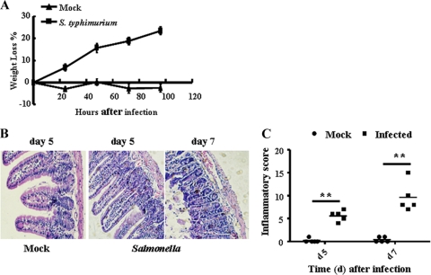
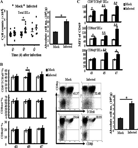
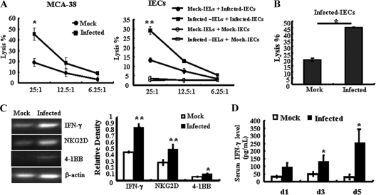
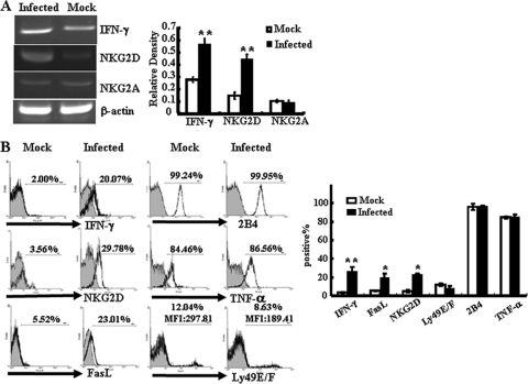
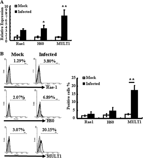
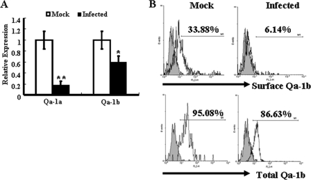
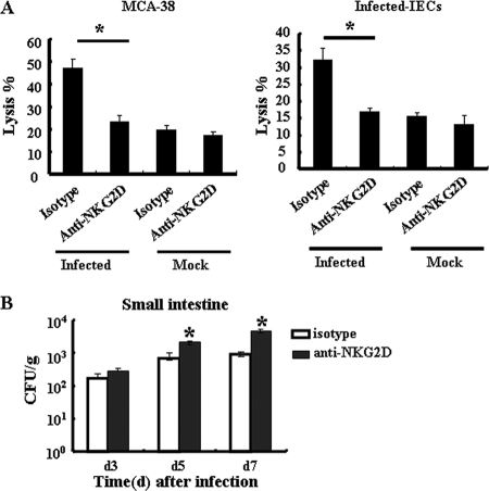
The intestinal immune system is crucial for the maintenance of mucosal homeostasis and has evolved under the dual pressure of protecting the host from pathogenic infection and coexisting with the dense and diverse commensal organisms in the lumen. Intestinal intraepithelial lymphocytes (iIELs) are the first element of the host T cell compartment available to respond to oral infection by pathogens. This study demonstrated that oral infection by Salmonella enterica serovar Typhimurium promoted the expansion of iIELs, particularly CD8(+) TCRγδ(+) IELs, enhanced expression of NKG2D on iIELs, increased expression of MULT1, and decreased expression of Qa-1 by intestinal epithelial cells (IECs), leading to activation of, particularly, CD8(+) TCRγδ(+) iIELs and cytolytic activity against S. Typhimurium-infected IECs. Blockade of NKG2D recognition or depletion of TCRγδ(+) cells using a depleting monoclonal antibody significantly attenuated the clearance of S. Typhimurium in the intestine and other tissues. This study suggests that iIELs, particularly CD8(+) TCRγδ(+) iIELs, play important roles in the detection of pathogenic bacteria and eradication of infected epithelial cells and, thus, provide protection against invading pathogens. These data further our understanding of the mechanisms by which the immune system of the intestinal mucosa discriminates between pathogenic and commensal organisms.


Figures

Fig 1
Fig 1
Induction of mouse enteritis by S. Typhimurium infection. B6 mice were infected orally with wild-type S. Typhimurium (5 × 10⁵ bcteria) or mock infected (LB broth). (A) Weight loss (referring to weight at 0 h) was evaluated at various time points postinfection (n = 5). (B) Representative photographs for hematoxylin and eosin-stained, paraffin-embedded sections from B6 mice (n = 5) at day 5 and day 7 after S. Typhimurium infection (original magnification, ×200). Note the presence of a diffuse enteritis associated with edema, which is caused by an infiltration that consists predominantly of mononuclear leukocytes in mouse small intestine infected with S. Typhimurium. (C) Histopathology scores in the small intestine from mice infected with S. Typhimurium. Data are aggregate scores of the pathology from five tissue sections from five individual mice per group. Each data point represents the result for an individual animal, and the scatter of data is shown, with horizontal lines representing the means. **, P < 0.01, compared with the mock-infected group.
Fig 2
Fig 2
Oral infection with S. Typhimurium causes changes in iIEL populations. At various time points after infection, the small intestine was removed and isolated iIELs were stained with fluorescently labeled MAbs for analysis by flow cytometry. (A) Total numbers of iIELs at different time points (left) and absolute numbers of iIELs at day 5 (right) after S. Typhimurium infection were determined by flow cytometry. (B) Statistical analysis of the percentage of CD8+ TCRγδ+ iIELs, CD8αα+ iIELs, and CD8αβ+ iIELs at days 3, 5, and 7 after S. Typhimurium infection (left), the distribution of CD8+ TCRγδ+ iIELs, CD8αα+ IELs, and CD8αβ+iIELs from the small intestine of mock-infected or S. Typhimurium-infected mice (middle), and the absolute numbers of CD8+ TCRγδ+ iIELs (right) at day 5 after oral infection. (C) Statistical analysis of CD69 expression (MFI, mean fluorescent intensity) by CD8αα+ iIELs, CD8αβ+ iIELs, and CD8+ TCRγδ+ iIELs. Data are expressed as the means ± standard deviations (SD) of results of at least three independent experiments. **, P < 0.01; *, P < 0.05, compared with the mock-infected group.
Fig 3
Fig 3
S. Typhimurium infection enhances the NK cell-like cytotoxicity of iIELs. Total iIELs were isolated from mock-infected or Salmonella-infected B6 mice at day 5 after infection. (A) The cytotoxicity of iIELs against MCA-38 cells or S. Typhimurium-infected IECs was measured by MTT assay at various E:T ratios. (B) The cytotoxicity of iIELs against S. Typhimurium-infected IECs was measured by FACS assay with CFSE/7-AAD double staining at the E:T ratio of 25:1. (C) The mRNA levels of NKG2D, IFN-γ, and 4-1BB in Salmonella- or mock-infected iIELs were detected by RT-PCR. The expression of mRNA was normalized to that of β-actin. (D) Serum IFN-γ levels were detected by ELISA. Data are expressed as the means ± SD of results of at least three separate experiments. **, P < 0.01; *, P < 0.05, compared with the mock-infected group.
Fig 4
Fig 4
S. Typhimurium infection increases the expression of NKG2D and cytotoxicity-related molecules on CD8+ TCRγδ+ iIELs. Total iIELs were isolated from mock-infected or Salmonella-infected B6 mice at day 5 after infection. TCRγδ+ iIELs were enriched to up to 90% purity from the iIELs by MACS. (A) The mRNA levels of NKG2D, IFN-γ, and NKG2A of TCRγδ+ iIELs were analyzed by RT-PCR. The expression levels of mRNA were normalized to that of β-actin. (B) The expression levels of IFN-γ, TNF-α, NKG2D, FasL, 2B4, and Ly49E/F of CD8+ TCRγδ+ iIELs after S. Typhimurium infection were examined by flow cytometry. Data shown are representative of three independent experiments (left) and of a statistical analysis of IFN-γ, TNF-α, NKG2D, FasL, 2B4, and Ly49E/F expression (right). Cells stained with isotype-matched control Ig demonstrated the specificity of MAb binding (filled histograms). **, P < 0.01; *, P < 0.05, compared with the mock-infected group. MFI, mean fluorescence intensity.
Fig 5
Fig 5
S. Typhimurium infection induces MULT1 expression on IECs. IECs were isolated from mock-infected or Salmonella-infected mice at day 5 after infection. (A) The mRNA levels of Rae1, H60, and MULT1 were determined by quantitative PCR. Results are shown as the increase in the expression of Rae1, H60, or MULT1 compared with that of β-actin. Gene expression values were then calculated based on the ΔΔCT method, using the mean of results for mock-infected IECs as a calibrator. Relative quantities (RQs) were determined using the equation RQ = 2−ΔΔCT. *, P < 0.05, compared with the mock-infected group. (B) The surface expression of Rae1, H60, and MULT1 on IECs was analyzed by flow cytometry. (Left) Data shown are representative of three independent experiments. (Right) A statistical analysis of Rae1, H60, and MULT1 expression is also displayed. **, P < 0.01, compared with the mock-infected group.
Fig 6
Fig 6
S. Typhimurium infection decreases Qa-1 expression on IECs. IECs were isolated from mock-infected or Salmonella-infected mice at day 5 after infection. (A) The mRNA levels of Qa-1a and Qa-1b were determined by quantitative PCR. Results are shown as the relative increase in the expression of Qa-1a or Qa-1b compared with the level of expression of β-actin. Gene expression values were then calculated based on the ΔΔCT method, using the mean of results with mock-infected IECs as a calibrator. Relative quantities (RQs) were determined using the equation RQ = 2−ΔΔCT. **, P < 0.01; *, P < 0.05, compared with the mock-infected group. (B) The surface expression and total expression of Qa-1b were analyzed by flow cytometry. The total expression of Qa-1b consisted of the surface and intracellular expression of Qa-1b. Data shown are representative of three independent experiments. *, P < 0.05, compared with the isotype-matched control group.
Fig 7
Fig 7
Blockade of NKG2D recognition inhibits the cytotoxicity of IELs against infected IECs and increases the numbers of S. Typhimurium bacteria in the small intestine. (A) TCRγδ+ iIELs were enriched to up to 90% purity from the iIELs by MACS. The cytotoxicity of TCRγδ+ iIELs against MCA-38 cells or S. Typhimurium-infected IECs was detected at an E:T ratio of 25:1 by MTT assay. Purified TCRγδ+ iIELs were cocultured with saturating concentrations of anti-NKG2D Ab (20 μg/ml) or isotype-matched control antibody and then washed for use as effector cells. (B) C57BL/6 mice (n = 3) were pretreated with anti-NKG2D MAb or control IgG MAb (400 μg i.v.) per mouse at 24 h before S. Typhimurium infection. At the indicated time points after infection, the small intestines, with PP excised, were removed and homogenized and numbers of CFU per gram of organ were determined. Data are expressed as the means ± SD of results from at least three separate experiments. *, P < 0.05, compared with the isotype-matched control group.
Fig 8
Fig 8
In vivo S. Typhimurium growth following oral infection. C57BL/6 mice (n = 3) were pretreated with anti-TCRγδ Ab or control IgG Ab (i.p.) at 200 μg per mouse 3 days before S. Typhimurium infection. At the indicated time points after infection, the small intestine, PP, spleens, and livers were removed and homogenized and numbers of CFU per gram of organ were determined. Data are expressed as the means ± SD of results from three separate experiments. *, P < 0.05, compared with the isotype-matched control group.

References

    1. Bao S, Beagley K, France M, Shen J, Husband A. 2000. Interferon-γ plays a critical role in intestinal immunity against Salmonella typhimurium infection. Immunology 99:464–472 - PMC - PubMed
    1. Bhagat G, et al. 2008. Small intestinal CD8+ TCRγδ + NKG2A+ intraepithelial lymphocytes have attributes of regulatory cells in patients with celiac disease. J. Clin. Invest. 118:281–293 - PMC - PubMed
    1. Carding SR, Egan PJ. 2002. γδT cells: functional plasticity and heterogeneity. Nat. Rev. Immunol. 2:336–345 - PubMed
    1. Coburn B, Li Y, Owen D, Vallance BA, Finlay BB. 2005. Salmonella enterica serovar Typhimurium pathogenicity island 2 is necessary for complete virulence in a mouse model of infectious enterocolitis. Infect. Immun. 73:3219–3227 - PMC - PubMed
    1. Das G, Janeway CA. 1999. Development of CD8α/α and CD8α/β T cells in major histocompatibility complex class I-deficient mice. J. Exp. Med. 190:881–884 - PMC - PubMed

Publication types

MeSH terms

Substances