. 2012 Jan 17;125(2):335-45.
doi: 10.1161/CIRCULATIONAHA.111.045096. Epub 2011 Dec 5.

Migraine mutations increase stroke vulnerability by facilitating ischemic depolarizations

Affiliations

Migraine mutations increase stroke vulnerability by facilitating ischemic depolarizations

Katharina Eikermann-Haerter et al. Circulation. .

Abstract

Background: Migraine is an independent risk factor for stroke. Mechanisms underlying this association are unclear. Familial hemiplegic migraine (FHM), a migraine subtype that also carries an increased stroke risk, is a useful model for common migraine phenotypes because of shared aura and headache features, trigger factors, and underlying glutamatergic mechanisms.

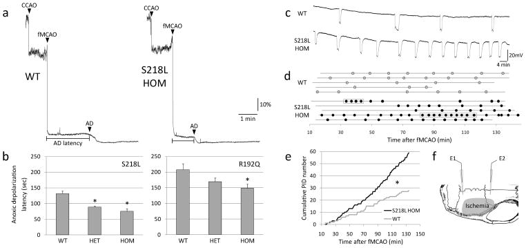
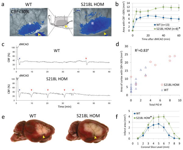
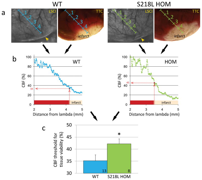
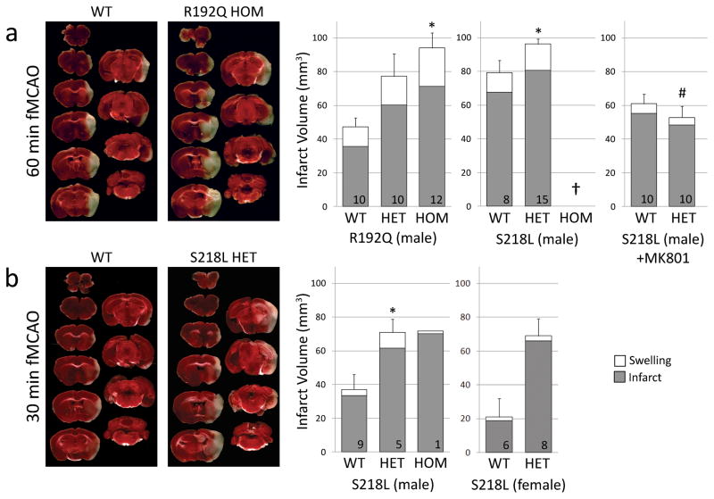
Methods and results: Here, we show that FHM type 1 (FHM1) mutations in Ca(V)2.1 voltage-gated Ca(2+) channels render the brain more vulnerable to ischemic stroke. Compared with wild-type mice, 2 FHM1 mutant mouse strains developed earlier onset of anoxic depolarization and more frequent peri-infarct depolarizations associated with rapid expansion of infarct core on diffusion-weighted magnetic resonance imaging and larger perfusion deficits on laser speckle flowmetry. Cerebral blood flow required for tissue survival was higher in the mutants, leading to infarction with milder ischemia. As a result, mutants developed larger infarcts and worse neurological outcomes after stroke, which were selectively attenuated by a glutamate receptor antagonist.

Conclusions: We propose that enhanced susceptibility to ischemic depolarizations akin to spreading depression predisposes migraineurs to infarction during mild ischemic events, thereby increasing the stroke risk.


Conflict of interest statement

Conflict of Interest Disclosures: None

Figures

Figure 1
Figure 1
Faster anoxic depolarization rates and more frequent peri-infarct depolarizations in FHM1 mutant mice. a) Representative laser Doppler tracings show CBF reduction upon common carotid artery occlusion (CCAO) followed by fMCAO. A further decline in CBF marks the onset of anoxic depolarization (AD), and is due to the vasoconstrictive effect of tissue depolarization on ischemic microvasculature. Scale bars: vertical 10%, horizontal 1 min. b) The latency to AD was shorter in S218L and R192Q mutants (p<0.001 and p=0.035, respectively), with a trend for allele-dosage effect. *p<0.05 vs. WT. c) Representative electrophysiological tracings show more frequent PIDs in S218L HOM compared to WT. Scale bars: vertical 20 mV, horizontal 4 min. d) PIDs (round symbols shown as a function of time) occurred in S218L HOM more frequently and sometimes in clusters (rectangular boxes). Horizontal lines indicate the time of onset and end of electrophysiological recordings in each mouse (n=5 each). When calculating the average PID frequency, these minor differences in recording duration were taken into account. e) Pooled cumulative PID numbers as a function of time after fMCAO was more than doubled in S218L HOM (*p<0.001; n=5 each). f) Experimental setup showing two intracortical glass micropipettes (E1, E2) placed outside the ischemic territory to detect PIDs after fMCAO. Shaded area indicates typical distribution of CBF deficit after fMCAO.
Figure 2
Figure 2
FHM1 mutant mice show accelerated lesion growth on MRI during hyperacute stroke. a) ADC lesion (i.e., ischemic core with restricted water diffusion, purple) was larger on diffusion-weighted MR images in S218L (left panel) and R192Q (right panel) mutants compared to WT during the hyperacute phase after fMCAO. b) ADC lesion volumes shown as a function of time after fMCAO were 40–50% larger in S218L and R192Q HOM compared to WT as early as 30 minutes after fMCAO suggesting faster growth of ischemic core (p=0.019 and p=0.018, respectively). The difference remained significant at 60 minutes. Vertical and horizontal error bars reflect the standard errors for total ADC lesion volume and timing of MRI scans, respectively. c) Thirty minutes after stroke onset, enlarged ADC lesion volumes in S218L HOM and R192Q HOM were primarily due to more severe cortical involvement (p=0.015 and p=0.023, respectively), although S218L HOM mutants showed hyperacute ADC changes in the hippocampus and thalamus as well (P=0.018 and P=0.117, respectively). Of note, the average regional ADC values in the center of ischemic core did not significantly differ between FHM1 mutants and their WT controls, suggesting that cytotoxic cell swelling is complete in ischemic core in all groups (60±7% vs. 56±6% in R192Q WT and HOM, and 57±5% vs. 57±6% of contralateral hemisphere in S218L WT and HOM, respectively, 60 minutes after stroke onset). *p<0.05 vs. WT.
Figure 3
Figure 3
FHM1 mutant mice develop larger areas of CBF deficit during dMCAO because of increased susceptibility to ischemic depolarizations. a) Representative laser speckle contrast images show the area of cortex with ≤30% residual CBF compared to pre-ischemic baseline (blue pixels), 60 min after dMCAO in WT and S218L HOM. Similar data were obtained using the R192Q strain (n=6 mutant and 6 WT; data not shown). Imaging was performed over the right hemisphere (light gray shaded rectangle in the inset), through intact skull. Arrowheads indicate clip occlusion. b) The area of CBF deficit expanded rapidly in S218L HOM throughout the 60 minute dMCAO (*p=0.004). c) Representative tracings show cortical blood flow reductions in penumbra (measured within the gray squares shown in ‘a’) after dMCAO. Anoxic depolarization triggers the first peri-infarct depolarization (blue arrowheads) marking a second abrupt reduction in perfusion. Each subsequent peri-infarct depolarization (red arrowheads) causes a characteristic blood flow transient. d) The frequency of PIDs was higher in S218L HOM, and correlated with the area of hypoperfused cortex 60 minutes after dMCAO (#p<0.001). Each symbol represents the PID frequency in individual mice. e) Representative TTC-stained whole brains show enlarged infarcts in S218L HOM 48 hours after 60 min dMCAO. f) The area of infarcts in 1 mm-thick coronal slices (0=anterior, 9=posterior) were larger in S218L HOM compared to WT (*p=0.016 S218L HOM vs. WT for infarct areas). Integrated total infarct volumes were also larger in the mutants (19±3 vs. 11±2 mm3, respectively; p=0.008).
Figure 4
Figure 4
Elevated blood flow threshold for tissue survival in FHM1 mutant mice. a) Representative laser speckle contrast images (LSCI) during dMCAO (left) and TTC-stained brain showing the infarct 48 hours after 60 min dMCAO (right) are shown for WT and S218L HOM mice. Imaging field was positioned as shown in Figure 3a. Images were spatially co-registered using surface landmarks. Line profiles (blue and green oblique lines, labeled in mm) were drawn between lambda and the clip occluding the middle cerebral artery branch (yellow arrowheads). b) For each animal, cortical blood flow (CBF) was plotted along these line profiles as a function of distance from lambda using laser speckle images, and the blood flow level corresponding to the infarct edge was determined (red dotted lines). This value represented the CBF threshold for viability, below which the tissue infarcted in each mouse. c) The average viability threshold was significantly higher in S218L HOM mutants compared to WT controls (p=0.048) indicating that FHM1 mutant brains are more vulnerable to ischemia and require higher blood flow to survive. Numbers of mice are shown on each bar. *p<0.05 vs. WT.
Figure 5
Figure 5
FHM1 mutant mice develop larger infarcts after experimental stroke selectively attenuated by NMDA receptor antagonist MK-801. Left panel shows representative infarcts (unstained white tissue) 24 hours after fMCAO. Right panel shows infarct and ischemic swelling volumes (grey and white bars, respectively). a) After 60 minute fMCAO, infarcts were larger in both R192Q and S218L mutants compared to WT (p=0.007 and p=0.019, respectively). One of 9 WT, 8 of 23 S218L HET, and all 8 S218L HOM (†) mice died within 24 hours (Table 1), and were excluded from infarct volume analysis. Because genetic backgrounds and infarct volumes significantly differed between WT controls of the two mutant strains, we did not directly compare S218L and R192Q mutants in this study. MK-801 (1 mg/kg, intraperitoneally 15 minutes before fMCAO) significantly reduced infarct volume in S218L HET mutants (P<0.001 vs. untreated S218L HET shown in ‘a’) but not in the WT (P=0.061 vs. untreated WT shown in ‘a’). Therefore, MK-801 was more efficacious in FHM1 mutants (P=0.026 for infarct reduction by MK-801 between WT and FHM1 mutants). As a result, after MK-801, infarct and swelling volumes were comparable between WT and S218L HET mice (P=0.367), as were neurological outcomes (Table 1). b) Thirty minute fMCAO also resulted in larger infarcts in male and female S218L mutants compared to WT (p=0.028). Despite shorter ischemia, 3 of 4 S218L HOM mutants died within 24 hours and were again excluded from infarct volume analysis; the data from the only surviving HOM mutant are shown. There was no mortality in WT and HET groups after 30 min fMCAO. Numbers of mice are shown on each bar. Standard error bars and p values refer to total volume (i.e., infarct plus swelling). *p<0.05 vs. WT, #p<0.05 vs. untreated S218L HET after 60 min fMCAO.

References

    1. Etminan M, Takkouche B, Isorna FC, Samii A. Risk of ischaemic stroke in people with migraine: Systematic review and meta-analysis of observational studies. BMJ. 2005;330:63. - PMC - PubMed
    1. Schurks M, Rist PM, Bigal ME, Buring JE, Lipton RB, Kurth T. Migraine and cardiovascular disease: Systematic review and meta-analysis. BMJ. 2009;339:b3914. - PMC - PubMed
    1. Kruit MC, van Buchem MA, Hofman PA, Bakkers JT, Terwindt GM, Ferrari MD, Launer LJ. Migraine as a risk factor for subclinical brain lesions. JAMA. 2004;291:427–434. - PubMed
    1. Scher AI, Gudmundsson LS, Sigurdsson S, Ghambaryan A, Aspelund T, Eiriksdottir G, van Buchem MA, Gudnason V, Launer LJ. Migraine headache in middle age and late-life brain infarcts. JAMA. 2009;301:2563–2570. - PMC - PubMed
    1. Thomsen LL, Eriksen MK, Roemer SF, Andersen I, Olesen J, Russell MB. A population-based study of familial hemiplegic migraine suggests revised diagnostic criteria. Brain. 2002;125:1379–1391. - PubMed

Publication types

Substances