. 2012 Feb 15;590(4):725-36.
doi: 10.1113/jphysiol.2011.220624. Epub 2011 Dec 5.

Repetitive activity slows axonal conduction velocity and concomitantly increases mechanical activation threshold in single axons of the rat cranial dura

Affiliations

Repetitive activity slows axonal conduction velocity and concomitantly increases mechanical activation threshold in single axons of the rat cranial dura

Roberto De Col et al. J Physiol. .

Abstract

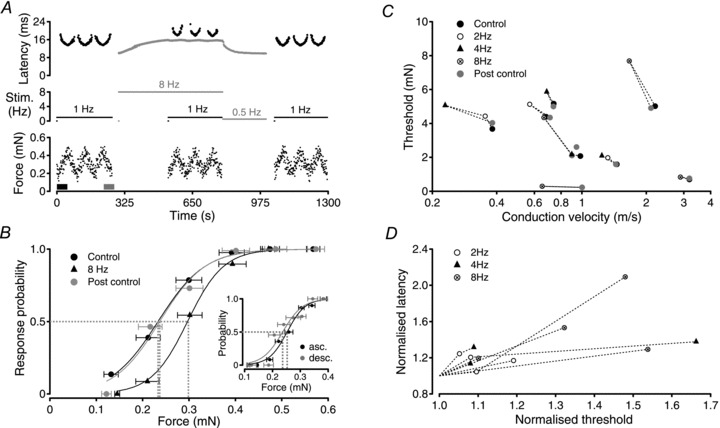
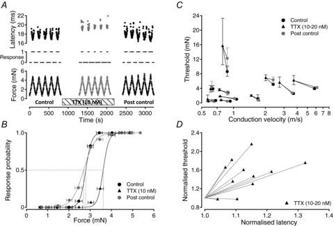
The passage of an action potential along a peripheral axon modulates the conduction velocity of subsequent action potentials. In C-neurones with unmyelinated axons repetitive activity progressively slows axonal conduction velocity and in microneurographic recordings from healthy human subjects the magnitude of this slowing can be used to predict the receptive properties of individual axons. Recently, a reduction in the number of available voltage-gated sodium channels (Na(V)) through inactivation has been implicated as the predominant factor responsible for the slowing of axonal conduction. Since Na(V)s are also responsible for the initiation of action potentials in sensory nerve terminals, changes in their availability may be expected to affect activation threshold for sensory stimuli. To examine this proposal, dynamic mechanical stimuli were used to make precise estimates of activation threshold in single unmyelinated axons innervating the rat cranial dura mater. Decreases in axonal conduction velocity induced by repetitive electrical stimulation were paralleled by an increase in mechanical activation threshold. Application of TTX (10-20 nM) also slowed axonal conduction velocity in all 11 fibres examined and in 9 of these this resulted in a parallel increase in mechanical activation threshold. We interpret this as indicating that a reduction in available Na(V) number contributes to both axonal conduction velocity slowing and the observed parallel increase in mechanical activation threshold. The slowing of axonal conduction velocity observed during repetitive activity thus represents a form of accommodation, i.e. self inhibition, which is likely to be decisive in limiting peripheral input to the spinal dorsal horn and thereby regulating processes that could otherwise lead to central sensitization.


Figures

Figure 1
Figure 1. Bimodal activation of a single sensory axon using a combined electrical and actuator-driven mechanical stimulator
Mechanical stimuli are delivered via an actuator with the armature suspended between rubber diaphragms and a strain gauge based force transducer incorporated into the axial shaft (A). The cathode is placed in contact with the tissue while the anode is attached to the shaft and immersed in the solution perfusing the bath. The stimulator allows electrical and mechanical stimuli to be applied independently in close spatial proximity to one another (A and see Methods) such that an action potential response can be evoked by either electrical or mechanical stimulation (B, grey and black spikes, respectively). Action potential responses to mechanical and electrical stimulation can be shown to arise from a single axon by exploiting the phenomena of refractoriness. This is illustrated by the series of traces in B. Traces are aligned to the onset (i.e. 0 ms) of the 20-ms-wide mechanical stimulus pulse. In each trace, the time of electrical stimulation is indicated by an arrow. The profile of mechanical stimulation is shown in the bottom panel. For clarity, electrical stimulus artifacts have been removed by subtraction. The action potential response to mechanical stimulation occurs approximately 14–15 ms after onset of the mechanical stimulus. The latency of the response to electrical stimulation is approximately 2 ms. Electrical stimulation in the period 8–12 ms after the onset of mechanical stimulation produces an electrically evoked action potential, the absolute refractory period of which prevents generation of an action potential response to mechanical stimulation. Similarly, an action potential response to mechanical stimulation precludes the generation of an electrically evoked action potential when the electrical stimulus is delivered 14–16 ms after the onset of mechanical stimulation. Since it is not possible to evoke an action potential response with either stimulus modality during the refractory period of an action potential evoked by the other stimulus modality, both stimulus modalities must activate the same sensory axon. For each sensory ending reported on here this functional cross-modality refractory period analysis was performed to verify the singular origin of action potential responses to both stimulus modalities.
Figure 2
Figure 2. Determination of mechanical activation threshold in a single axon (c.v. = 2 m s−1)
The mechanical activation threshold was established by determining the likelihood of evoking a single action potential in response to brief mechanical stimuli of varying amplitude. Single action potential responses could be evoked using a wide range of mechanical stimulus pulse widths as illustrated in A. Both the latency to the action potential response (B) and the threshold of mechanical activation (D) increase as the width of mechanical stimulus pulse increases. The mechanical activation threshold was determined at 0.5 Hz. At each pulse width the amplitude of the mechanical stimulus was varied systematically (C, lower trace) and the action potential response monitored (C, centre trace). In C, data from mechanical stimuli delivered with a pulse width of 10 ms is shown. The force at which an action potential response is evoked in 50% of trials (P = 0.5) is considered to be the threshold of mechanical activation (D). The force at P = 0.5 was determined from a Boltzmann fit of response probability on stimulus force. The horizontal error bars in D indicate the standard deviation of values contributing to each force bin. Each bin contained no less than 10 values.
Figure 3
Figure 3. Repetitive action potential activity increases the threshold of mechanical activation in sensory axons
For the single C-fibre (c.v. = 1.0 m s−1) shown in A and B, the threshold of mechanical activation was determined using the method of action potential response likelihood (see Fig. 2). Using mechanical stimuli alone at 1 Hz (A, black markers) the control threshold of mechanical activation was 0.227 ± 0.11 mN (B). Electrical stimulation (A, grey markers) of the axon at 8 Hz begins at 300 s and produces a characteristic increase in response latency, i.e. slowing of axonal conduction velocity, to a stable latency of approximately 15.6 ms (A, grey trace). Using the bimodal stimulator, mechanical stimuli were interleaved instead of every eighth electrical stimulus between 534 and 790 s (A, black markers). The slowed conduction velocity was determined from the average latency to 1776 action potential responses to electrical stimulation. Monitoring the likelihood of responses to these mechanical stimuli gave a threshold for mechanical activation of 0.29 ± 0.09 mN during stimulation at 8 Hz (B). The small fluctuations in electrical response latency observed during determination of mechanical threshold results from mechanical stimulus trials in which no action potential response was evoked, allowing axonal conduction velocity to recover partially from its slowed state. Similarly, the latency of action potential responses to mechanical stimulation (A, black markers) increases when the axon is subject to electrical stimulation at 8 Hz, further confirming that both stimulus modalities activate the same axon. A reduction in the frequency of electrical stimulation from 8 Hz to 0.5 Hz at 796 s and subsequent cessation of electrical stimulation at 1000 s allows the axonal conduction velocity to return to the non-conditioned value. Re-determination of the threshold for mechanical threshold (A, beginning at 1043 s) gives a value of 0.229 ± 0.13 mN (B). To examine potential hysteresis during each sequence of mechanical stimulation, a comparison was made between threshold values determined from responses during the first ascending series (A, thick black bar in force trace) and the last descending series (A, thick grey bar in force trace) of mechanical stimuli. Thresholds calculated from these 2 subsets of mechanical stimuli are shown in the inset in B. Under control conditions, mechanical threshold determined from only those stimuli presented during the first ascending (B, inset asc.) and last descending series (B, inset desc.) are not significantly different (0.252 ± 0.007 mN ascending; 0.237 ± 0.01 mN descending). The reversible increase in mechanical activation threshold with increasing firing rate was confirmed in 8 individual axons (C and D). In each axon, the threshold for mechanical activation returned to within 10% of the control value after the period of high frequency electrical stimulation (C). Horizontal error bars represent forces at 10% and 90% response probability. In D, relative changes in mechanical activation threshold are plotted against relative changes in conduction latency. Mechanical activation threshold determined during the period of electrical stimulation was normalised to the mechanical threshold determined in the control period before electrical stimulation. Similarly, the steady-state conduction latency during the period of electrical stimulation was normalised to the control conduction latency determined from the first response after a 300 s pause.
Figure 4
Figure 4. TTX (10–20 nm) increases the threshold of mechanical activation in single axons
This is shown by example for a single C-fibre (c.v. = 0.9 m s−1) in A and B. The threshold of mechanical activation was determined using mechanical stimuli alone at 0.5 Hz (A). To do this, the absence or presence (A, centre trace, 0 or 1) of an action potential response to the peak force of each mechanical stimulus (A, lower trace) was determined. Response likelihood was calculated for all stimuli within each force bin (B). Threshold was determined from the inflection point of a sigmoid fit to the resulting plot of response probability on force (B). The threshold of mechanical activation determined in this way was 2.8 mN under control conditions (B). Application of TTX (10 nm) increased the latency of action potential responses to mechanical stimulation (A, centre) and the threshold of mechanical activation to 3.6 mN (B). Following washout of TTX, the latency of the response to mechanical activation returned to control values and the threshold of mechanical activation returned to 2.7 mN (A, right; B). For 9 of 11 fibres examined, the mechanical activation threshold increased in the presence of TTX (10–20 nm, C and D). In two axons, however, TTX (20 mm) was without significant effect on the threshold of mechanical activation despite producing a slowing of conduction velocity (D). In each axon, the threshold of mechanical activation returned to within 40% of the control threshold after washout of TTX (C). For visual comparison across fibres, mechanical activation thresholds and conduction latencies were normalised to their control values (D).

Comment in

References

    1. Abrahamsen B, Zhao J, Asante CO, Cendan CM, Marsh S, Martinez-Barbera JP, Nassar MA, Dickenson AH, Wood JN. The cell and molecular basis of mechanical, cold, and inflammatory pain. Science. 2008;321:702–705. - PubMed
    1. Akopian AN, Sivilotti L, Wood JN. A tetrodotoxin-resistant voltage-gated sodium channel expressed by sensory neurons. Nature. 1996;379:257–262. - PubMed
    1. Blair NT, Bean BP. Role of tetrodotoxin-resistant Na+ current slow inactivation in adaptation of action potential firing in small-diameter dorsal root ganglion neurons. J Neurosci. 2003;23:10338–10350. - PMC - PubMed
    1. Brock JA, McLachlan EM, Belmonte C. Tetrodotoxin-resistant impulses in single nociceptor nerve terminals in guinea-pig cornea. J Physiol. 1998;512:211–217. - PMC - PubMed
    1. Carr RW, Pianova S, Brock JA. The effects of polarizing current on nerve terminal impulses recorded from polymodal and cold receptors in the guinea-pig cornea. J Gen Physiol. 2002;120:395–405. - PMC - PubMed

Publication types

LinkOut - more resources