. 2012 Feb;32(3):689-703.
doi: 10.1128/MCB.06455-11. Epub 2011 Dec 5.

Conserved molecular interactions within the HBO1 acetyltransferase complexes regulate cell proliferation

Affiliations

Conserved molecular interactions within the HBO1 acetyltransferase complexes regulate cell proliferation

Nikita Avvakumov et al. Mol Cell Biol. 2012 Feb.

Abstract

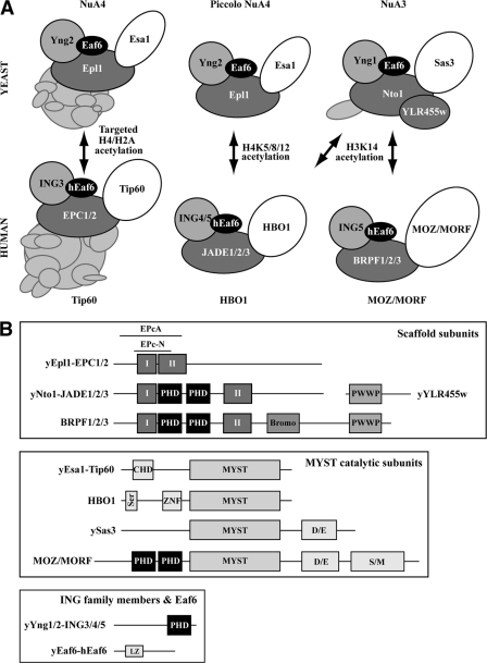
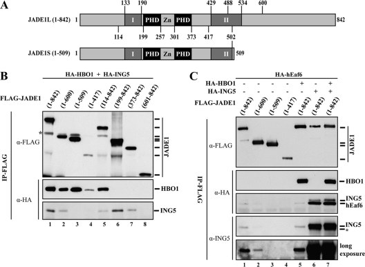
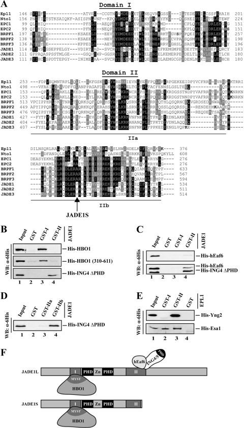
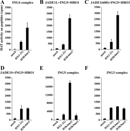
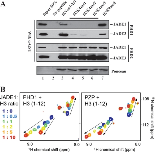
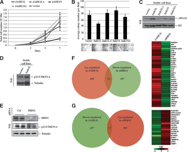
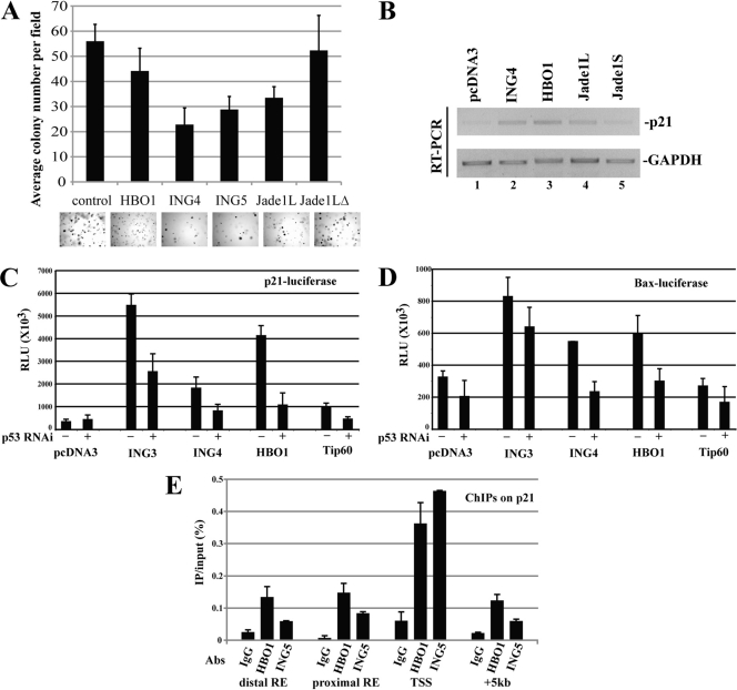
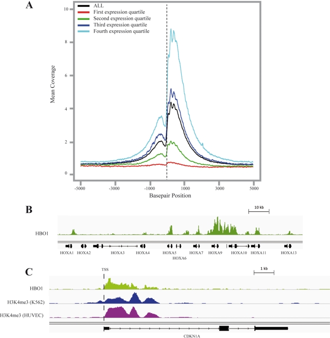
Acetyltransferase complexes of the MYST family with distinct substrate specificities and functions maintain a conserved association with different ING tumor suppressor proteins. ING complexes containing the HBO1 acetylase are a major source of histone H3 and H4 acetylation in vivo and play critical roles in gene regulation and DNA replication. Here, our molecular dissection of HBO1/ING complexes unravels the protein domains required for their assembly and function. Multiple PHD finger domains present in different subunits bind the histone H3 N-terminal tail with a distinct specificity toward lysine 4 methylation status. We show that natively regulated association of the ING4/5 PHD domain with HBO1-JADE determines the growth inhibitory function of the complex, linked to its tumor suppressor activity. Functional genomic analyses indicate that the p53 pathway is a main target of the complex, at least in part through direct transcription regulation at the initiation site of p21/CDKN1A. These results demonstrate the importance of ING association with MYST acetyltransferases in controlling cell proliferation, a regulated link that accounts for the reported tumor suppressor activities of these complexes.


Figures

Fig 1
Fig 1
Common features of the MYST-ING histone acetyltransferase complexes. (A) Structural and functional conservation of MYST-ING HAT complexes from yeast to human. (B) Homologous core subunits of all MYST-ING complexes and their associated protein domains. Domains not mentioned in the text: Bromo, bromodomain; CHD, chromodomain; Ser, serine-rich; ZNF, zinc finger; D/E, acidic; S/M, serine/methionine-rich; LZ, leucine zipper. The previously identified domains EPcA in EPC, E(Pc) and Epl1, Epc-N and I/II in Epl1, Nto1, EPC, JADE, and BRPF are also identified (8, 42, 62).
Fig 2
Fig 2
Molecular determinants for assembly of HBO1 HAT complexes. (A) Schematic representation of JADE1 protein isoforms produced by mRNA splice variants. The numbers are amino acids. (B) Distinct regions of JADE1 are required for HBO1 and ING5 interaction. Indicated N-terminal and C-terminal deletion mutants of FLAG-JADE1 were affinity purified after cotransfection with HA-HBO1 and HA-ING5 and analyzed by Western blotting. JADE1(1-509) corresponds to the JADE1S natural isoform. The numbers within parentheses above the lanes are the amino acid positions [e.g., (1-842), amino acids 1 to 842]. α-FLAG, anti-FLAG antibody. (C) Stable association of hEaf6 with JADE1 requires ING5. FLAG-JADE1 coimmunoprecipitations were analyzed as described after cotransfections with the indicated vectors. Asterisks indicate residual HB01 (B) and hEaf6 (c) signals.
Fig 3
Fig 3
Molecular dissection of MYST-ING HAT complexes identifies two conserved domains essential for assembly. (A) Sequence alignment of conserved domains in yeast and human proteins associated with ING/MYST-containing HAT complexes. Domain II can be further subdivided into two parts based on concentrations of homology. The location of the frameshift removing the C-terminal portion of domain II in JADE1S is shown. Sites of posttranslational modifications are indicated in boldface type. (B to D) ING4 binds JADE1 domain IIb in vitro, while HBO1 interacts with domain I, and hEaf6 needs ING4 to efficiently associate with JADE1 domain II. GST pulldown assays with the indicated recombinant proteins were analyzed by Western blotting (WB). HBO1 amino acids (aa) 310 to 611 [HBO1 (310-611)] represents the carboxyl terminus of the protein containing the MYST domain. ING4 ΔPHD includes aa 1 to 169 of the protein. (E) The functions of domains I and II are conserved in other MYST-ING complexes. GST pulldowns as described above for panel D using homologous domains in yeast Epl1 with Esa1 MYST and Yng2 ING proteins. (F) Model of interactions within HBO1 complexes and the effect of the JADE1S shorter isoform.
Fig 4
Fig 4
Domain II-dependent association of ING subunits stimulates MYST HAT activity toward histone H3 peptides methylated on lysine 4. (A) Native ING4 complex preferentially acetylates histone H3 tails that are methylated on lysine 4. Purified native ING4 complex (ING4, JADE, HBO1, and hEaf6 [Fig. 1A]) was tested in HAT assays with the indicated histone H3 peptides. Reactions were spotted on membranes and counted by liquid scintillation. (B to D) Domain IIb-dependent physical association of the ING subunit is required for stimulation of HBO1 HAT activity on H3K4me tails. HBO1 HAT was affinity purified after cotransfection with the indicated cDNAs and tested in HAT assays on peptides as described above for panel A. (E and F) Stimulation of H3 acetylation by H3K4me is seen only with H3-specific HATs. Purified native ING5 (a mix of HBO1 and MOZ/MORF complexes [Fig. 1A]) and ING3 (Tip60) acetyltransferase complexes were tested as described above for panel A.
Fig 5
Fig 5
The first PHD finger of the JADE1 PZP domain dictates the specificity of the entire domain toward histone H3K4 methylation status. (A) JADE PHD finger domains interact with histone H3 tail peptides in vitro, but with different effect of lysine 4 methylation. Peptide pulldown assays with the indicated biotinylated peptides and recombinant PHD fingers fused to GST were analyzed by Western blotting with anti-GST (WB:α-GST). JADE PHD1s prefer the nonmethylated form, while PHD2s bind H3 peptides in a methylation-independent manner. H3 K9 methylation has limited effect on the binding of these PHD fingers. (B) NMR analysis indicates that JADE1 PHD1 alone binds the H3 tail similarly to the full PZP domain. Superimposed 1H,15N HSQC spectra of JADE1 PHD1 and PZP, recorded during the addition of the indicated H3 peptides. The spectra are color coded according to the protein-peptide ratio.
Fig 6
Fig 6
JADE1 interaction with ING is essential for tumor suppressor activity which implicates HBO1-dependent upregulation of the p21 CDK inhibitor. (A) Expression of JADE1L strongly inhibits HeLa cell growth, while JADE1S or PHD-deleted mutants do not. Indicated transduced cell lines were seeded at the same density, and cells were counted over a period of 7 days. (B) JADE1L inhibits anchorage-independent cell growth, while JADE1S or PHD-deleted mutants do not. HeLa cell clones expressing the different constructs were grown in soft agar for 3 weeks, and colony formation was measured. Examples of image fields are shown below the bars in the graph. (C) JADE1L-expressing cells contain higher levels of γH2AX, a marker of DNA damage response, cell cycle checkpoint activation, and senescence. Whole-cell extracts of the indicated JADE1-expressing clones were analyzed by Western blotting with the indicated antibodies. (D and E) JADE1L expression increases p21 protein level, while HBO1 depletion decreases it. Western blot as in panel C, using p21 and tubulin antibodies on the indicated transduced or knocked-down HeLa cells. (F and G) Venn diagrams showing the number of genes significantly up- or downregulated by JADE1L expression or HBO1 depletion, and the genes commonly affected but in opposite ways (fold change of >1.5 with P value < 0.05).
Fig 7
Fig 7
HBO1-JADE-ING complexes directly bind and stimulate p53-regulated genes to control cell proliferation. (A) HBO1-JADE1L-ING4/5 inhibits anchorage-independent cell growth. HeLa clones expressing the different constructs were incubated in soft agar, and colony formation was measured as described in the legend to Fig. 6B. (B) Overexpression of HBO1, JADE1L, and ING4 increases endogenous p21 transcription. RT-PCR analysis of HeLa cells transfected with the indicated expression vectors. GAPDH, glyceraldehyde-3-phosphate dehydrogenase. (C and D) HBO1 and ING4 proteins stimulate p53-dependent transcription. Luciferase assays on RKO cells transfected with the indicated expression vectors. The cells were also transfected with a shRNA expression vector against p53 or control (empty). Luciferase reporters tested were under the control of the p21 (C) or BAX (D) promoters. Data from triplicate cultures are presented with standard deviations (error bars). RLU, relative light units; RNAi, RNA interference. (E) HBO1-ING complex is bound to the p21 promoter in vivo, near the transcription start site (TSS). ChIP analysis of HBO1 and ING5 enrichment on the p21/CDKN1A gene. Values representing direct IP over the input ratio are based on three different cultures with standard errors. RE, response element (shows the locations of known p53 binding sites on the p21 promoter).
Fig 8
Fig 8
HBO1 is mainly located in the 5′ end of transcribed genes and colocalizes with H3K4me3. (A) HBO1 ChIP-seq signal intensity around the transcription start site (TSS) of genes in human RKO cells correlates globally with gene expression levels. The global profiles were produced using the UCSC genome browser gene definition and the Python package HTseq (see Materials and Methods). The binning of genes by their function or expression levels in RKO cells (70) was performed by subdividing genes in four equal categories (the fourth quartile includes the most highly expressed genes). (B) Enrichment of HBO1 signal over the HOXA cluster in the RKO cell line. (C) HBO1 signal is enriched in the 5′ region of the p21 coding region and colocalizes with the H3K4me3 signal. Our ChIP-seq HBO1 data from RKO cells aligned with the H3K4me3 signal from HUVEC and K562 cells obtained from the Broad Institute/ENCODE project (11).

References

    1. Acosta JC, et al. 2008. Chemokine signaling via the CXCR2 receptor reinforces senescence. Cell 133:1006–1018 - PubMed
    1. Avvakumov N, Cote J. 2007. The MYST family of histone acetyltransferases and their intimate links to cancer. Oncogene 26:5395–5407 - PubMed
    1. Bonner WM, et al. 2008. GammaH2AX and cancer. Nat. Rev. Cancer 8:957–967 - PMC - PubMed
    1. Boudreault AA, et al. 2003. Yeast enhancer of polycomb defines global Esa1-dependent acetylation of chromatin. Genes Dev. 17:1415–1428 - PMC - PubMed
    1. Champagne KS, et al. 2008. The crystal structure of the ING5 PHD finger in complex with an H3K4me3 histone peptide. Proteins 72:1371–1376 - PMC - PubMed

Publication types

MeSH terms