. 2012 Jan 20;287(4):2437-45.
doi: 10.1074/jbc.M111.315051. Epub 2011 Dec 5.

Amyloid precursor protein revisited: neuron-specific expression and highly stable nature of soluble derivatives

Affiliations

Amyloid precursor protein revisited: neuron-specific expression and highly stable nature of soluble derivatives

Qinxi Guo et al. J Biol Chem. .

Abstract

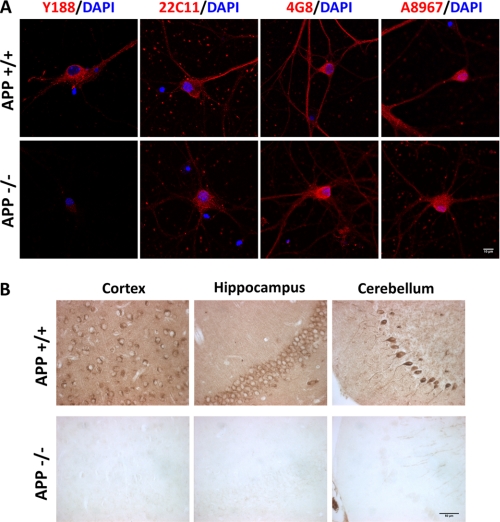
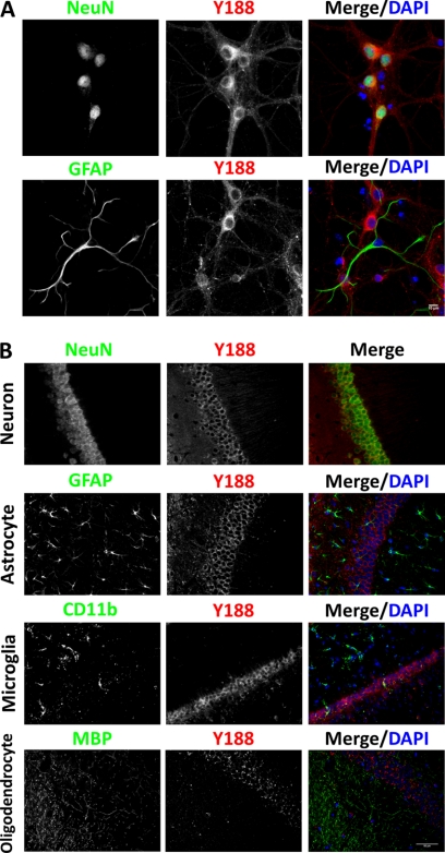
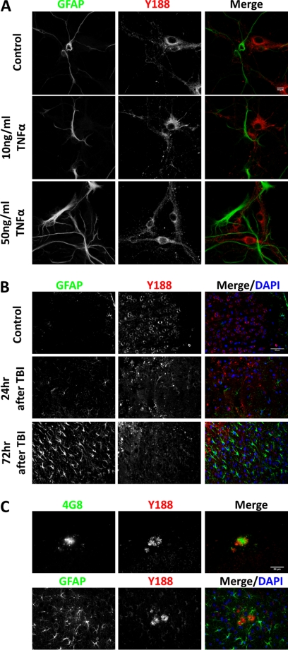
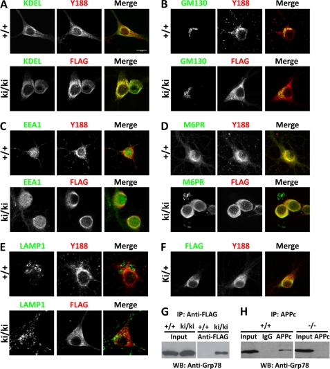
APP processing and amyloid-β production play a central role in Alzheimer disease pathogenesis. APP has been considered a ubiquitously expressed protein. In addition to amyloid-β, α- or β-secretase-dependent cleavage of APP also generates soluble secreted APP (APPsα or APPsβ, respectively). Interestingly, APPsβ has been shown to be subject to further cleavage to create an N-APP fragment that binds to the DR6 death receptor and mediates axon pruning and degeneration under trophic factor withdrawal conditions. By performing APP immunocytochemical staining, we found that, unexpectedly, many antibodies yielded nonspecific staining in APP-null samples. Screening of a series of antibodies allowed us to identify a rabbit monoclonal antibody Y188 that is highly specific for APP and prompted us to re-examine the expression, localization, and stability of endogenous APP and APPsβ in wild-type and in APPsβ knock-in mice, respectively. In contrast to earlier studies, we found that APP is specifically expressed in neurons and that its expression cannot be detected in major types of glial cells under basal or neuroinflammatory conditions. Both APPsα and APPsβ are highly stable in the central nervous system (CNS) and do not undergo further cleavage with or without trophic factor support. Our results clarify several key questions with regard to the fundamental properties of APP and offer critical cellular insights into the pathophysiology of APP.


Figures

FIGURE 1.
FIGURE 1.
Specificity of the Y188 antibody in vitro and in vivo. A, representative immunocytochemical staining images of 14 DIV wild-type (APP+/+) and APP KO (APP−/−) primary hippocampal cultures stained with Y188, 22C11, 4G8, and A8967 antibodies. Blue, DAPI; Red, APP. Scale bar, 10 μm. B, immunohistochemical staining of wild-type (APP+/+) and APP KO (APP−/−) cortex, hippocampus, and cerebellum sections using the Y188 antibody. Scale bar, 50 μm.
FIGURE 2.
FIGURE 2.
Endogenous APP is a neuron-specific protein in vitro and in vivo. A, co-staining of NeuN (upper) or GFAP (lower) with Y188 antibodies of 14 DIV primary hippocampal cultures showed that APP is only expressed in NeuN-positive, but not GFAP-positive, cells. Blue, DAPI; Green, NeuN or GFAP; Red, Y188. Scale bar, 10 μm. B, co-staining of adult mouse hippocampus sections using Y188 and neuron marker NeuN, astrocyte marker GFAP, microglia marker CD11b, or oligodendrocyte marker MBP revealed that although all NeuN-positive neurons express APP, APP expression in major types of glial cells is below detection. Blue, DAPI; Green, NeuN, GFAP, CD11b, and MBP; Red, Y188. Scale bar, 50 μm.
FIGURE 3.
FIGURE 3.
APP expression is not induced in activated astrocytes in vitro and in vivo. A, 12 DIV primary hippocampal cultures were treated with PBS (Control), 10 ng/ml TNFα, and 50 ng/ml TNFα followed by immunocytochemical staining for APP. Blue, DAPI; Green, GFAP; Red, Y188. Scale bar, 10 μm. B, controlled cortical impact-induced TBI was performed on adult mouse brain. APP immunostaining was carried out in uninjured controls (Control), 24 or 72 h after TBI. Blue, DAPI; Green, GFAP; Red, Y188. Scale bar, 50 μm. C, immunostaining of 20 month-old APP/hAβ/PS1 brains. Upper panel, APP detected by Y188 (red) was found around 4G8-positive Aβ plaques (green). Lower panel, GFAP-positive reactive astrocytes (green) do not express APP (red). Scale bar, 50 μm.
FIGURE 4.
FIGURE 4.
Subcellular localization of FL-APP and APPsβ. A–E, 14 DIV hippocampal neurons cultured from wild-type (+/+) or homozygous APPsβ knock-in (ki/ki) mice were co-stained with APP (red) and organelle markers (green). A, ER (anti-KDEL); B, Golgi (GM130); C, early endosome (EEA1); D, late endosome (M6PR); E, lysosome (LAMP1). APP and APPsβ were recognized by Y188 or anti-FLAG antibody, respectively. F, co-staining of FL-APP and APPsβ in APPsβ ki/+ neurons. Green, FLAG; Red, Y188. G, interaction between Grp78 and the APP soluble domain. Wild-type (+/+) or homozygous APPsβ knock-in (ki/ki) brain lysates were immunoprecipitated (IP) using the anti-FLAG antibody conjugated to magnetic beads followed by elution with 3×FLAG peptide and Western blotting (WB) using an antibody against Grp78. Input, total lysates. H, interaction between Grp78 and FL-APP. Wild-type (+/+) or APP-null (−/−) brain lysates were immunoprecipitated using the APPc antibody or rabbit IgG plus protein-G beads followed by Western blotting using an antibody against Grp78.
FIGURE 5.
FIGURE 5.
APPsβ is a highly stable protein. A and B, Western blot analysis using the 22C11 antibody on conditioned medium collected from APPsβ ki/ki (A) or wild-type (B) cultures under normal neuronal culture conditions (day 1, 5, 9, 13, 17, and 21), at day 7 immediately before TFD, or 24, 72, or 120 h after TFD, showing the intact nature of APPs recovered from residual surviving neurons. C, anti-V5 Western blot of fresh medium containing APPsβ-V5/His recombinant protein incubated with TFD-treated neurons or cell-free culture for 24 h, 72 h or 120 h.

References

    1. Zheng H., Koo E. H. (2011) Biology and pathophysiology of the amyloid precursor protein. Mol. Neurodegener. 6, 27. - PMC - PubMed
    1. Chauvet N., Apert C., Dumoulin A., Epelbaum J., Alonso G. (1997) Mab22C11 antibody to amyloid precursor protein recognizes a protein associated with specific astroglial cells of the rat central nervous system characterized by their capacity to support axonal outgrowth. J. Comp. Neurol. 377, 550–564 - PubMed
    1. LeBlanc A. C., Papadopoulos M., Bélair C., Chu W., Crosato M., Powell J., Goodyer C. G. (1997) Processing of amyloid precursor protein in human primary neuron and astrocyte cultures. J. Neurochem. 68, 1183–1190 - PubMed
    1. Rohan de Silva H. A., Jen A., Wickenden C., Jen L. S., Wilkinson S. L., Patel A. J. (1997) Cell-specific expression of β-amyloid precursor protein isoform mRNAs and proteins in neurons and astrocytes. Brain Res. Mol. Brain Res. 47, 147–156 - PubMed
    1. Li H., Wang B., Wang Z., Guo Q., Tabuchi K., Hammer R. E., Südhof T. C., Zheng H. (2010) Soluble amyloid precursor protein (APP) regulates transthyretin and Klotho gene expression without rescuing the essential function of APP. Proc. Natl. Acad. Sci. U.S.A. 107, 17362–17367 - PMC - PubMed

Publication types

MeSH terms

Substances