

Role of mast cells in pathogenesis of oral lichen planus
- PMID: 22144827
- PMCID: PMC3227251
- DOI: 10.4103/0973-029X.86674
Role of mast cells in pathogenesis of oral lichen planus
Abstract
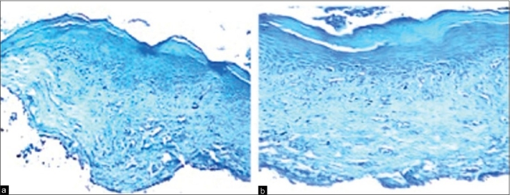
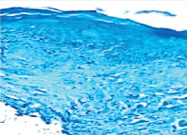
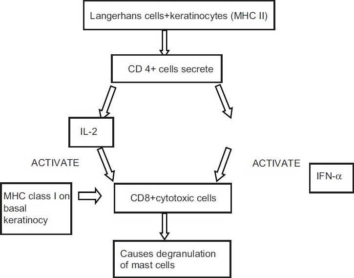
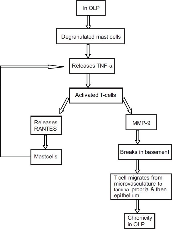
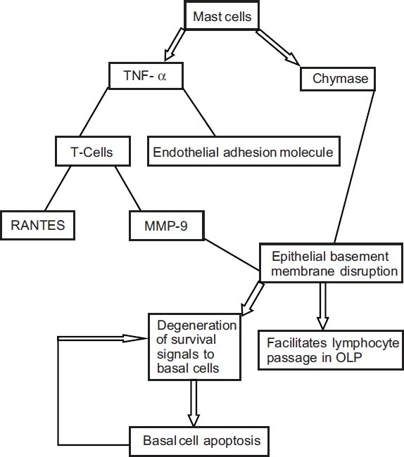
Recent attention has been directed toward the role of mast cells in the pathogenesis of oral lichen planus. Mast cells are responsible for trafficking inflammatory cells into the connective tissue that in turn helps in progression and maintenance of chronicity of oral lichen planus (OLP). OLP is a T-cell-mediated chronic inflammatory oral mucosal disease of unknown etiology, and lesions contain few B-cells or plasma cells and minimal deposits of immunoglobulin or complement. Hence, OLP is ideally positioned for the study of human T-cell-mediated inflammation and autoimmunity. This study was done to evaluate the mast cell count using toluidine blue stain in OLP and compares it with oral lichenoid reaction (OLR), and to propose the possible role of mast cells in the pathogenesis of OLP and OLR. Ten cases each of OLP and OLR and five cases of normal buccal mucosa were taken from the archives of Department of Oral Pathology. The samples were stained with toluidine blue using standard toluidine blue method by Wolman 1971. An increase in mast cell count was observed in OLP and OLR in comparison to normal oral mucosa. However, no significant differences in mast cell count were noted between OLP and OLR.
Keywords: Lichen planus; lichenoid reaction; mast cells.
Conflict of interest statement
Figures
Similar articles
-
To Investigate the Involvement of Mast Cells in the Pathogenesis of Oral Lichen Planus and Oral Lichenoid Reactions.J Pharm Bioallied Sci. 2024 Dec;16(Suppl 5):S4755-S4759. doi: 10.4103/jpbs.jpbs_913_24. Epub 2025 Jan 30. J Pharm Bioallied Sci. 2024. PMID: 40061733 Free PMC article.
-
A histochemical and immunohistochemical study of mast cells in differentiating oral lichen planus from oral lichenoid reactions.Quintessence Int. 2010 Mar;41(3):221-7. Quintessence Int. 2010. PMID: 20213023
-
Role of Mast Cells in Oral Lichen Planus and Oral Lichenoid Reactions.Autoimmune Dis. 2018 Jan 17;2018:7936564. doi: 10.1155/2018/7936564. eCollection 2018. Autoimmune Dis. 2018. PMID: 29593898 Free PMC article.
-
ORAL LICHEN PLANUS AND ORAL LICHENOID REACTION--AN UPDATE.Acta Clin Croat. 2015 Dec;54(4):516-20. Acta Clin Croat. 2015. PMID: 27017728 Review.
-
Oral lichen planus and lichenoid reactions: etiopathogenesis, diagnosis, management and malignant transformation.J Oral Sci. 2007 Jun;49(2):89-106. doi: 10.2334/josnusd.49.89. J Oral Sci. 2007. PMID: 17634721 Review.
Cited by
-
Identification of the Nerve-Cancer Cross-Talk-Related Prognostic Gene Model in Head and Neck Squamous Cell Carcinoma.Front Oncol. 2021 Nov 29;11:788671. doi: 10.3389/fonc.2021.788671. eCollection 2021. Front Oncol. 2021. PMID: 34912722 Free PMC article.
-
Degranulated mast cells and TNF-α in oral lichen planus and oral lichenoid reactions diseases.Adv Biomed Res. 2012;1:52. doi: 10.4103/2277-9175.100161. Epub 2012 Aug 28. Adv Biomed Res. 2012. PMID: 23326783 Free PMC article.
-
Submucosal Fibrotic Bands in Oral Lichen Planus: A Clinico-Pathological Investigation of a Newly Described Phenomenon.Head Neck Pathol. 2021 Jun;15(2):395-401. doi: 10.1007/s12105-020-01203-6. Epub 2020 Jul 23. Head Neck Pathol. 2021. PMID: 32705486 Free PMC article.
-
Mast cells and oral pathologies: A Review.J Nat Sci Biol Med. 2015 Jan-Jun;6(1):35-9. doi: 10.4103/0976-9668.149075. J Nat Sci Biol Med. 2015. PMID: 25810632 Free PMC article. Review.
-
Histopathologic evaluation of oral lichen planus and oral lichenoid reaction: A comparative analysis based on basement membrane thickness and the distribution of mast cells.J Oral Maxillofac Pathol. 2021 Sep-Dec;25(3):549-550. doi: 10.4103/jomfp.JOMFP_220_20. Epub 2022 Jan 11. J Oral Maxillofac Pathol. 2021. PMID: 35281137 Free PMC article.
References
-
- Riley JF. In: Mast cells. 2nd ed. Livingston E, Livingston S, editors. Edinburg h London: 1959.
-
- Ham WA, Cormack HD, editors. Histology. 8th ed. Philadelphia: J.B. Lippincott Company; 1979. The origins, morphologies and functions of the cells of the loose connective tissue; pp. 225–59.
-
- Lawrence B. Haemolymphoid system. In: Lawrence B, Martin BM, Patricia C, Mary D, Juhan D, editors. Gray's Anatomy. 38th ed. Edinburgh: Churchill Livingston Harcourt Publishers Ltd; 2000. pp. 1399–451.
-
- Walsh LJ, Kaminer MS, Lazarus GS, Lavker RM, Murphy GF. Role of laminin in localization of human dermal mast cells. Labs Invest. 1991;65:433–40. - PubMed
-
- Walsh LJ, Davis MF, Xu LJ, Savege NW. Relationship between mast cell degranulation and inflammation in the oral cavity. J Oral Pathol Med. 1995;24:266–72. - PubMed
