

"Hybrid" lesion of the maxilla
- PMID: 22144833
- PMCID: PMC3227257
- DOI: 10.4103/0973-029X.86693
"Hybrid" lesion of the maxilla
Abstract
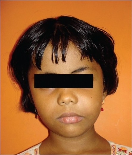
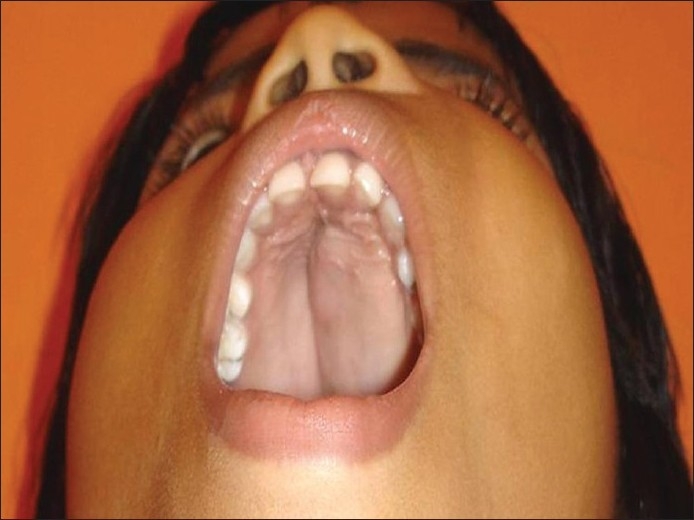
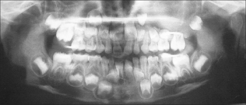
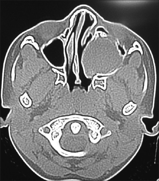
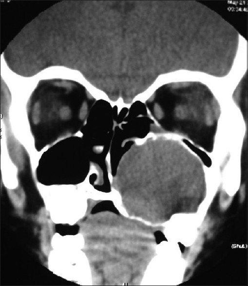
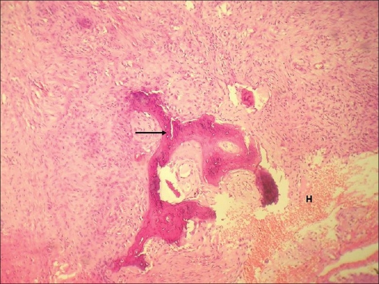
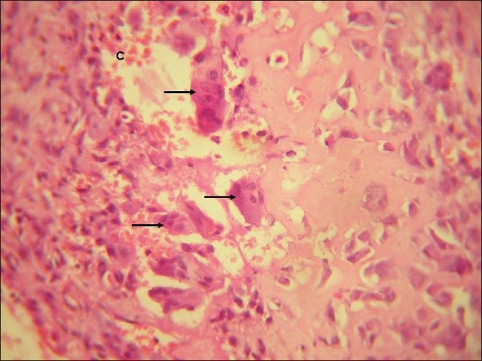
Juvenile ossifying fibroma is an uncommon benign but aggressive fibroosseous lesion that affects the craniofacial skeleton. Their distinct clinical and histopathological features warrant the lesion to be considered as a separate entity from other fibro-osseous group of lesions such as fibrous dysplasia and cemento ossifying fibroma. Concomitant development of secondary aneurysmal bone cyst may rarely occur, which makes the lesion more aggressive and difficult to treat. We report a case of a 6 year old girl who was diagnosed with aneurysmal bone cyst during her earlier presentation at a private hospital and was treated for the same. The lesion recurred within 6 months. The second incisional biopsy specimen revealed features of trabecular variant of juvenile ossifying fibroma along with areas of aneurysmal bone cyst.
Keywords: Hybrid lesion; aneurysmal bone cyst; juvenile ossifying fibroma; trabecular variant.
Conflict of interest statement
Figures

Similar articles
-
Recurrent juvenile psammomatoid ossifying fibroma with secondary aneurysmal bone cyst of the maxilla: a case report and review of literature.Clin Pract. 2018 Jul 24;8(3):1085. doi: 10.4081/cp.2018.1085. eCollection 2018 Jul 10. Clin Pract. 2018. PMID: 30090219 Free PMC article.
-
A Rare Case of Cemento-Ossifying Fibroma: A Case Report.Cureus. 2023 May 7;15(5):e38685. doi: 10.7759/cureus.38685. eCollection 2023 May. Cureus. 2023. PMID: 37292559 Free PMC article.
-
Trabecular juvenile ossifying fibroma with aneurysmal bone cyst: a rare presentation.Pediatr Dent. 2011 Sep-Oct;33(5):388-91. Pediatr Dent. 2011. PMID: 22104705
-
Psammomatoid and trabecular juvenile ossifying fibroma of the craniofacial skeleton: two distinct clinicopathologic entities.Oral Surg Oral Med Oral Pathol Oral Radiol Endod. 2002 Mar;93(3):296-304. doi: 10.1067/moe.2002.121545. Oral Surg Oral Med Oral Pathol Oral Radiol Endod. 2002. PMID: 11925539 Review.
-
Juvenile psammomatoid ossifying fibroma of maxillary sinus: case report with review of literature.J Maxillofac Oral Surg. 2014 Jun;13(2):109-14. doi: 10.1007/s12663-013-0479-6. Epub 2013 Feb 7. J Maxillofac Oral Surg. 2014. PMID: 24822000 Free PMC article. Review.
Cited by
-
Simple and aneurysmal bone cyst: Aspects of jaw pseudocysts based on an experience of Brazilian pathology service during 53 years.Med Oral Patol Oral Cir Bucal. 2017 Jan 1;22(1):e64-e69. doi: 10.4317/medoral.21551. Med Oral Patol Oral Cir Bucal. 2017. PMID: 27918745 Free PMC article.
-
Aneurysmal Bone Cyst Plus Lesions: A Case Report and a Literature Review.Cureus. 2022 Aug 12;14(8):e27912. doi: 10.7759/cureus.27912. eCollection 2022 Aug. Cureus. 2022. PMID: 36120211 Free PMC article.
-
Management of fibro-osseous lesions of the craniofacial area. Presentation of 19 cases and review of the literature.Med Oral Patol Oral Cir Bucal. 2013 May 1;18(3):e479-85. doi: 10.4317/medoral.18289. Med Oral Patol Oral Cir Bucal. 2013. PMID: 23524411 Free PMC article. Review.
-
Aneurysmal Bone Cyst of the Jaws: Clinicopathological Study.J Maxillofac Oral Surg. 2014 Dec;13(4):458-63. doi: 10.1007/s12663-013-0552-1. Epub 2013 Jul 25. J Maxillofac Oral Surg. 2014. PMID: 26225011 Free PMC article.
-
Juvenile trabecular ossifying fibroma associated with central giant cell granuloma and aneurysmal bone cyst like changes - A triple hybrid tumour? Or a pathologic sequelae?J Oral Maxillofac Pathol. 2024 Apr-Jun;28(2):337-342. doi: 10.4103/jomfp.jomfp_554_23. Epub 2024 Jul 11. J Oral Maxillofac Pathol. 2024. PMID: 39157848 Free PMC article.
References
-
- Sun G, Chen X, Tang E, Li Z, Li J. Juvenile ossifying fibroma of the maxilla. Int J Oral Maxillofac Surg. 2007;36:82–5. - PubMed
-
- El-Mofty S. Psammomatoid and trabecular juvenile ossifying fibroma of the craniofacial skeleton: Two distinct clinicopathologic entities. Oral Surg Oral Med Oral Pathol Oral Radiol Endod. 2002;93:296–304. - PubMed
-
- Struthers PJ, Shear M. Aneurysmal bone cyst of jaws. II. Pathogenesis. Int J Oral Surg. 1984;13:92–100. - PubMed
-
- Blayney AW, el Tayeb AA. The hybrid fibro osseous lesions. J Laryngol Otol. 1986;100:291–302. - PubMed
