

Juvenile hemangioma: A case report with an emphasis on its clinical phases (evolution and involution), and immunohistochemically distinctive physiologic differences
- PMID: 22144837
- PMCID: PMC3227261
- DOI: 10.4103/0973-029X.86705
Juvenile hemangioma: A case report with an emphasis on its clinical phases (evolution and involution), and immunohistochemically distinctive physiologic differences
Abstract
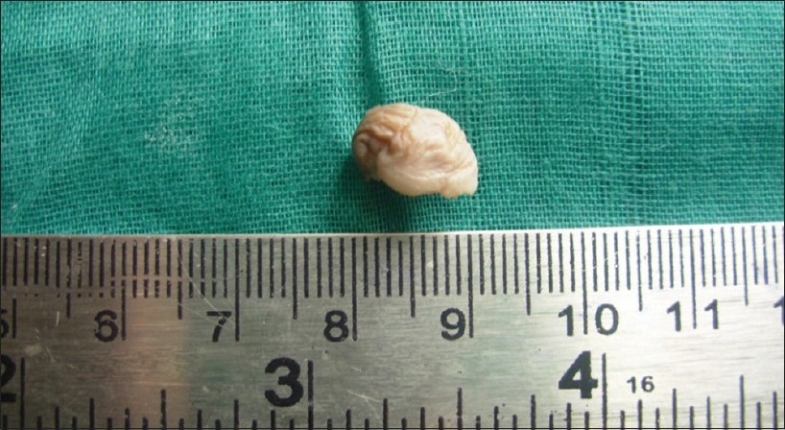
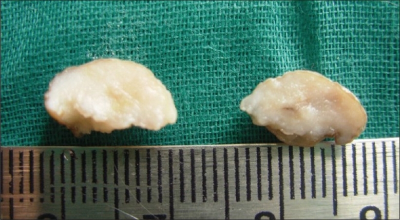
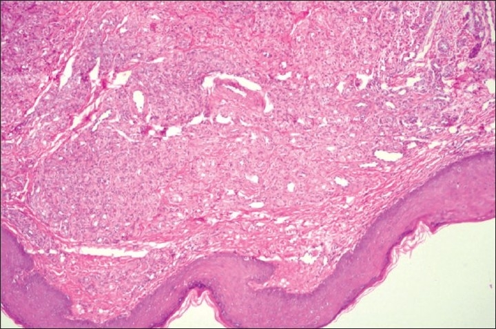
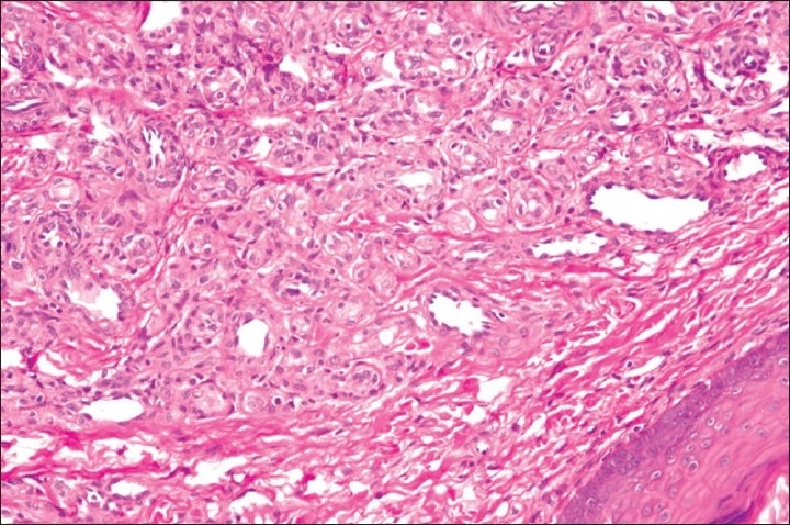
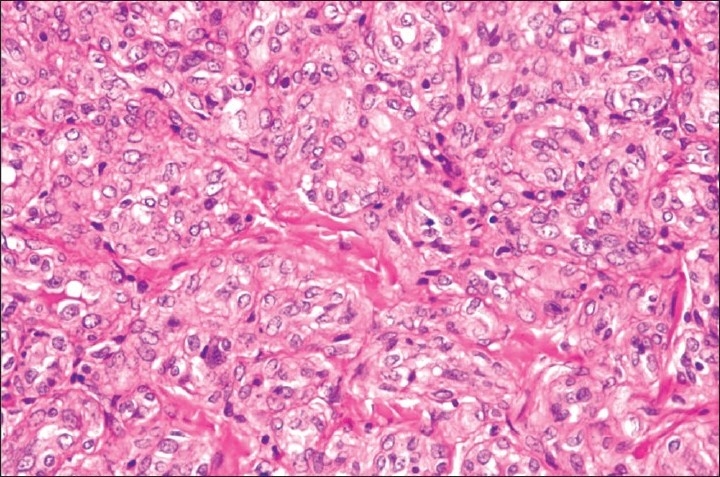
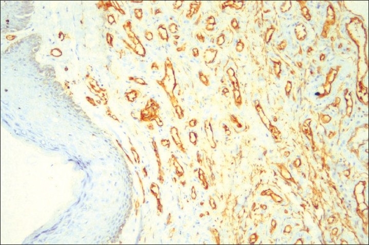
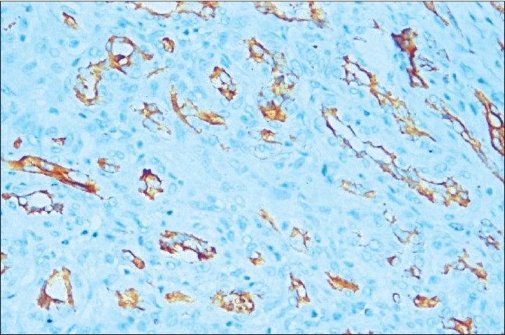
Hemangiomas occupy a grey zone between hamartomatous malformations and true neoplasms. They are frequently designated and regarded as neoplasms because of their usually localized nature and mass effect. Although clearly benign, they can become very large and unsightly, and can even be fatal if they affect vital structures. They almost never become malignant, although a few documented examples of this complication are on record. A high percentage occur in children, manifesting within the first month of life. One half of these cases are in the head and neck area. Hemangiomas have been classified according to their clinical appearance and the caliber of vessel involved, namely, capillary, cavernous and venous. Capillary hemangiomas are made up of small vessels of capillary caliber. One such capillary hemangioma, the juvenile hemangioma (JH), is usually present at birth or appears during the first month and enlarges rapidly during the first few months of life (infancy), only to stop growing when the child is approximately 6 years old. We present one such JH, seen in a 3 year old male child, which appeared when the child was 2 months old. Routine histopathological (H and E) and immunohistochemical analysis (CD 34, CD 31) was done on biopsy received.
Keywords: Capillary hemangioma; infancy; juvenile hemangioma.
Conflict of interest statement
Figures
Similar articles
-
[Cervico-cephalic hemangiomas and vascular malformations. Histopathological appearance and classification].J Mal Vasc. 1992;17(1):20-5. J Mal Vasc. 1992. PMID: 1588229 Review. French.
-
Results of argon laser exposure of capillary hemangiomas of infancy--preliminary report.Plast Reconstr Surg. 1981 Feb;67(2):188-93. Plast Reconstr Surg. 1981. PMID: 7465667
-
Cutaneous vascular proliferation. Part II. Hyperplasias and benign neoplasms.J Am Acad Dermatol. 1997 Dec;37(6):887-919; quiz 920-2. doi: 10.1016/s0190-9622(97)70065-3. J Am Acad Dermatol. 1997. PMID: 9418757 Review.
-
Periocular Capillary Infantile Hemangiomas.2024 Dec 11. In: StatPearls [Internet]. Treasure Island (FL): StatPearls Publishing; 2025 Jan–. 2024 Dec 11. In: StatPearls [Internet]. Treasure Island (FL): StatPearls Publishing; 2025 Jan–. PMID: 30855837 Free Books & Documents.
-
[Hemangiomas and vascular malformations of the head and neck].Otolaryngol Pol. 2006;60(5):663-74. Otolaryngol Pol. 2006. PMID: 17263237 Review. Polish.
Cited by
-
Capillary hemangioma in the ileum: Obscure small-bowel bleeding in an elderly person.Turk J Gastroenterol. 2018 Jul;29(4):520-521. doi: 10.5152/tjg.2018.17612. Turk J Gastroenterol. 2018. PMID: 30249572 Free PMC article. No abstract available.
-
Juvenile capillary hemangioma.Indian Dermatol Online J. 2016 Mar-Apr;7(2):125-6. doi: 10.4103/2229-5178.178079. Indian Dermatol Online J. 2016. PMID: 27057499 Free PMC article. No abstract available.
-
Vascular tumors of the mediastinum.Mediastinum. 2020 Sep 30;4:25. doi: 10.21037/med-20-40. eCollection 2020. Mediastinum. 2020. PMID: 35118293 Free PMC article. Review.
References
-
- Walsh TS, Tompkins VN. Some observations on the strawberry nevus of infancy. Cancer. 1956;9:869. - PubMed
-
- Jang YC, Arumugam S, Ferguson M, Gibran NS, Isik FF. Changes in matrix composition during the growth and regression of human hemangiomas. J Surg Res. 1998;80:9–15. - PubMed
-
- North PE, Waner M, Mizerack A, Mrak RE, Nicholas R, Kincannon J, et al. A unique microvascular phenotype shared by juvenile hemangiomas and human placenta. Arch Dermatol. 2001;137:559. - PubMed
-
- Barnes L, Eveson JW, Reichart P, Sidransky D. World Health Organisation Classification of Tumors: Pathology and genetics of head and neck tumors. Lyon, France: IARC Press; 2005.
