. 2011 Dec;7(12):e1002343.
doi: 10.1371/journal.ppat.1002343. Epub 2011 Dec 1.

Feeding cells induced by phytoparasitic nematodes require γ-tubulin ring complex for microtubule reorganization

Affiliations

Feeding cells induced by phytoparasitic nematodes require γ-tubulin ring complex for microtubule reorganization

Mohamed Youssef Banora et al. PLoS Pathog. 2011 Dec.

Abstract

Reorganization of the microtubule network is important for the fast isodiametric expansion of giant-feeding cells induced by root-knot nematodes. The efficiency of microtubule reorganization depends on the nucleation of new microtubules, their elongation rate and activity of microtubule severing factors. New microtubules in plants are nucleated by cytoplasmic or microtubule-bound γ-tubulin ring complexes. Here we investigate the requirement of γ-tubulin complexes for giant feeding cells development using the interaction between Arabidopsis and Meloidogyne spp. as a model system. Immunocytochemical analyses demonstrate that γ-tubulin localizes to both cortical cytoplasm and mitotic microtubule arrays of the giant cells where it can associate with microtubules. The transcripts of two Arabidopsis γ-tubulin (TUBG1 and TUBG2) and two γ-tubulin complex proteins genes (GCP3 and GCP4) are upregulated in galls. Electron microscopy demonstrates association of GCP3 and γ-tubulin as part of a complex in the cytoplasm of giant cells. Knockout of either or both γ-tubulin genes results in the gene dose-dependent alteration of the morphology of feeding site and failure of nematode life cycle completion. We conclude that the γ-tubulin complex is essential for the control of microtubular network remodelling in the course of initiation and development of giant-feeding cells, and for the successful reproduction of nematodes in their plant hosts.


Conflict of interest statement

The authors have declared that no competing interests exist.

Figures
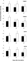
Figure 1
Figure 1. Analysis of Expression Levels of TUBG1, TUBG2, GCP3 and GCP4 in Galls.
Relative amount of transcripts of TUBG1, TUBG2, GCP3 and GCP4 genes in Arabidopsis galls 7, 14 and 21 DAI (white bars) with Meloidogyne incognita by quantitative RT-PCR in comparison to uninfected condition (black bars). All values were normalized according to qBase with the two reference genes At5g62050 and At5g10790, and expressed as normalized relative transcript quantities. The bars are means ±SD of three independent biological replicates.
Figure 2
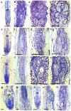
Figure 2. Histological Analysis of Galls and Roots in γ-Tubulin Mutant and Wild-Type Arabidopsis seedlings.
Bright-field images of sections stained with toluidine blue. (A) Uninfected root of wild-type seedlings 40 DAS. (B) Gall in wild type roots 7 DAI. (C) Gall in wild-type roots 14 DAI. (D) Gall in wild-type roots 21 DAI. (E) Uninfected root of the γ-tubulin mutant tubg1-1 seedlings 40 DAS. (F) Gall in tubg1-1 mutant 7 DAI. (G) Gall in tubg1-1 mutant 14 DAI. (H) Gall in tubg1-1 mutant 21 DAI. (I) Uninfected root of the γ-tubulin mutant tubg2-1 seedlings 40 DAS. (J) Gall in tubg2-1 mutant 7 DAI. (K) Gall in tubg2-1 mutant 14 DAI (L) Gall in tubg2-1 mutant 21 DAI. (M) Uninfected root of γ-tubulin double mutant tubg1-1 tubg2-2. (N) Gall in tubg1-1 tubg2-2 3 DAI. (O) Gall in tubg1-1 tubg2-2 7 DAI. (P) Gall in amiR-GCP4-9 7 DAI. (Q) Gall in amiR-GCP4-9 14 DAI. UR, uninfected root; Asterisks, giant cell; G, gall; n, nematode. Bars = 100 µm (A) to (D); 50 µm (E) to (L); 20 µm (M) to (Q).
Figure 3
Figure 3. Nematode Infection Test of γ-Tubulin Mutants tubg1-1 and tubg2-1 Compared to Wild-Type WS or Col-0.
The number of galls (white bars) and egg-masses (lined bars) are significantly decreased in nematode infected roots of both mutant lines compared to wild-type. Data shown represent means±SD from at least two experiments in which a minimum of 60 seedlings of each line were evaluated for nematode infection. Statistically significant differences were determined by the one-way-ANOVA using the SPSS for Windows statistical data analysis package (P≤0.05).
Figure 4
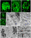
Figure 4. Immunofluorescence Detection of γ-Tubulin on Galls and Roots in Mutants and Wild-Type Arabidopsis seedlings.
Galls 14 DAI of wild-type (A), of tubg1-1 (B), of tubg2-1 (C). Uninfected root of wild-type seedling (D). Gall 7 DAI of tubg1-1 tubg2-2 (E). Dissected galls (14 DAI) were sectioned and processed for double immunoelectron microscopy with anti-γ- and α-tubulin primary antibodies, followed by secondary 10 and 5 nm gold-conjugated antibody respectively. (F) γ-tubulin (white arrows) is localized with cortical MT (black arrow) and associated with α-tubulin (blue arrows). (G) γ-tubulin binds to cytoplasmic MT. (H), (H') and (H'') γ-tubulin is dispersed throughout the misaligned phragmoplast MTs and co-localizes with α-tubulins. Fragments of cell wall are visible at MT ends as dark patches (red arrows) possibly inducing failure in giant cell cytokinesis. Asterisks, giant cell; n, nematode, UR, uninfected root; NC, neighboring cells; CMT, cortical microtubule; Cp* giant cell cytoplasm; CW, cell wall; MT, microtubule; Nu, nucleus. Bars = 50 µm in (A) to (E), 500 nm in (F) and (G) and 5 µm in (H).
Figure 5
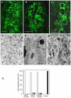
Figure 5. γ-Tubulin Complex Protein 3 (GCP3) is Present in Giant Cells and Co-localizes with γ-Tubulin.
(A) to (C) Immunostaining of GCP3 (green) in galls 14DAI of wild-type and mutant lines. Gall in wild-type roots (A). Galls in tubg1-1 roots (B). Galls in tubg2-1 roots (C). Dissected wild type galls (14 DAI) were sectioned and processed for double immunoelectron microscopy with anti-GCP3 and γ-tubulin primary antibodies, followed by secondary 10 and 5 nm gold-conjugated antibody respectively. GCP3 (black arrows) and γ-tubulin (white arrows) co-localize in the cytoplasm (D) and at the nuclear surface (E, E'). Both proteins are also present as monomers in the cytoplasm. (F) Histogram illustrating distances between gold particles showing that GCP3 and γ-tubulin are often in proximity less than 10 nm suggesting their interaction. White bars are for distances less then 10 nm, grey for distances between 10 and 50 nm, and black bars for distances above 50 nm. Asterisks, giant cell; n, nematode; Nu, nucleus; Cp*, giant cell cytoplasm. Bars = 50 µm (A) to (C); 100 nm (D); 300 nm (E).
Figure 6
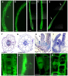
Figure 6. Ectopic Expression of γ-Tubulin in Arabidopsis thaliana Seedlings Causes Root Twisting and Leaf Curling.
γ-Tubulin overexpressing roots show twisted phenotype (A) and (B). Roots treated with either oryzalin (C) or propyzamide (D) did not show root twisting as observed (arrows) in the γ-tubulin overexpressing untreated roots (E). Cross section of a γ-tubulin overexpressing root stained with toluidine blue showed miss-shaping of the root cells and disorganization of the root tissues (F) compared to the wild-type (G). Longitudinal section of a shoot apical meristem stained with toluidine blue showing a leaf curling phenotype (black arrow) of γ-tubulin overexpressing seedlings (H) compared to the wild-type (I). γ-tubulin localization (green) in the root elongation zone (J) and lateral root meristem showing a patchy expression pattern (K). γ-tubulin localization along a spindle (L) and a phragmoplast (M). Bars = 100 µm in (A) to (E); 50 µm (F) to (I), (J) and (K); 20 µm (L) and (M).
Figure 7
Figure 7. γ-Tubulin Localization and Overexpression in Giant Cells of Nematode Infected Roots of the TUBG1-GFP line.
(A) γ-Tubulin localization in an uninfected root. (B) γ-Tubulin localization in a whole gall. (C) γ-Tubulin localization in young giant cells 3 DAI (D) and 5 DAI. (E) γ-Tubulin localization in a whole gall 7 DAI and (E') detail of a giant cell showing accumulation of γ-tubulin protein close to the nematode head (arrow). (F) γ-Tubulin localization in a giant cell 10 DAI. (G) γ-Tubulin localization around the nuclei of a giant cell 7 DAI. (H) Giant cell overexpressing γ-tubulin 7 DAI. (I) Mitotic events in a giant cell (white arrows) overexpressing γ-tubulin 14 DAI. (J) Giant cell overexpressing γ-tubulin 21 DAI. (K) Giant cell in a wild-type gall 14 DAI. UR, uninfected root; Asterisks, giant cell; G, gall; NC, neighbouring cells; n, nematode, Nu, nucleus. Bars = 50 µm in (A) and (B), 20 µm in (C), (E), (H) and (K), 10 µm in (D), (G), (I) and (J), 5 µm (F).
Figure 8
Figure 8. Giant Cell Area, Number of Nuclei and Infection Tests of γ-Tubulin Mutants Compared to Wild-Type.
Giant cell area (A) and number of nuclei (B) in roots under ectopic expression of γ-tubulin compared to wild-type and nematode infection test (C) of roots under ectopic expression of γ-tubulin. Area was measured on 60 giant cells. Number of nuclei was counted on 60 giant cells. The number of galls (white bars) and egg-masses (lined bars) are significantly decreased under ectopic expression of γ-tubulin compared to wild-type. Data shown represent means±SD from at least two experiments in which a minimum of 60 seedlings of each line were evaluated for nematode infection. Statistically significant differences were determined by the one-way-ANOVA using the SPSS for Windows statistical data analysis package (P≤0.05).
Figure 9
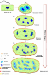
Figure 9. Microtubule Organization in Giant Cells.
The model of giant cell cytoskeleton reorganisation is based on observations of a large number of gall sections. The illustrations do not precisely reflect the total number of chromosomes or nuclei effectively present per giant-cell. Root-knot nematodes invade root cells and induce vascular cells (A) to become giant-feeding cells (B) to (F). The first visible symptom of nematode infection on the microtubule cytoskeleton of a young giant cell is the increase in density of tubulins in the cytoplasm. At this stage, the first nuclear division results into two enlarged nuclei with outsized nucleoli (B). Giant cells contain a dense cytoplasm and scarce cytoplasmic microtubules which co-localize with GCP3 protein. Young giant cells contain a dense network of randomly distributed cortical microtubules bound to γ-tubulins, and GCP3 (A) to (F). During giant cell expansion the cytoplasm contain scarce microtubules, and γ-tubulins and GCP3 proteins. The cytoplasmic microtubules remain disarrayed throughout giant cell development. Mitotic giant cells harbour nuclei containing a large number of condensed chromosomes often dividing in synchrony (C) to (E). During prophase nuclei are often clearly separated containing their packed chromosomes (C). In the course of metaphase to telophase spindles are large and malformed (D). Accumulation of γ-tubulin and GCP3 occurs mainly around the chromosomes and on the spindles. γ-tubulin and GCP3 localize to giant cell phragmoplasts which are misaligned and fail to expand centrifugally resulting in aborted cytokinesis (E). Some nuclei appear to show incomplete division or to have fused (F). The density of cortical microtubules is reduced and and cytoplasmic microtubules are sparse. Mature giant cells finally present multiple lobed nuclei which recurrently appear connected to each other, are surrounded by γ-tubulin and GCP3 proteins which are often co-localized suggesting the presence of MTOCS at these sites of giant cells. Our data suggests that γ-tubulin and GCP3 recruitment contributes to microtubule nucleation in mitotic and cortical arrays in root-knot nematode feeding cells.

Similar articles

Cited by

References

    1. Jansky SH, Simon R, Spooner DM. A test of taxonomic predictivity: Resistance to early blight in wild relatives of cultivated potato. Phytopathology. 2008;98:680–687. - PubMed
    1. Wyss U, Grundler FMW, Munch A. The parasitic behavior of second-stage juveniles of Meloidogyne incognita in roots of Arabidopsis thaliana. Nematologica. 1992;38:98–111.
    1. Von Mende N. Invasion and migration behaviour of sedentary nematodes. In: Fenoll C, Grundler FMW, Ohl SA, editors. Cellular and molecular aspects of plant-nematodes interactions. Vol. 21. Dordrecht, The Netherlands: Kluwer Academics Publishers; 1997. pp. 1129–1140.
    1. Jones MGK. The development and function of plant cells modified by endoparasitic nematodes. In: Zuckerman BM, Rhode RA, editors. Plant Parasitic Nematodes. New York: Academic Press; 1981. pp. 225–279.
    1. Wiggers RJ, Starr JL, Price HJ. DNA content and variation in chromosome number in plant cells affected by Meloidogyne incognita and M. arenaria. Phytopathology. 1990;80:1391–1395.

MeSH terms