. 2011 Dec;7(12):e1002402.
doi: 10.1371/journal.ppat.1002402. Epub 2011 Dec 1.

Follicular dendritic cell-specific prion protein (PrP) expression alone is sufficient to sustain prion infection in the spleen

Affiliations

Follicular dendritic cell-specific prion protein (PrP) expression alone is sufficient to sustain prion infection in the spleen

Laura McCulloch et al. PLoS Pathog. 2011 Dec.

Abstract

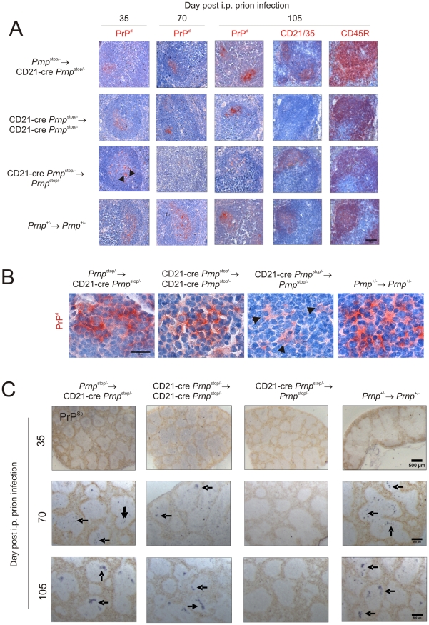
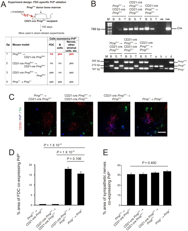
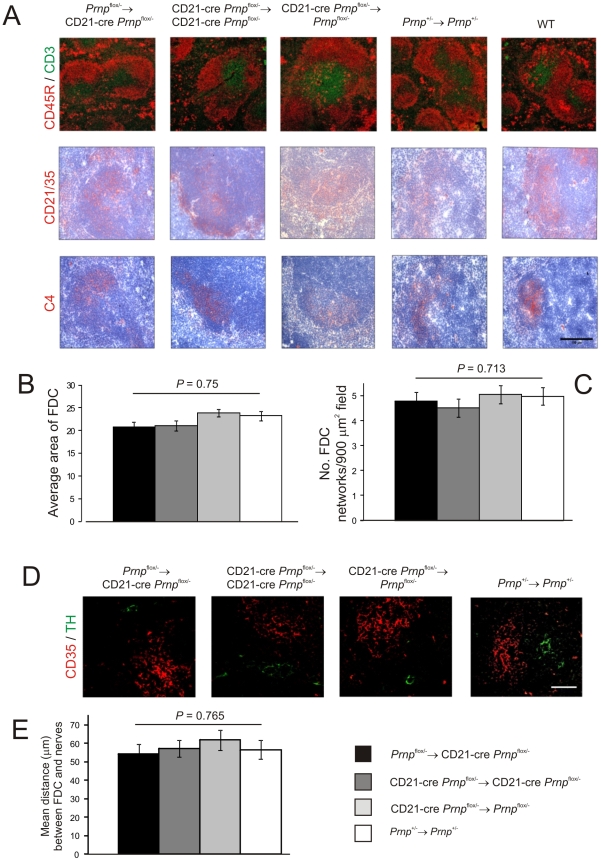
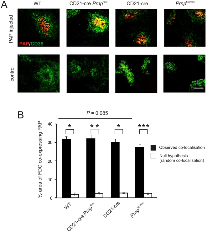
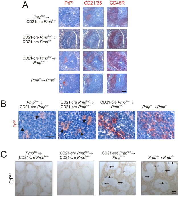
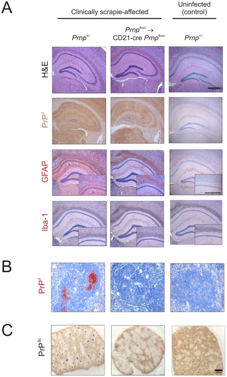
Prion diseases are characterised by the accumulation of PrP(Sc), an abnormally folded isoform of the cellular prion protein (PrP(C)), in affected tissues. Following peripheral exposure high levels of prion-specific PrP(Sc) accumulate first upon follicular dendritic cells (FDC) in lymphoid tissues before spreading to the CNS. Expression of PrP(C) is mandatory for cells to sustain prion infection and FDC appear to express high levels. However, whether FDC actively replicate prions or simply acquire them from other infected cells is uncertain. In the attempts to-date to establish the role of FDC in prion pathogenesis it was not possible to dissociate the Prnp expression of FDC from that of the nervous system and all other non-haematopoietic lineages. This is important as FDC may simply acquire prions after synthesis by other infected cells. To establish the role of FDC in prion pathogenesis transgenic mice were created in which PrP(C) expression was specifically "switched on" or "off" only on FDC. We show that PrP(C)-expression only on FDC is sufficient to sustain prion replication in the spleen. Furthermore, prion replication is blocked in the spleen when PrP(C)-expression is specifically ablated only on FDC. These data definitively demonstrate that FDC are the essential sites of prion replication in lymphoid tissues. The demonstration that Prnp-ablation only on FDC blocked splenic prion accumulation without apparent consequences for FDC status represents a novel opportunity to prevent neuroinvasion by modulation of PrP(C) expression on FDC.


Conflict of interest statement

The authors have declared that no competing interests exist.

Figures

Figure 1
Figure 1. Cre-mediated gene recombination in FDC in the spleens, lymph nodes and Peyer's patches of CD21-Cre ROSA26flox/Flox mice.
A) Analysis of the cellular sites of LacZ expression (blue) in the spleens, inguinal lymph nodes, Peyer's patches and mesenteric lymph nodes of CD21-Cre ROSA26flox/flox mice shows Cre-mediated recombination in a focus of cells within the B cell follicles. Sections were counterstained with nuclear fast red (red). B) IHC analysis of FDC (CD35+ cells, upper row, red) and B cells (CD45R+ cells, lower row, red) confirmed that Cre-mediated LacZ expression (blue) was associated with FDC in the spleens of WT→CD21-Cre ROSA26flox/flox mice. No LacZ expression was associated with FDC in spleens from ROSA26flox/flox mice that lacked Cre. C) IHC analysis of the status of FDC (CD35+ and C4-binding cells; red) and B cells expressing CD45R, CD19, and CD1d (red) in spleens from WT, CD21-Cre ROSA26flox/flox, WT→CD21-Cre ROSA26flox/flox and ROSA26flox/flox mice. Scale bars 100 µm. n = 6 mice/group.
Figure 2
Figure 2. FDC-restricted PrPc expression in the spleens of Prnp stop/-→CD21-Cre Prnp stop/- mice.
A) The anticipated distribution of PrPC expression on FDC and B cells in tissues from each mouse group. B) The detection of Cre in the tail and spleen but not blood of the Prnp stop/-→CD21-Cre Prnp stop/- mice confirmed the restriction of the Cre-expression to the stromal but not haematopoietic compartments of these mice (upper panel). Efficient Cre-mediated recombination of Prnpstop (Prnpstop (R)) was restricted to the FDC-containing stromal compartment of the spleens of Prnp stop/-→CD21-Cre Prnp stop/- mice when compared to control mice. Cre-mediated recombination by CD21-expressing lymphocytes was efficiently prevented in these mice by the irradiation and transfer of Prnp stop/- bone marrow as demonstrated by the lack of a Prnp stop(R) band in DNA extracted from blood (lower panel). B, blood; S, spleen; T, tail; M, DNA size markers; a, b, c, control DNA samples for each transgene combination tested which were (a) Prnp -/-, (b) Prnp stop/- and (c) complete recombination of the stop cassette within the Prnp stop/- allele. C) IHC analysis of PrPC expression (blue) by FDC (CD35+ cells; red) and sympathetic nerves (TH+ cells, green) confirmed PrPC expression was restricted to FDC in spleens of Prnp stop/-→CD21-Cre Prnp stop/- mice. Scale bar, 100 µm. D) Morphometric analysis confirmed that the magnitude of the PrPC expression co-localized upon the surfaces of FDC in the spleens of Prnp stop/-→CD21-Cre Prnp stop/- mice was similar to that observed upon FDC in spleens from Prnp +/-Prnp +/- control mice (p<0.690, n = 48 FDC networks/group). In contrast, in the absence of Cre-recombinase expression by FDC in CD21-Cre Prnp stop/-Prnp stop/- mice, PrPC expression was substantially lower than that observed upon FDC in spleens from Prnp +/-Prnp +/- control mice (p<1×10-25, n = 48 FDC networks/group). E) Morphometric analysis confirmed that PrPC expression upon the surfaces of sympathetic nerves in the spleens of Prnp stop/-→CD21-Cre Prnp stop/-, CD21-Cre Prnp stop/-→ CD21-Cre Prnp stop/- and CD21-Cre Prnp stop/-Prnp stop/- mice was significantly ablated when compared to that observed upon sympathetic nerves in spleens from Prnp +/-Prnp +/- control mice (p<1×10-25, n = 48 sympathetic nerves/group). For all panels n = 6 mice/group.
Figure 3
Figure 3. Effect of FDC-restricted PrPc expression on the spleens of Prnp stop/-→CD21-Cre Prnp stop/- mice.
A) IHC analysis of the status of FDC networks (C4-binding cells and CD21/CD35+ cells; red), B cells expressing CD45R (red), and CD3+ T cells (green). Morphometric analysis confirmed that there were no significant difference in the size (B) and number (C) of the CD35+ FDC networks in spleens from Prnp stop/-→CD21-Cre Prnp stop/-, CD21-Cre Prnp stop/-→CD21-Cre Prnp stop/-, CD21-Cre Prnp stop/-Prnp stop/- mice and Prnp +/-Prnp +/- control mice (n = 32 FDC networks/group). D and E), Comparison of the sympathetic innervation in spleens from each mouse group. D) IHC detection of TH-positive sympathetic nerves (green) and FDCs (CD35+ cells; red). Scale bar, 50 µm. E) Quantitative analysis of the relative positioning of the FDC and sympathetic nerves showed there were no significant differences in average distance between these cell populations in spleens from each mouse group (P = 0.932, n = 48 FDC networks/group). For all panels n = 6 mice/group.
Figure 4
Figure 4. Effect of FDC-restricted PrPc expression on PrPSc accumulation in the spleen.
Mice were injected i.p. with the ME7 scrapie agent and tissues collected 35, 70 days and 105 days after exposure. A and B) High levels of PrPd were detected in association with FDC (CD21/35 positive cells) in the B cell follicles (CD45R positive cells) of spleens of mice with PrPC-expressing FDC: Prnp stop/-→CD21-Cre Prnp stop/- mice, CD21-Cre Prnp stop/-→CD21-Cre Prnp stop/- mice and Prnp +/-Prnp +/- control mice. B) High magnification images of the sites of PrPd accumulation (red) at 70 days post-injection with scrapie. Arrowheads show PrP-accumulation within tingible body macrophages. C) Analysis of adjacent sections by PET-immunoblot analysis confirmed the presence of PK-resistant PrPSc (blue/black). In contrast, no PrPd or PrPSc was detected in spleens of CD21-Cre→Prnp stop/-mice that lacked PrPC-expressing FDC. Arrows indicate PrPSc accumulation upon FDC. A, scale bar  = 100 µm. B, scale bar  = 20 µm. C, scale bar  = 500 µm. For all panels n = 4 mice/group.
Figure 5
Figure 5. FDC-restricted PrPc-ablation in the spleens of Prnp flox/-→CD21-Cre Prnp flox/- mice.
A) The anticipated distribution of PrPC expression on FDC and B cells in tissues from each mouse group. B) PCR analysis of DNA isolated from the spleens, blood and tails of Prnp flox/-→CD21-Cre Prnp flox/- mice confirmed that efficient Cre-mediated DNA recombination and Prnp-ablation (Prnp deflox) was restricted to the FDC-containing stromal compartment of the spleen. Cre-mediated recombination of CD21-expressing lymphocytes was efficiently prevented in these mice by the irradiation and transfer of Prnp flox/- bone marrow as demonstrated by the lack of a Prnp deflox band in DNA extracted from blood (lower panel). B, blood; S, spleen; T, tail; M, DNA size markers; a, b, c, d control DNA samples for each transgene combination tested which were (a) Prnp flox/flox,(b) Prnp flox/-, (c) Prnp -/- and (d) Prnp flox/- with complete recombination of the floxed exon 3. C) IHC analysis of PrPC expression (blue) by FDC (CD35+ cells; red) and sympathetic nerves (TH+ cells, green) confirmed the PrPC-ablation was restricted to FDC in spleens of Prnp flox/-→CD21-Cre Prnp flox/- mice. Scale bar, 100 µm. D) Morphometric analysis confirmed that the magnitude of the PrPC expression co-localized upon the surfaces of FDC in the spleens of Prnp flox/-→CD21-Cre Prnp flox/- mice was significantly lower than that observed upon FDC from Prnp +/-Prnp +/- control mice (P<1.0×10⁻²⁴, n = 48 FDC networks/group). In contrast, in the absence of Cre-recombinase expression by FDC in CD21-Cre Prnp flox/-Prnp flox/-mice, PrPC expression was similar to that observed upon FDC in spleens from Prnp +/-Prnp +/- control mice (P = 0.106, n = 48 FDC networks/group). E) Morphometric analysis confirmed that the magnitude of the PrPC expression co-localized upon the surfaces sympathetic nerves in the spleens of Prnp flox/-→CD21-Cre Prnp flox/-, CD21-Cre Prnp flox/-→ CD21-Cre Prnp flox/- and CD21-Cre Prnp flox/-Prnp flox/- mice was not significantly different when compared to that observed upon sympathetic nerves in spleens from Prnp +/-Prnp +/- control mice (p = 0.400, n = 48 sympathetic nerves/group). For all panels n = 6 mice/group.
Figure 6
Figure 6. Effect of FDC-restricted PrPC-ablation on FDC status.
A) IHC analysis of the status of FDC (C4-binding cells and CD21/CD35+ cells; red), B cells expressing CD45R (red), and CD3+ T cells (green). Morphometric analysis confirmed that there were was no significant difference in the size (B) and number (C) of the CD35+ FDC networks in spleens of mice from each mouse group (n = 32 FDC networks/group). D and E), Comparison of the sympathetic innervation in spleens from Prnp flox/-→CD21-Cre Prnp flox/-, CD21-Cre Prnp flox/-→CD21-Cre Prnp flox/-, CD21-Cre Prnp flox/-Prnp flox/-mice and Prnp +/-Prnp +/- control mice. D) IHC detection of TH-positive sympathetic nerves (green) and FDCs (CD35+ cells; red). Scale bar, 50 µm. E) Quantitative analysis of the relative positioning of the FDC networks and sympathetic nerves showed there was no significant difference in the average distance between these cell populations in spleens from each mouse group (P = 0.765, n = 48 FDC networks/group). For all panels n = 6 mice/group.
Figure 7
Figure 7. Effect of FDC-restricted PrPC-ablation on immune complex trapping.
A) Mice were passively immunized with preformed PAP immune complexes, and 24 h later, the presence of immune complexes (red) upon FDC (CD35+ cells, green) assessed by IHC. Scale bar, 100 µm. B) Morphometric analysis confirmed that the magnitude of the immune complex-trapping co-localized upon the surfaces of FDC from Prnp-ablated Prnp flox/-→CD21-Cre Prnp flox/- mice was not significantly different from that observed in spleens from control mice. This analysis also confirmed that the immune complexes were preferentially associated with FDC in these tissues and significantly greater than the null hypothesis that the pixels were randomly distributed. *, P<1×10⁻²¹; **, P<1×10⁻³²; *** P<9×10⁻²⁸; n = 40 FDC networks/group. For all panels n = 6 mice/group.
Figure 8
Figure 8. Effect of FDC-restricted PrPC-ablation on PrPSc accumulation in the spleen.
Mice were injected i.p. with the ME7 scrapie agent and tissues collected 70 days after exposure. A) High levels of PrPd (red, left-hand column) were detected in association with FDC (red, middle column) in the B cell follicles (red, right-hand column) of spleens from CD21-Cre Prnp flox/-Prnp flox/-mice and Prnp +/-Prnp +/- control mice that contained PrPC-expressing FDC. B) High magnification images of the sites of PrPd accumulation (red) at 70 days post-injection with scrapie. C) PET blot analysis of analysis of adjacent sections by PET-immunoblot analysis confirmed presence of PK-resistant PrPSc (blue/black). In contrast, no PrPSc was detected in spleens of Prnp flox/-→CD21-Cre Prnp flox/- and CD21-Cre Prnp flox/-→CD21-Cre Prnp flox/- mice that lacked PrPC-expressing FDC. In the spleens of some of these mice, low levels of PrPd were occasionally localised within tingible body macrophages (B, arrowheads). A, scale bar  = 100 µm. B, scale bar  = 20 µm. C, scale bar  = 500 µm. Arrows indicate PrPSc accumulation upon FDC. For all panels n = 4 mice/group.
Figure 9
Figure 9. Effect of FDC-restricted PrPC-ablation on PrPSc accumulation in the brains and spleens of scrapie-affected mice.
Control mice (Prnp +/- mice) and Prnp flox/-→CD21-Cre Prnp flox/- mice that lacked PrPC-expressing FDC were injected i.c. with the scrapie agent directly into the CNS. Brains and spleens were collected from clinically scrapie-affected mice to compare the neuropathology and cellular sites of PrPSc accumulation. A) High levels of spongiform pathology (H&E, upper row), heavy accumulations of PrPd (brown, second row), reactive astrocytes expressing GFAP (brown, third row) and active microglia expressing Iba-1 (brown, bottom row) were detected in the hippocampi of the brains of all clinically scrapie-affected mice. Scale bars, 500 µm. B) High levels of PrPd (red) were detected in association with FDC in spleens from clinically scrapie-affected control mice that contained PrPC-expressing FDC. C) PET blot analysis of analysis of adjacent sections by PET-immunoblot analysis confirmed the presence of PK-resistant PrPSc (blue/black). In contrast, no PrPd or PrPSc was detected in spleens of Prnp flox/-→CD21-Cre Prnp flox/- that lacked PrPC-expressing FDC. Scale bars  = 500 µm.

Similar articles

Cited by

References

    1. Mabbott NA, Young J, McConnell I, Bruce ME. Follicular dendritic cell dedifferentiation by treatment with an inhibitor of the lymphotoxin pathway dramatically reduces scrapie susceptibility. J Virol. 2003;77:6845–6854. - PMC - PubMed
    1. Prinz M, Huber G, Macpherson AJS, Heppner FL, Glatzel M, et al. Oral prion infection requires normal numbers of Peyer's patches but not of enteric lymphocytes. Am J Pathol. 2003;162:1103–1111. - PMC - PubMed
    1. Horiuchi M, Furuoka H, Kitamura N, Shinagawa M. Alymphoplasia mice are resistant to prion infection via oral route. Jap J Vet Res. 2006;53:149–157. - PubMed
    1. Glaysher BR, Mabbott NA. Role of the GALT in scrapie agent neuroinvasion from the intestine. J Immunol. 2007;178:3757–3766. - PubMed
    1. Andreoletti O, Berthon P, Marc D, Sarradin P, Grosclaude J, et al. Early accumulation of PrPSc in gut-associated lymphoid and nervous tissues of susceptible sheep from a Romanov flock with natural scrapie. J Gen Virol. 2000;81:3115–3126. - PubMed

Publication types