. 2011 Dec;7(12):e1002419.
doi: 10.1371/journal.ppat.1002419. Epub 2011 Dec 1.

Genesis of mammalian prions: from non-infectious amyloid fibrils to a transmissible prion disease

Affiliations

Genesis of mammalian prions: from non-infectious amyloid fibrils to a transmissible prion disease

Natallia Makarava et al. PLoS Pathog. 2011 Dec.

Abstract

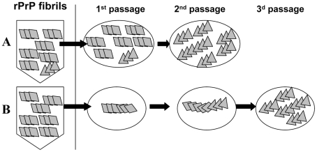
The transmissible agent of prion disease consists of a prion protein in its abnormal, β-sheet rich state (PrP(Sc)), which is capable of replicating itself according to the template-assisted mechanism. This mechanism postulates that the folding pattern of a newly recruited polypeptide chain accurately reproduces that of a PrP(Sc) template. Here we report that authentic PrP(Sc) and transmissible prion disease can be generated de novo in wild type animals by recombinant PrP (rPrP) amyloid fibrils, which are structurally different from PrP(Sc) and lack any detectable PrP(Sc) particles. When induced by rPrP fibrils, a long silent stage that involved two serial passages preceded development of the clinical disease. Once emerged, the prion disease was characterized by unique clinical, neuropathological, and biochemical features. The long silent stage to the disease was accompanied by significant transformation in neuropathological properties and biochemical features of the proteinase K-resistant PrP material (PrPres) before authentic PrP(Sc) evolved. The current work illustrates that transmissible prion diseases can be induced by PrP structures different from that of authentic PrP(Sc) and suggests that a new mechanism different from the classical templating exists. This new mechanism designated as "deformed templating" postulates that a change in the PrP folding pattern from the one present in rPrP fibrils to an alternative specific for PrP(Sc) can occur. The current work provides important new insight into the mechanisms underlying genesis of the transmissible protein states and has numerous implications for understanding the etiology of neurodegenerative diseases.


Conflict of interest statement

The authors have declared that no competing interests exist.

Figures

Figure 1
Figure 1. Serial passages of rPrP fibrils and de novo generation of PrPSc in Syrian hamsters.
(A) Western blotting of brain homogenate (BH) from the animal inoculated with BSA-annealed rPrP amyloid fibrils. Western blots were stained with 3F4 (left panel) or R1 (right panel) antibody. Undigested samples were loaded at 1/10th the amount of the digested samples. (B) Western blotting of BHs of animals from the 1st, 2nd, or 3rd passages of BSA-annealed rPrP fibrils (lanes 1, 2 and 3, respectively) stained with 3F4 (top panel) or SAF-84 (bottom panel) antibody. The new prion strain produced as a result of inoculation of BSA-annealed rPrP fibrils will be designated as LOTSS. The 1st, 2nd, or 3rd passages of SSLOW, the strain that was previously produced as a result of inoculation of normal BH (NBH)-annealed rPrP fibrils (lanes 4, 5 and 6 respectively) , or 263 K are provided as references. (C) sPMCAb of brain material from the 1st passage of LOTSS. 10% BH from the animal inoculated with BSA-annealed rPrP amyloid fibrils was diluted 10-fold into 10% RNA-depleted (NBH(-RNA)) or NBH and subjected to sPMCAb. Each PMCAb round consisted of 48 cycles, 30 min each; 10-fold dilutions were used for each subsequent sPMCAb round. Western blots were stained with 3F4 (top panel) or SAF-84 (bottom panel) antibody. Undigested 10% NBH is provided as a reference.
Figure 2
Figure 2. Analysis of PrP deposition in LOTSS-inoculated animals from the 2d passage.
(A, B) PrP plaques in the subependymal regions and minor synaptic immunoreactivity in the basal ganglia and thalamus. (C, D) PrP plaques in the subependymal regions in hippocampus and frontal cortex. (E, F) Very minor focal PrP deposition in cerebellum. (G, H) Plaques around the aqueductus and synaptic PrP immunoreactivity in the posterior and anterior horns of spinal cord.
Figure 3
Figure 3. Histopathological analysis of brains of LOTSS-inoculated animals.
Lesion profile (A) and PrP immunopositivity score (B) for hamsters from the 2nd (o, red lines) or 3rd passages (Δ, black lines) of LOTSS. The histopathological profiles for SSLOW-inoculated hamsters (2nd passage, ◊ blue line) were previously described in and are provided as a reference. The lesion profile was obtained by averaging the scores for spongiform change, neuronal loss and gliosis for three animals within each group. (C) Comparison of spongiform changes in the hippocampus, frontal cortex, thalamus, and cerebellum stained with Hematoxylin and Eosin (H&E; upper panels) or anti-PrP 3F4 antibody (lower panels) in hamsters from 2nd or 3rd passages of LOTSS, as indicated. Insets in H&E stained images of the frontal cortex and thalamus from LOTSS 2nd animals represent GFAP immunostaining, and enlargement of the PrP immunostaining. (D) Brain subependymal region stained with H&E and 3F4 antibody in LOTSS-inoculated animals from 2nd (left panel) or 3rd (right panel) passages. Scale bar = 00 µm in C and = 60 µm in D.
Figure 4
Figure 4. Analysis of epitope-specific conformational stability.
Conformational stability profiles for 263 K (A), SSLOW (B), or LOTSS (C) monitored in GdnHCl-induced denaturation assay using 3F4 (•, blue), D18 (▪, red) or SAF-84 (▴, green) antibodies. Data represent average ± SD of four denaturation experiments. Brain materials from SSLOW- or LOTSS-inoculated animals from the 2nd passages were used. 96-well dot blot was used instead of Western blot for quantitative analysis of multiple replicas in a single blot assay.
Figure 5
Figure 5. Evolution of PrPres during LOTSS serial transmission.
(A) Western blotting of BHs of animals from the 1st, 2nd, or 3rd passages of LOTSS stained with SAF-84 antibodies. Animals from the 1st and 2nd were euthanized at 661 days postinoculation in the absence of clinical disease, whereas animals from the 3rd passage were euthanized at the beginning of first clinical signs, i.e. 307 days postinoculation (lanes 4, 5 and 6), or at the end of the terminal stage (lanes 7, 8). Overexposed Western blot with 13 kDa atypical PrPres fragment is shown in bottom panel. Dashed line marks the central position of the atypical 16 kDa band. Undigested 10% NBH is provided as a reference. (B) Schematic representation of the dynamics in PrPres profile during serial transmission, where gray boxes represent atypical PrPres, whereas black boxes represent standard PrPres.
Figure 6
Figure 6. Comparison of PrP deposition at the early and late stages of the disease during the 3rd passage.
PrP deposition in frontal cortex, cerebellum or subependymal areas in two animals euthanized at 307 days postinoculation (A, B), and three animals euthanized at the terminal stage of the disease at 494, 521, and 494 days postinoculation (C, D, and E, respectively). Scale bar upper left represents 40 µm for all except for the right panels of the frontal cortex and subependymal images where it represents 5 µm. Arrowheads indicate perineuronal deposits, double arrowhead indicates fine synaptic immunoreactivity and arrows indicate granular immunoreactivity around ependymal cells.
Figure 7
Figure 7. Preparations of rPrP fibrils have no detectible PrPSc.
PMCAb titration of LOTSS brain material (A and B). (A) LOTSS brain material was serially diluted to up to 1013-fold and each dilution was subjected to six rounds of sPMCAb. Representative results are shown on Western blot stained with 3F4 antibody. Undigested 10% NBH is provided as a reference. (B) For each dilution of brain material, the fractions of sPMCAb reactions with a positive signal on Western blot were plotted against the logarithm of dilution (•) and non-linear least-square regression to a sigmoidal function (black solid line, R = 0.9999) was used to calculate PMCAb50 titer. (C) sPMCAb reactions were seeded with two independent preparations of BSA-annealed rPrP amyloid fibrils (lanes 2–7 with amyloid I or lanes 16–20 with amyloid II), 109-fold diluted LOTSS brain material (lanes 9, 10), or with amyloid fibrils II and 109-fold diluted LOTSS brain material (lanes 11–14), then six rounds of sPMCAb were conducted for each condition. The final concentraton of rPrP amyloid fibrils in sPMCAb reaction was 10 µg/ml for amyloid I or 5 µg/ml for amyloid II. Two non-seeded reactions are shown as negative control. Brains from LOTSS-inoculated animals from the 2nd passages were used for all experiments. Undigested 10% NBH is provided as a reference. Western blots were stained with 3F4.
Figure 8
Figure 8. Analysis of the C-terminal PrPres fragments.
Western blot of PK-digested BH from the animal inoculated with BSA-annealed rPrP amyloid fibrils (lanes 1, 2) or BSA-annealed rPrP amyloid fibrils (lanes 3 and 4) stained with SAF-84 (panel A) or D18 (panel B) antibodies. BH in lane 2 was treated with PNGase F. Before PK digestion, rPrP fibrils were mixed with 10% NBH to balance the condition for PK digestion. Samples in lanes 1, 2 and 3 were treated with 20 µg/ml of PK, and the sample in lane 4 – with 10 µg/ml of PK. Western blot was stained with SAF-84.
Figure 9
Figure 9. Schematic representation of two mechanisms responsible for generating transmissible prion diseases de novo.
According to the first mechanism (A), the preparations of rPrP amyloid fibrils contain very small amounts of PrPSc. The silent stage of the disease is attributed to the long time required for amplification of this extremely small amount of PrPSc. A second mechanism referred to as deformed templating postulates that there are no PrPSc particles in the preparations of amyloid fibrils (B). Instead, when inoculated into animals, amyloid fibrils can seed conversion of PrPC into PrPres, although with a low efficiency. PrPres undergoes slow transformation, a process that might require long silent stage, before authentic PrPSc emerges.

Similar articles

Cited by

References

    1. Prusiner SB. Prion diseases. In: Nathanson N, Ahmed R, Gonzalez-Scarano F, Griffin D, Holmes K, et al., editors. Viral Pathogenesis. New York: Raven Press; 1996. pp. 855–911.
    1. Prusiner SB. Novel proteinaceous infectious particles cause scrapie. Science. 1982;216:136–144. - PubMed
    1. Griffith JS. Self-replication and scrapie. Nature. 1967;215:1043–1044. - PubMed
    1. Legname G, Baskakov IV, Nguyen H-OB, Riesner D, Cohen FE, et al. Synthetic mammalian prions. Science. 2004;305:673–676. - PubMed
    1. Colby DW, Giles K, Legname G, Wille H, Baskakov IV, et al. Design and construction of diverse mammalian prion strains. Proc Acad Natl Sci U S A. 2009;106:20417–20422. - PMC - PubMed

Publication types