. 2011 Dec;7(12):e1002421.
doi: 10.1371/journal.ppat.1002421. Epub 2011 Dec 1.

Wall teichoic acids of Staphylococcus aureus limit recognition by the drosophila peptidoglycan recognition protein-SA to promote pathogenicity

Affiliations

Wall teichoic acids of Staphylococcus aureus limit recognition by the drosophila peptidoglycan recognition protein-SA to promote pathogenicity

Magda L Atilano et al. PLoS Pathog. 2011 Dec.

Abstract

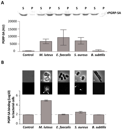
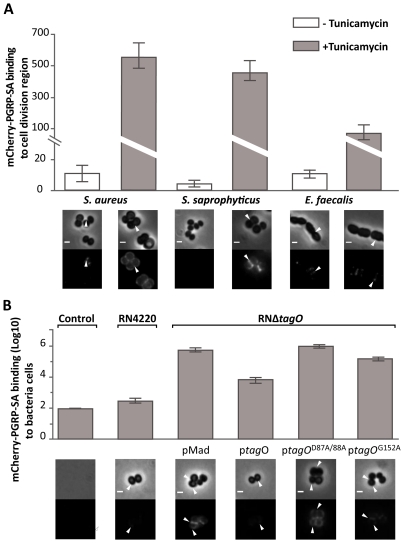
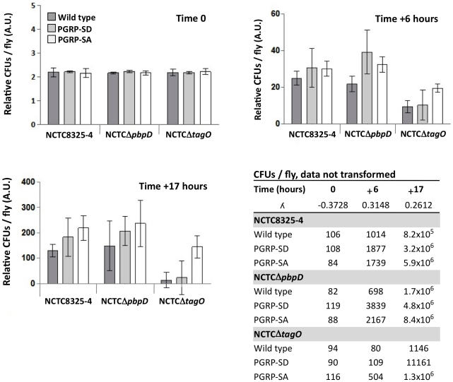
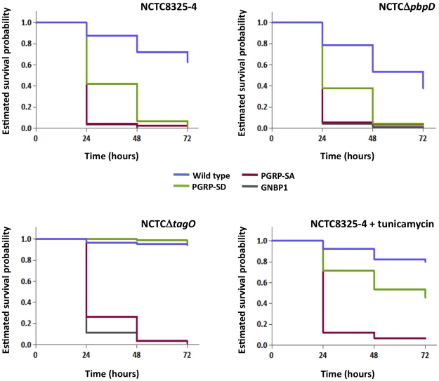
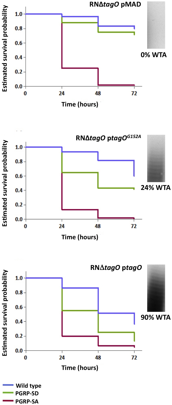
The cell wall of gram-positive bacteria is a complex network of surface proteins, capsular polysaccharides and wall teichoic acids (WTA) covalently linked to Peptidoglycan (PG). The absence of WTA has been associated with a reduced pathogenicity of Staphylococcus aureus (S. aureus). Here, we assessed whether this was due to increased detection of PG, an important target of innate immune receptors. Antibiotic-mediated or genetic inhibition of WTA production in S. aureus led to increased binding of the non-lytic PG Recognition Protein-SA (PGRP-SA), and this was associated with a reduction in host susceptibility to infection. Moreover, PGRP-SD, another innate sensor required to control wild type S. aureus infection, became redundant. Our data imply that by using WTA to limit access of innate immune receptors to PG, under-detected bacteria are able to establish an infection and ultimately overwhelm the host. We propose that different PGRPs work in concert to counter this strategy.


Conflict of interest statement

The authors have declared that no competing interests exist.

Figures

Figure 1
Figure 1. Differential binding of PGRP-SA to the surface of live Gram-positive bacteria.
(A) PGRP-SA and PG co-precipitation assay. Lys-type PG from M. luteus, E. faecalis, S. aureus, and DAP-type PG from B. subtilis (this acts as a negative substrate control for PGRP-SA binding, which recognizes Lys-type PG), was incubated with rPGRP-SA for 30 minutes. Unbound rPGRP-SA remained in the supernatant fraction upon centrifugation (S). rPGRP-SA bound to the insoluble PG was co-precipitated and found in the pellet fraction (P). Quantified data (performed using ImageJ software) was plotted as mean values with 95% confidence limits: very little co-precipitation of rPGRP-SA occurred in the absence of PG (labelled Control) or in the presence of B. subtilis DAP-type PG; however, PGRP-SA was co-precipitated similarly (One-way ANOVA, P>0.05) and at higher levels with the PG from M. luteus, E. faecalis, or S. aureus. The data shown (mean with 95% confidence intervals) was obtained from 4 independent co-precipitation experiments. (B) mCherry-PGRP-SA was incubated with bacteria cells harvested in exponential phase, washed with PBS and visualized using fluorescence microscopy. Grey panels are phase-contrast images of bacterial cells (white scale bar represents 1 µm), and black panels mCherry-PGRP-SA binding: white arrowheads highlight binding to the lateral cell surface or the region of cell division. The total fluorescence of mCherry-PGRP-SA bound to a bacterium (covering all lateral and cell division regions, and including background) was quantified for each species (n = 50), and represented as the median (with 25% and 75% inter-quartile range). Dashed-line indicates the level of the background signal, control. Kruskal-Wallis analysis with Dunn's multiple comparison post-test did not reveal significant differences (P>0.05) between mCherry-PGRP-SA binding to E. faecalis and B. subtilis, which were indistinguishable from the control. However, the protein bound more to S. aureus and M. luteus relative to the control, with the latter exhibiting highest binding (P<0.05 in all cases).
Figure 2
Figure 2. WTA reduce PGRP-SA binding at the bacterial cell surface.
Grey panels are phase-contrast images of bacterial cells (white scale bar represents 1 µm), and black panels mCherry-PGRP-SA binding; white arrowheads highlight binding to the lateral cell surface or region of cell division. The binding of mCherry-PGRP-SA to individual bacterial cells (n = 50) was quantified, and represented as the median (with 25% and 75% inter-quartile range). (A) mCherry-PGRP-SA binding to Gram-positive bacteria grown with or without tunicamycin, an inhibitor of WTA synthesis. mCherry-PGRP-SA binding to the cell division region, rather than total binding, was measured because binding at the former was consistently enhanced for all treated bacteria species. Mann-Whitney U tests were used to compare differences for treated and untreated between each type of bacteria (P<0.05 in all cases). (B) RNΔtagO mutant background rescued with variants of the tagO gene – expressed from a replicative pMAD vector – produce varying levels of WTA, given as a% relative to the wild type RN4220: pMAD vector (0%), ptagO (90%), ptagOD87A/D88A (0%), ptagOG152A (22%). Total binding of mCherry-PGRP-SA to the surface of live bacteria increases as the levels of WTA are reduced. Kruskal-Wallis analysis followed by Dunn's multiple comparison post-test, revealed significant differences for all comparisons (P<0.05) except for that between PGRP-SA binding to pMAD and ptagOD87A/D88A.
Figure 3
Figure 3. PGRP-SA is fundamental for controlling bacterial numbers in flies infected with a S. aureus mutant that lacks WTA.
Wild type flies, and those lacking PGRP-SD or PGRP-SA, were infected with different S. aureus strains: NCTC8325-4 is the wild type; NCTCΔpbpD is a mutant that produces WTA but has a PG similar to NCTCΔtagO, both exhibiting reduced cross-linking; NCTCΔtagO lacks WTA. The table gives the mean CFUs per fly (from 3 independent experiments). For each time point, the CFUs per fly data set was transformed via a Box-Cox transformation (which returns a λ number, where data-point  =  data-pointλ – 1/λ) and represented as means with 95% confidence intervals. Flies were inoculated with a low (∼100 CFUs per fly) and comparable number of bacteria (Time 0; Two-way ANOVA did not reveal significant differences, P>0.05), and CFUs per fly were determined at 6 and 17 hours post-infection. In contrast to NCTC8325-4 and NCTCΔpbpD, the number of NCTCΔtagO bacteria did not significantly increase in the wild type or PGRP-SD mutant background during the period of infection (Table); however, in the PGRP-SA mutant the number of bacteria increased significantly for all strains (P<0.05, Repeated Measures One-way ANOVA). Two-way ANOVA of the CFUs data at Time +17 hours revealed a significant interaction (P<0.05) between the bacteria and fly strains, which was due to the large increase of NCTCΔtagO CFUs in the PGRP-SA mutant, whilst differences in CFUs were similar for NCTC8325-4 and NCTCΔpbpD. One-way ANOVA and 95% Tukey's HSD intervals were used to look for factor differences at this time. For each fly background NCTC8325-4 and NCTCΔpbpD CFUs were equivalent (P>0.05). NCTCΔtagO CFUs in the wild type and PGRP-SD backgrounds were similar (P>0.05), but separated from all other values (P<0.05). In the PGRP-SA mutant, NCTCΔtagO CFUs reached levels seen with the other bacteria in wild type and PGRP-SD flies. The negative error bars for the NCTCΔtagO infection occur because of large variation of the biological repeats. This is consistent with the fact that NCTCΔtagO occasionally causes a lethal infection in both the wild type and PGRP-SD backgrounds.
Figure 4
Figure 4. PGRP-SA and not PGRP-SD is required to control infection by S. aureus mutant lacking WTA.
Flies assayed for survival were injected concurrently with those for determining CFUs. The survival of infected flies (n = 90) was monitored at 24-hour intervals for three days, and estimates of survival plotted (for clarity, 95% confidence intervals have been omitted). For each fly background – except wild type – survival curves were statistically inseparable for flies infected with NCTC8325-4 or NCTCΔpbpD (log-rank test, P>0.05). PGRP-SD, PGRP-SA and GNBP1 mutant flies succumbed strongly to infection by 72 hours, whereas wild type survived up to ∼60%. In contrast, wild type and PGRP-SD mutant flies were barely susceptible to infection with NCTCΔtagO, however, PGRP-SA and GNBP1 flies succumbed strongly; a similar survival trend was seen when flies were infected with tunicamycin-treated NCTC8325-4 (GNBP1 mutant flies were not infected for this experiment).
Figure 5
Figure 5. The levels of WTA modulate the requirement for PRRs.
Survival of infected flies (n = 60) was monitored at 24 hours intervals for three days, and estimates of survival constructed from the raw data. Flies were infected with S. aureus mutants that produce different levels of WTA (percentage of WTA produced by each strain was quantified as the signal intensity of bands of WTA in the native gels, and it was normalized against the corresponding value for the wild type – considered as 100%): RNΔtagO pMAD lacks WTA; RNΔtagO ptagO produces 90% WTA relative to the parental RN4220; and RNΔtagO ptagOG152A produces 24% WTA relative to the parental RN4220 strain. Wild type flies succumb successively to infection as the levels of WTA increase (log-rank test, P<0.05), likewise for the PGRP-SD mutant. In addition, survival of wild type and PGRP-SD mutant flies increasingly separates for each of the bacterial mutants: wild type versus PGRP-SD, P = 0.2452 (log-rank test, RNΔtagO pMAD); P = 0.0053 (RNΔtagO ptagOG152A); P = 0.0001 (RNΔtagO ptagO). For all infections, PGRP-SA mutant flies succumb equally to infection (log-rank test, P>0.05).

Similar articles

Cited by

References

    1. Foster TJ. Immune evasion by staphylococci. Nat Rev Microbiol. 2005;3:948–958. - PubMed
    1. Chaput C, Boneca IG. Peptidoglycan detection by mammals and flies. Microbes Infect. 2007;9:637–647. - PubMed
    1. Vollmer W, Blanot D, de Pedro MA. Peptidoglycan structure and architecture. FEMS Microbiol Rev. 2008;32:149–167. - PubMed
    1. Schleifer KH, Kandler O. Peptidoglycan types of bacterial cell walls and their taxonomic implications. Bacteriol Rev. 1972;36:407–477. - PMC - PubMed
    1. Scott JR, Barnett TC. Surface proteins of gram-positive bacteria and how they get there. Annu Rev Microbiol. 2006;60:397–423. - PubMed

Publication types

MeSH terms