. 2011 Dec;7(12):e1002407.
doi: 10.1371/journal.pgen.1002407. Epub 2011 Dec 1.

The major roles of DNA polymerases epsilon and delta at the eukaryotic replication fork are evolutionarily conserved

Affiliations

The major roles of DNA polymerases epsilon and delta at the eukaryotic replication fork are evolutionarily conserved

Izumi Miyabe et al. PLoS Genet. 2011 Dec.

Abstract

Coordinated replication of eukaryotic genomes is intrinsically asymmetric, with continuous leading strand synthesis preceding discontinuous lagging strand synthesis. Here we provide two types of evidence indicating that, in fission yeast, these two biosynthetic tasks are performed by two different replicases. First, in Schizosaccharomyces pombe strains encoding a polδ-L591M mutator allele, base substitutions in reporter genes placed in opposite orientations relative to a well-characterized replication origin are strand-specific and distributed in patterns implying that Polδ is primarily involved in lagging strand replication. Second, in strains encoding a polε-M630F allele and lacking the ability to repair rNMPs in DNA due to a defect in RNase H2, rNMPs are selectively observed in nascent leading strand DNA. The latter observation demonstrates that abundant rNMP incorporation during replication can be tolerated and that they are normally removed in an RNase H2-dependent manner. This provides strong physical evidence that Polε is the primary leading strand replicase. Collectively, these data and earlier results in budding yeast indicate that the major roles of Polδ and Polε at the eukaryotic replication fork are evolutionarily conserved.


Conflict of interest statement

The authors have declared that no competing interests exist.

Figures
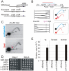
Figure 1
Figure 1. Polδ L591M shows mutagenic strand bias.
A. Schematic of the wild type ura4+ locus and the two versions of the modified ura4+:ura5+ loci. Forward: the transcribed strand of both ura4 and ura5 corresponds to the lagging strand when replicated from ars3003/3004. Reverse: the orientation of the ura4:ura5 sequences are switched so that the transcribed strand corresponds to the leading strand when replicated from ars3003/3004. Loss of either ura4 or ura5 function results in 5-FOA resistance. B. The direction of replication at the modified ura4 + locus. Top: Schematic of the ura4 +:ura5 + locus. Bottom: the principle of directional 2D-gel analysis. Asymmetric digestion of the HindIII-BlpI fragment with SpeI between the running of the first and second dimensions will result in a shift of the Y-arc. The position of the Y arc is indicated by an arrow. The direction of shift is dependent on the direction of replication. C. Comparison of replication intermediates within the HindIII-BlpI region either without (undigested) or after SpeI digestion between dimensions (SpeI). The shifted Y arc following SpeI digestion is indicated with a red arrow, the equivalent of which is also shown in the top frame of panel B. The majority of replication forks run from right to left, corresponding with the efficient initiation from ars3003/ars3004 on the right. D. Cell growth of wild type, polδ+ (wild-type polδ flanked by lox sites) and polδ-L591M. Serial dilutions of cells were spotted on YEA plates, incubated (30°C) for 2 or 4 days and photographed. E. Spontaneous mutation rates in each of the indicated backgrounds for polδ+ and polδ-L591M. Error bars show standard deviation.
Figure 2
Figure 2. Strand bias for polδ-L591M.
A. Schematic of replication through the Forward and Reverse ura4+:ura5+ loci. Leading strand synthesis shown in red, lagging strand synthesis is shown in green. The transcribed strand is shown in blue for reference. B. Top: the relative numbers of AT>GC and GC>AT mutations identified, classified as resulting from either A:dC or T:dG mispairing (AT>GC) and G:dT or C:dA mispairing (GC>AT). Bottom. Schematic illustration of replication of a specific A:T base pair in both orientations. If the lagging strand polymerase, but not the leading strand polymerase, is prone to mispairing dG opposite T during incorporation, but not dC opposite A, then this A:T base pair will mutate to G:C more frequently in the forward orientation than the reverse orientation.
Figure 3
Figure 3. Mutation spectra.
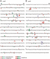
A. ura4 and B. ura5. The promoter region of ura5 (lower case) and the ORF's of both rad4 and ura5 (upper case) are shown. The position of each mapped mutation is indicated. Mutations arising in the “forward” background (transcribed strand replicated by lagging strand synthesis) are shown above the sequence, whereas the mutations arising in the “reverse” background (transcribed strand replicated by leading strand synthesis) are shown below the sequence.
Figure 4
Figure 4. Mutation rate and dNMP incorporation by polε-M630F.
A. Cell growth of wild type cells and polε+ (wild-type polε flanked by lox sites) and polε-M630F strains. Serial dilutions of cells were spotted on YEA plates, incubated (30°C) for 2 or 3 days and photographed. B. Schematic of the wild type ura4+ locus and the two versions of the modified ura4+:ura5+ loci. C. Spontaneous mutation rates for polε+, msh2Δ, polε-M630F and polε-M630F msh2Δ double mutant cells in the ura4+ (wild type), Forward and Reverse backgrounds. Error bars are standard deviations.
Figure 5
Figure 5. Strand bias for rNMP incorporation.
A. Alkali sensitivity of genomic DNA. Genomic DNA from the indicated strains was either digested with EcoRI or left undigested, then treated with alkali and separated by alkaline agarose gel electrophoresis. DNA was revealed with ethidium bromide following neutralization and then processed for Southern analysis and probed with a ura4 probe that reveals both DNA strands. B. Schematic of the loci either side of ars3003/3004 indicating the positions of the EcoRI sites, plus the location and strand specificity of the probes used. C. Alkali sensitivity of each strand, either on the left of ars3003/3004 (probes A and B) or on the right (probes C and D). Strains were either polε+ (+) or polε-M630F (−) with or without concomitant deletion of rnh201, as indicated. The membrane from Figure 3D was stripped and hybridized with the indicated single stranded probes. Probe A and C hybridize with the top strand, probes B and D hybridize with the bottom strand.

Similar articles

Cited by

References

    1. Garg P, Burgers PM. DNA polymerases that propagate the eukaryotic DNA replication fork. Crit Rev Biochem Mol Biol. 2005;40:115–128. - PubMed
    1. Johnson A, O'Donnell M. Cellular DNA replicases: components and dynamics at the replication fork. Annu Rev Biochem. 2005;74:283–315. - PubMed
    1. Kunkel TA. Evolving views of DNA replication (in)fidelity. Cold Spring Harb Symp Quant Biol. 2009;74:91–101. - PMC - PubMed
    1. Kunkel TA, Bebenek K. DNA replication fidelity. Annu Rev Biochem. 2000;69:497–529. - PubMed
    1. Nick McElhinny SA, Gordenin DA, Stith CM, Burgers PM, Kunkel TA. Division of labor at the eukaryotic replication fork. Mol Cell. 2008;30:137–144. - PMC - PubMed

Publication types

MeSH terms