

Genetically induced cholinergic hyper-innervation enhances taste learning
- PMID: 22144949
- PMCID: PMC3227857
- DOI: 10.3389/fnsys.2011.00097
Genetically induced cholinergic hyper-innervation enhances taste learning
Abstract
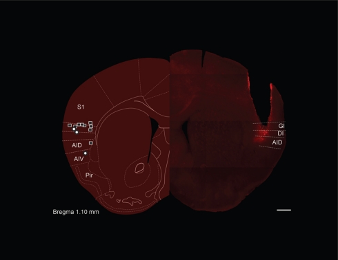
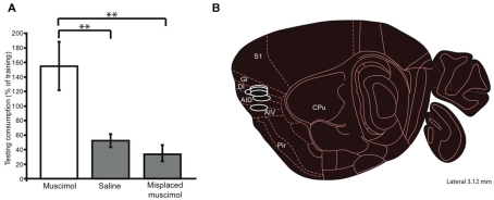
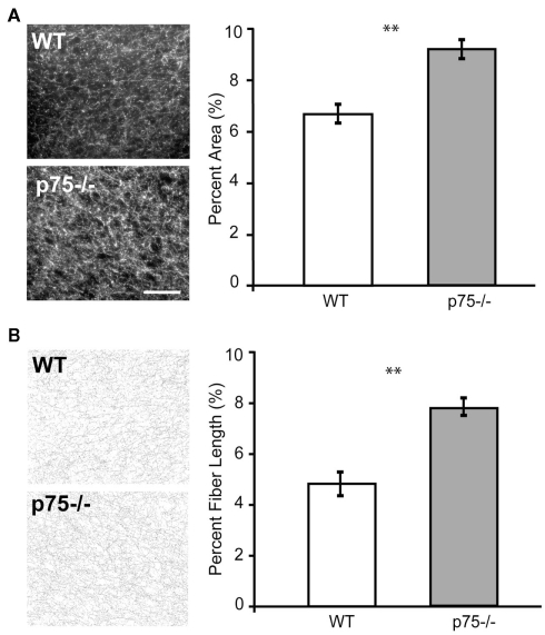
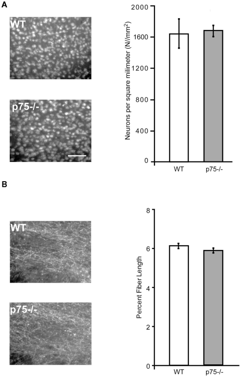
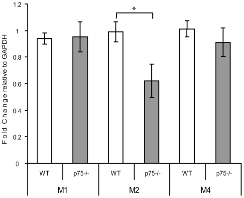
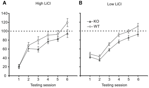
Acute inhibition of acetylcholine (ACh) has been shown to impair many forms of simple learning, and notably conditioned taste aversion (CTA). The most adhered-to theory that has emerged as a result of this work - that ACh increases a taste's perceived novelty, and thereby its associability - would be further strengthened by evidence showing that enhanced cholinergic function improves learning above normal levels. Experimental testing of this corollary hypothesis has been limited, however, by side-effects of pharmacological ACh agonism and by the absence of a model that achieves long-term increases in cholinergic signaling. Here, we present this further test of the ACh hypothesis, making use of mice lacking the p75 pan-neurotrophin receptor gene, which show a resultant over-abundance of cholinergic neurons in sub-regions of the basal forebrain (BF). We first demonstrate that the p75-/- abnormality directly affects portions of the CTA circuit, locating mouse gustatory cortex (GC) using a functional assay and then using immunohistochemisty to demonstrate cholinergic hyper-innervation of GC in the mutant mice - hyper-innervation that is unaccompanied by changes in cell numbers or compensatory changes in muscarinic receptor densities. We then demonstrate that both p75-/- and wild-type (WT) mice learn robust CTAs, which extinguish more slowly in the mutants. Further testing to distinguish effects on learning from alterations in memory retention demonstrate that p75-/- mice do in fact learn stronger CTAs than WT mice. These data provide novel evidence for the hypothesis linking ACh and taste learning.
Keywords: cholinergic system; conditioned taste aversion; p75 knockout mouse; taste learning.
Figures
Similar articles
-
Impaired hippocampal acetylcholine release parallels spatial memory deficits in Tg2576 mice subjected to basal forebrain cholinergic degeneration.Brain Res. 2014 Jan 16;1543:253-62. doi: 10.1016/j.brainres.2013.10.055. Epub 2013 Nov 11. Brain Res. 2014. PMID: 24231553
-
Temporally-precise basolateral amygdala activation is required for the formation of taste memories in gustatory cortex.J Physiol. 2020 Dec;598(23):5505-5522. doi: 10.1113/JP280213. Epub 2020 Sep 18. J Physiol. 2020. PMID: 32857870
-
Effects of acetylcholine on coding of taste information in the primary gustatory cortex in rats.Exp Brain Res. 2007 May;179(1):97-109. doi: 10.1007/s00221-006-0772-4. Epub 2006 Nov 16. Exp Brain Res. 2007. PMID: 17109107
-
The cholinergic system and spatial learning.Behav Brain Res. 2011 Aug 10;221(2):389-411. doi: 10.1016/j.bbr.2010.11.036. Epub 2010 Nov 23. Behav Brain Res. 2011. PMID: 21108971 Review.
-
Neural substrates for conditioned taste aversion in the rat.Behav Brain Res. 1994 Dec 15;65(2):123-37. doi: 10.1016/0166-4328(94)90097-3. Behav Brain Res. 1994. PMID: 7718144 Review.
Cited by
-
The role of the gustatory cortex in incidental experience-evoked enhancement of later taste learning.Learn Mem. 2018 Oct 15;25(11):587-600. doi: 10.1101/lm.048181.118. Print 2018 Nov. Learn Mem. 2018. PMID: 30322892 Free PMC article.
-
Preexposure to salty and sour taste enhances conditioned taste aversion to novel sucrose.Learn Mem. 2016 Apr 15;23(5):221-8. doi: 10.1101/lm.040360.115. Print 2016 May. Learn Mem. 2016. PMID: 27084929 Free PMC article.
-
Impact of a deletion of the full-length and short isoform of p75NTR on cholinergic innervation and the population of postmitotic doublecortin positive cells in the dentate gyrus.Front Neuroanat. 2015 May 27;9:63. doi: 10.3389/fnana.2015.00063. eCollection 2015. Front Neuroanat. 2015. PMID: 26074780 Free PMC article.
-
Single and population coding of taste in the gustatory cortex of awake mice.J Neurophysiol. 2019 Oct 1;122(4):1342-1356. doi: 10.1152/jn.00357.2019. Epub 2019 Jul 24. J Neurophysiol. 2019. PMID: 31339800 Free PMC article.
-
The differential role of cortical protein synthesis in taste memory formation and persistence.NPJ Sci Learn. 2016;1:16001. doi: 10.1038/npjscilearn.2016.1. Epub 2016 May 11. NPJ Sci Learn. 2016. PMID: 27721985 Free PMC article.
References
-
- Barrett G. L., Reid C. A., Tsafoulis C., Zhu W., Williams D. A., Paolini A. G., Trieu J., Murphy M. (2010). Enhanced spatial memory and hippocampal long-term potentiation in p75 neurotrophin receptor knockout mice. Hippocampus 20, 145–152 - PubMed
Grants and funding
LinkOut - more resources
Full Text Sources
Research Materials
Miscellaneous