

Application for proteomic techniques in studying osteoarthritis: a review
- PMID: 22144964
- PMCID: PMC3228966
- DOI: 10.3389/fphys.2011.00090
Application for proteomic techniques in studying osteoarthritis: a review
Abstract
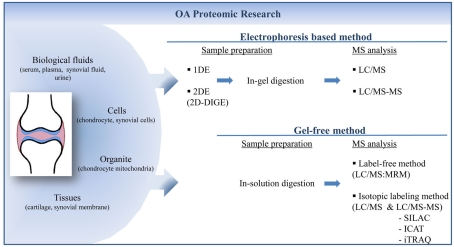
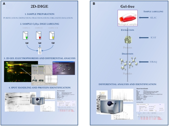
After the genomic era, proteomic corresponds to a wide variety of techniques that study the protein content of cells, tissue, or organism and that allow the isolation of protein of interest. It offers the choice between gel-based and gel-free methods or shotgun proteomics. Applications of proteomic technology may concern three principal objectives in several biomedical or clinical domains of research as in osteoarthritis: (i) to understand the physiopathology or underlying mechanisms leading to a disease or associated with a particular model, (ii), to find disease-specific biomarker, and (iii) to identify new therapeutic targets. This review aimed at gathering most of the data regarding the proteomic techniques and their applications to osteoarthritis research. It also reported technical limitations and solutions, as for example for sample preparation. Proteomics open wide perspectives in biochemical research but many technical matters still remain to be solved.
Keywords: osteoarthritis; proteomic.
Figures
Similar articles
-
Applications of proteomics to osteoarthritis, a musculoskeletal disease characterized by aging.Front Physiol. 2011 Dec 23;2:108. doi: 10.3389/fphys.2011.00108. eCollection 2011. Front Physiol. 2011. PMID: 22207853 Free PMC article.
-
Proteomic reactors and their applications in biology.FEBS J. 2011 Oct;278(20):3796-806. doi: 10.1111/j.1742-4658.2011.08292.x. Epub 2011 Sep 6. FEBS J. 2011. PMID: 21824287 Review.
-
Mass spectrometry for translational proteomics: progress and clinical implications.Genome Med. 2012 Aug 31;4(8):63. doi: 10.1186/gm364. eCollection 2012. Genome Med. 2012. PMID: 22943415 Free PMC article. Review.
-
Proteomics role in the search for improved diagnosis, prognosis and treatment of osteoarthritis.Osteoarthritis Cartilage. 2010 Apr;18(4):500-9. doi: 10.1016/j.joca.2009.11.012. Epub 2009 Dec 23. Osteoarthritis Cartilage. 2010. PMID: 20060947 Review.
-
Clinical proteomics: present and future prospects.Clin Biochem Rev. 2006 May;27(2):99-116. Clin Biochem Rev. 2006. PMID: 17077880 Free PMC article.
Cited by
-
Applications of proteomics to osteoarthritis, a musculoskeletal disease characterized by aging.Front Physiol. 2011 Dec 23;2:108. doi: 10.3389/fphys.2011.00108. eCollection 2011. Front Physiol. 2011. PMID: 22207853 Free PMC article.
-
Serum proteomic biomarkers diagnostic of knee osteoarthritis.Osteoarthritis Cartilage. 2024 Mar;32(3):329-337. doi: 10.1016/j.joca.2023.09.007. Epub 2023 Sep 19. Osteoarthritis Cartilage. 2024. PMID: 37734705 Free PMC article.
-
Biochemical markers of aging for longitudinal studies in humans.Epidemiol Rev. 2013;35(1):132-51. doi: 10.1093/epirev/mxs011. Epub 2013 Feb 4. Epidemiol Rev. 2013. PMID: 23382477 Free PMC article. Review.
-
Prediction and analysis of osteoarthritis hub genes with bioinformatics.Ann Transl Med. 2023 Jan 31;11(2):66. doi: 10.21037/atm-22-6450. Ann Transl Med. 2023. PMID: 36819525 Free PMC article.
-
Clinical and omics biomarkers in osteoarthritis diagnosis and treatment.J Orthop Translat. 2025 Jan 22;50:295-305. doi: 10.1016/j.jot.2024.12.007. eCollection 2025 Jan. J Orthop Translat. 2025. PMID: 39911590 Free PMC article. Review.
References
-
- Baillet A., Trocme C., Berthier S., Arlotto M., Grange L., Chenau J., Quetant S., Seve M., Berger F., Juvin R., Morel F., Gaudin P. (2010). Synovial fluid proteomic fingerprint: S100A8, S100A9 and S100A12 proteins discriminate rheumatoid arthritis from other inflammatory joint diseases. Rheumatology (Oxford) 49, 671–68210.1093/rheumatology/kep452 - DOI - PubMed
LinkOut - more resources
Full Text Sources