. 2011:5:51-61.
doi: 10.2174/1875397301005010051. Epub 2011 Aug 22.

Drug discovery toward antagonists of methyl-lysine binding proteins

Affiliations

Drug discovery toward antagonists of methyl-lysine binding proteins

J Martin Herold et al. Curr Chem Genomics. 2011.

Abstract

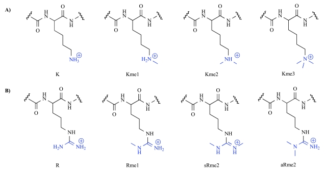
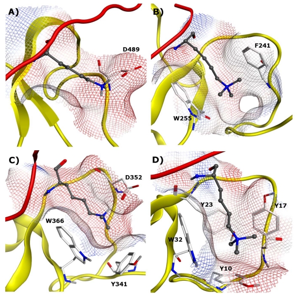
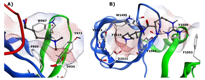
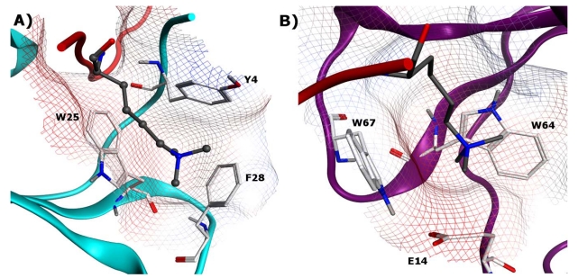
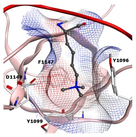
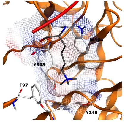
The recognition of methyl-lysine and -arginine residues on both histone and other proteins by specific "reader" elements is important for chromatin regulation, gene expression, and control of cell-cycle progression. Recently the crucial role of these reader proteins in cancer development and dedifferentiation has emerged, owing to the increased interest among the scientific community. The methyl-lysine and -arginine readers are a large and very diverse set of effector proteins and targeting them with small molecule probes in drug discovery will inevitably require a detailed understanding of their structural biology and mechanism of binding. In the following review, the critical elements of methyl-lysine and -arginine recognition will be summarized with respect to each protein family and initial results in assay development, probe design, and drug discovery will be highlighted.

Keywords: Histones; chemical probes; chromatin; drug discovery.; methyl-arginine; methyl-lysine; pi-cation interactions; post-translational modifications; reader domains.


Figures

Fig. (1)
Fig. (1)
Different methylation states of lysine (A) and arginine (B).
Fig. (2)
Fig. (2)
Example of tandem repeat of MBT domains: A) L3MBTL1 bound to H4K20me2 (PDB: 2RJF) and B) SCML2 bound to a mono-methylated lysine (PDB: 2VYT). Protonated methyl-lysines are displayed in ball and stick model with gray carbon atoms, key binding site residues are displayed in stick model with white carbon atoms.
Fig. (3)
Fig. (3)
A) Binding cavity of L3MBTL1 bound to H4K20me2 (PDB:2RJF) and B) Binding cavity of SCML2 bound to a mono-methylated lysine (PDB: 2VYT). Protonated methyl-lysines are displayed in ball and stick model with gray carbon atoms, key binding site residues are displayed in stick model with white carbon atoms.
Fig. (4)
Fig. (4)
Nicotinamide ligand (4) shown in the co-crystal structure with L3MBTL1. PDB: 3P8H.
Fig. (5)
Fig. (5)
Binding pockets of four representative PHD fingers: A) PHF21A (BHC80) bound to unmethylated H3K4 (PDB: 2PUY). B) PHF13 bound to H3K4me3 (PDB: 3O7A). C) PYGO1 bound to H3K4me2 (PDB: 2VPE). D) BPTF bound to H3K4me3 (PDB: 2F6J). In all four structures, PHD domains are shown in yellow ribbon and histone tails are shown in red ribbon. Protonated methyl-lysines (unmethylated H3K4 in 5A) and trimethyl-lysines are displayed in ball and stick model with gray carbon atoms. Key binding site residues of the PHD fingers are displayed in stick model with white carbon atoms.
Fig. (6)
Fig. (6)
A) Double tudor domain JMJD2A with the aromatic cavity bound to H3K9me2 (PDB: 2OX0) B) Binding pocket of 53BP1 tandem tudor domain bound to H4K20me2 (PDB: 2IG0). In both structures, the two tudor domains are shown in blue and green, respectively. The trimethyl-lysine in A and the protonated dimethyl-lysine and the neighboring H4R19 residue in B are displayed in ball and stick model with gray carbon atoms. Key binding site residues are displayed in stick model with white carbon atoms.
Fig. (7)
Fig. (7)
A) Aromatic binding pocket of CBX5 (human HP1α) bound to H3K9me3 (PDB: 3FDT) B) Chromodomain CHD1 binding pocket with H3K4me3 (PDB: 2B2W). In both structures, the trimethylated lysines are displayed in ball and stick model with gray carbon atoms, key binding site residues are displayed in stick model with white carbon atoms.
Fig. (8)
Fig. (8)
PWWD domain Brpf1 Binding Pocket with H3K36me3 (PDB: 2X4Y). The trimethylated lysine is displayed in ball and stick model with gray carbon atoms, key binding site residues are displayed in stick model with white carbon atoms.
Fig. (9)
Fig. (9)
EED as an example of a WD40 domain binding to H3K27me3 (PDB: 3JZG). The trimethylated lysine is displayed in ball and stick model with gray carbon atoms, key binding site residues are displayed in stick model with white carbon atoms.

Similar articles

Cited by

References

    1. Bhaumik SR, Smith E, Shilatifard A. Covalent modifications of histones during development and disease pathogenesis. Nat Struct Mol Biol. 2007;14:1008–16. - PubMed
    1. Wang GG, Allis CD, Chi P. Chromatin remodeling and cancer, Part I: Covalent histone modifications. Trends Mol Med. 2007;13:363–72. - PubMed
    1. Jenuwein T, Allis CD. Translating the histone code. Science. 2001;293:1074–80. - PubMed
    1. Strahl BD, Allis CD. The language of covalent histone modifications. Nature. 2000;403:41–5. - PubMed
    1. Zhang Y, Reinberg D. Transcription regulation by histone methylation: interplay between different covalent modifications of the core histone tails. Genes Dev. 2001;15:2343–60. - PubMed

LinkOut - more resources