. 2011;6(12):e28306.
doi: 10.1371/journal.pone.0028306. Epub 2011 Dec 1.

LKB1 is an essential regulator of spermatozoa release during spermiation in the mammalian testis

Affiliations

LKB1 is an essential regulator of spermatozoa release during spermiation in the mammalian testis

Fiona C Denison et al. PLoS One. 2011.

Abstract

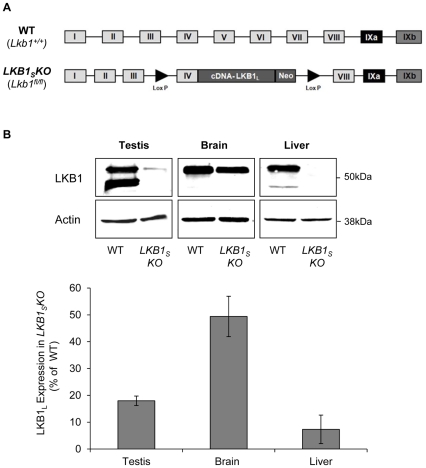
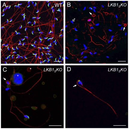
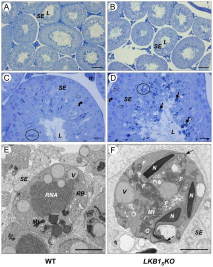
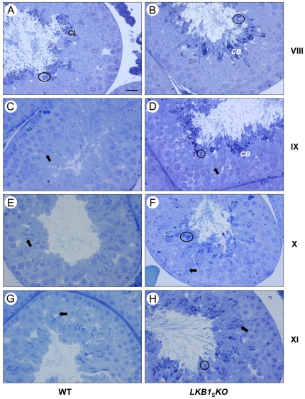
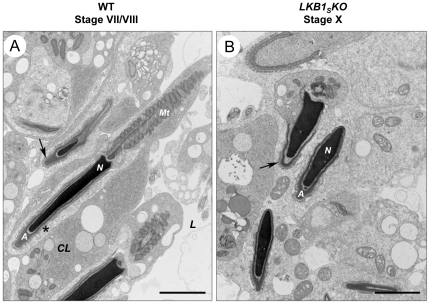
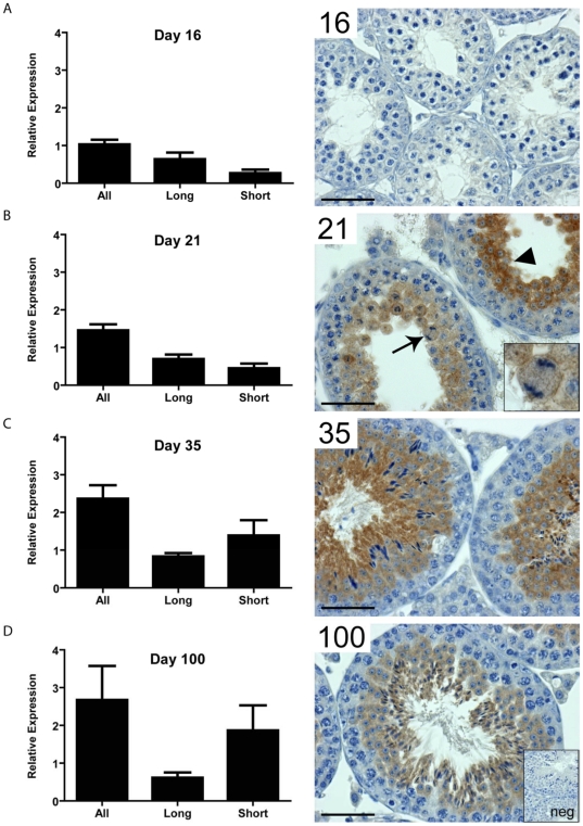
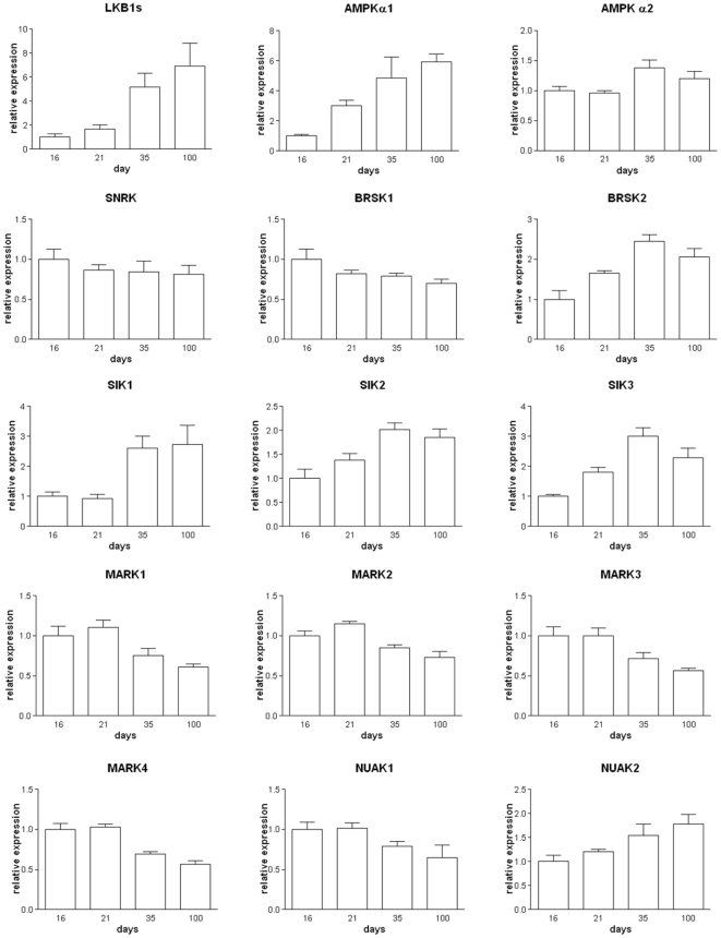
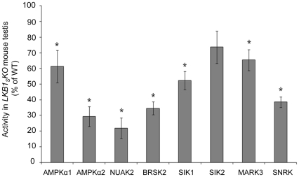
LKB1 acts as a master upstream protein kinase regulating a number of kinases involved in diverse cellular functions. Recent studies have suggested a role for LKB1 in male fertility. Male mice with reduced total LKB1 expression, including the complete absence of the major splice variant in testis (LKB1(S)), are completely infertile. We sought to further characterise these mice and determine the mechanism underlying this infertility. This involved expression studies of LKB1 in developing germ cells, morphological analysis of mature spermatozoa and histological studies of both the testis and epididymis using light microscopy and transmission electron microscopy. We conclude that a defect in the release of mature spermatids from the seminiferous epithelium (spermiation) during spermatozoan development is a major cause of the infertility phenotype. We also present evidence that this is due, at least in part, to defects in the breakdown of the junctions, known as ectoplasmic specialisations, between the sertoli cells of the testis epithelium and the heads of the maturing spermatids. Overall this study uncovers a critical role for LKB1 in spermiation, a highly regulated, but poorly understood process vital for male fertility.


Conflict of interest statement

Competing Interests: The authors have read the journal's policy and have the following conflicts: FCD was funded by a studentship from Unilever Discover, UK. This does not alter the authors' adherence to all the PLoS ONE policies on sharing data and materials.

Figures

Figure 1
Figure 1. LKB1 expression in LKB1SKO mice.
A) Generation of LKB1SKO mice, adapted from . Exons IV–VII (encoding the kinase domain) of the wild-type (WT) Lkb1 gene were replaced by a cassette encompassing exon IV plus cDNA encoding the rest of LKB1L. This cassette was flanked by lox-P excision sites (▸) and contained a neomycin (Neo) gene for selection. Mice homozygous for expression of this cassette (Lkb1fl/fl) no longer express LKB1S and are de facto LKB1SKO mice. B) Mouse tissues homogenates were analysed by western blotting with a monoclonal antibody raised against total LKB1. An anti-actin antibody was used to show equal loading. Representative blots are shown and the migration of molecular mass standards are as indicated. C) Quantification of the relative intensity of the LKB1L band between wild-type and LKB1SKO mice is shown in the bar graph below. Results are plotted as the percentage expression of LKB1L in LKB1SKO mice compared to wild-type and are the means±S.E.M. of three separate blots from three individual mice.
Figure 2
Figure 2. Fluorescence microscopy images of mature spermatozoa.
Spermatozoa were taken from the cauda epididymis of wild-type (A) and LKB1SKO mice (BD), and visualised by immunofluorescence. Tails were stained with an anti-tubulin antibody (red), acrosomes with an anti-acrosomal monoclonal antibody (green) and nuclei with DAPI (blue). Sperm from LKB1SKO mice frequently show coiled tails (filled white arrows). In (D), the tail has formed a ‘lasso’-type structure around an abnormally shaped nucleus. Sperm nuclei from LKB1SKO mice often lack acrososmes (open white arrow in B) as shown by the lack of green fluorescence at the anterior nuclear surface. Abnormal cellular debris is visible in LKB1SKO samples, as indicated by asterisks in (C). Slides were viewed on a Leica TCS SP1 confocal microscope. Images are representative of at least three mice (Scale bar  = 20 µM).
Figure 3
Figure 3. Histology of the cauda and caput epididymis.
Cauda epididymis (AB) and caput epididymis (CD) sections were visualised by light microscopy. Representative images are shown from observations of three mice per genotype. Epididymal epithelia (E) and tubule lumens (L) are labelled. (A) and (C) show sections through wild-type epididymis showing an abundance of spermatozoa within the lumen. (B) and (D) show sections through the epididymis of LKB1SKO mice showing abnormal structures within the lumen and very few spermatozoa. (EF) TEM images of the cauda epididymal lumen from a wild-type mouse (E) and a LKB1SKO mouse (F). The wild-type section shows numerous cross-sections through sperm heads (N) and tails (arrows). The section from a LKB1SKO mouse shows dense luminal fluid (as indicated by the darker background to WT), cellular debris, abnormal round structures (asterisk) and an absence of recognisable spermatozoa cross sections (A and B, scale bar  = 20 µm; C and D, scale bar  = 50 µm; E and F, scale bar  = 5 µm).
Figure 4
Figure 4. Testis histology.
Cross-sections of seminiferous tubules from wild-type (left-hand panel, A and C) and LKB1SKO (right-hand panel, B and D) mice. The seminiferous epithelium (SE), which is made up of sertoli cells and developing germ cells, is indicated. This surrounds the lumen (L) of the tubules. (C) and (D) show sections from tubules at approximately stage V. Circles have been used to show the nuclei of elongating spermatids. Spermatogonia (open curved arrow), spermatocytes (closed curved arrow) and round spermatids (open arrow) are also indicated. A number of dense, round bodies of varying sizes can be seen around the lumens from LKB1SKO mice. Examples of these are indicated in (D) (closed black arrows). Representative images are shown (A and B, scale bar  = 100 µm; C and D, scale bar  = 20 µm). (E) and (F) show TEM images of a residual body (RB) within the seminiferous epithelium (SE) from a wild-type mouse (E), and an abnormal cytoplasmic body from a LKB1SKO mouse (F). Normal residual often contain such structures as vacuoles (V), RNA and mitochondria (Mt). The abnormal cytoplasmic bodies seen in (F) are similar to residual bodies but often contain at least one condensed spermatid nuclei (N) and several cross sections of flagella (F) as indicated. Detached acrosomes, identified as the deeply staining crescent shape structures close to the anterior nuclear surface, are indicated with an arrow. Granular material is also indicated (G), (E and F, scale bar  = 2 µm).
Figure 5
Figure 5. LM images showing ‘failure of spermiation’ in LKB1SKO mice.
Representative images of seminiferous tubules are shown at stage VIII (A,B), when spermiation normally occurs; and stages IX, X, and XI (CH), after spermiation has normally taken place. The stage numbers are shown to the right of the images. Elongated spermatids are identifiable by their darkly-staining, condensed nuclei. The nuclei of immature round spermatids (open arrows) and elongating spermatids (closed arrows) are less deeply stained and can be seen embedded within the epithelium at the relevant stages of both WT and LKB1SKO sections. There is a progressive condensation and elongation of the nucleus of the elongating spermatids from stage IX to stage XI. Sections from wild-type mice are displayed on the left. At stage VIII, elongated spermatids (examples circled) and cytoplasmic lobes (CL) are visible around the lumen. There are no elongated spermatids present around the lumen after stage VIII in wild-type sections. In contrast, in tubules from LKB1SKO mice, shown on the right, elongated spermatids are visible around the lumen at all stages displayed (examples circled). In addition, abnormal deeply staining cytoplasmic bodies (CB) can be seen around the lumen, (scale bar  = 20 µm).
Figure 6
Figure 6. TEM images of cell junctions between sertoli cells and spermatids.
A) Elongated spermatids are shown around the lumen from a wild-type stage VII tubule before spermiation. The residual spermatid cytoplasm can be seen around the spermatid nucleus (N) as a cytoplasmic lobe (CL). Although the actin bundles of ectoplasmic specialisations are still visible around some spermatids (arrow), they are beginning to break down around others as indicated by an asterisk. Mitochondria aligned along the spermatozoan flagellum are labelled (Mt). Spermatid acrosomes (A) and the tubule lumen (L) are indicated. B) Elongated spermatids are shown around the lumen from a stage X LKB1SKO tubule. These spermatids would normally have been released at stage VIII. Areas of ectoplasmic specialisations are still visible around the retained spermatid heads as indicated by an arrow, (scale bar  = 2 µm).
Figure 7
Figure 7. LKB1 splice variant relative gene expression and testicular localisation.
A) At day 16, prior to meiosis, testicular LKB1 is expressed at relatively low levels, with LKB1 protein below the detectable limits of colourimetric immunohistochemistry. B) At day 21 total LKB1 expression has increased, predominantly through increased expression of LKB1S. LKB1 protein can be detected in meiotic spermatocytes (inset and arrow), and post-meiotic spermatids (arrowhead). At day 35 (C) and day 100 (D), the predominant transcript is LKB1S with LKB1 protein localised to the cytoplasm of elongated spermatids in addition to round spermatids and spermatocytes, (scale bar  = 50 µm). The insert labelled ‘neg’ shows a negative control in which no primary antibody was incubated with the tissue.
Figure 8
Figure 8. Expression of AMPK and AMPK-related kinases in testis.
The mRNA expression levels of LKB1 and downstream kinases in developing testis at post-partum days 16–100. Values are shown relative to the expression levels at day 16 and shown as the mean +/- SEM, n = 5.
Figure 9
Figure 9. Activity of AMPK and AMPK-related kinases in LKB1SKO testis.
Activity measured in immune-complexes using antibodies specific for AMPK and AMPK-related proteins are plotted as a percentage of the activity measured in wild type testis and shown as the mean±S.E.M. from three individual mice. * indicates a statistically significant difference in activity in LKB1SKO compared to wild-type samples (p<0.05 with Student's unpaired t test).

References

    1. Alessi DR, Sakamoto K, Bayascas JR. LKB1-dependent signaling pathways. Annu Rev Biochem. 2006;75:137–163. - PubMed
    1. Jenne DE, Reimann H, Nezu J, Friedel W, Loff S, et al. Peutz-Jeghers syndrome is caused by mutations in a novel serine threonine kinase. Nat Genet. 1998;18:38–43. - PubMed
    1. Hemminki A, Markie D, Tomlinson I, Avizienyte E, Roth S, et al. A serine/threonine kinase gene defective in Peutz-Jeghers syndrome. Nature. 1998;391:184–187. - PubMed
    1. Ylikorkala A, Rossi DJ, Korsisaari N, Luukko K, Alitalo K, et al. Vascular abnormalities and deregulation of VEGF in Lkb1-deficient mice. Science. 2001;293:1323–1326. - PubMed
    1. Boudeau J, Baas AF, Deak M, Morrice NA, Kieloch A, et al. MO25alpha/beta interact with STRADalpha/beta enhancing their ability to bind, activate and localize LKB1 in the cytoplasm. Embo J. 2003;22:5102–5114. - PMC - PubMed

Publication types

MeSH terms

Substances