doi: 10.4103/2230-8210.86984.
Radiological imaging in endocrine hypertension
Affiliations
- PMID: 22145144
- PMCID: PMC3230092
- DOI: 10.4103/2230-8210.86984
Item in Clipboard
Radiological imaging in endocrine hypertension
Indian J Endocrinol Metab.
2011 Oct.
Abstract
While different generations of assays have played important role in elucidating causes of different endocrine disorders, radiological techniques are instrumental in localizing the pathology. This statement cannot be truer in any disease entity other than endocrine hypertension. This review makes an effort to highlight the role of different radiological modalities, especially ultrasonography, computed tomography and magnetic resonance imaging, in the evaluation of different causes of endocrine hypertension.
Keywords: Adrenal imaging; endocrine hypertension; pituitary imaging.
Conflict of interest statement
Figures

Multi-detector computed tomography showing large heterogeneously enhancing necrotic left supra renal mass which was confirmed to be malignant pheochromocytoma at histopathology
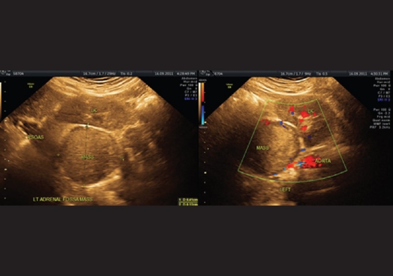
Large, well-circumscribed, homogenous mass in the left adrenal; biochemical tests confirmpheochromocytoma
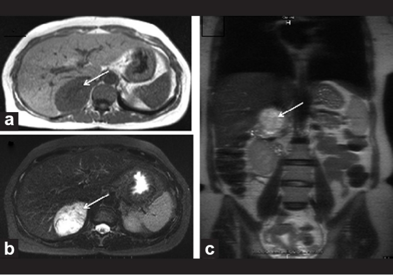
Pheochromocytoma: The mass is hypointense (arrow) on (a) axial T1-weighted image. The mass shows bright signal – “light bulb sign” (arrow) on (b) T2 axial and (c) coronal mages

Adrenal adenoma: Axial CT image in (a) shows hypodense fat containing mass (arrow) in the right adrenal. Axial CT image in (b) shows a lipid-poor adenoma in left adrenal (arrow) in a patient with Conn's syndrome

Adrenal adenoma: Axial CT image in (a) shows hypodense fat containing mass (arrow) in the right adrenal. (b) The mass is hyperintense (arrow) on fat-saturated axial T2-weighted images although the rest of the fat gets suppressed. (c) The mass shows signal drop (arrow) on out-of-phase T1 axial image

Adrenal carcinoma: Axial T1W image in (a) shows well-defined, large suprarenal mass and axial T2 image in (b) shows the mass being hyperintense. The mass is separate from left kidney (LK) and spleen (S)

Thyroid sonography in a Graves’ disease patient. The gland appears enlarged and diffusely heteroechoic (upper panel). On Doppler, there is significant increase in vascularity, also described classically as thyroid inferno (lower panel)

Parathyroid adenoma (M) detected by neck ultrasound. Note the oval shape, high vascularity (a) and hypoechoic appearance (b)

Pituitary macroadenoma in a patient with acromegaly: Coronal T1 (a) and axial T2 (b) showing a mass in sella with suprasellar extension having “figure of 8” appearance. The mass shows avid enhancement in post-contrast coronal T1 (c) and axial T1 (d) images with central nonenhancing area suggesting necrosis
References
-
- Bravo EL, Gifford RW. Current concepts: Pheochromocytoma – diagnosis localization and management. N Engl J Med. 1984;15(311):1298–303. - PubMed
-
- Witteles RM, Kaplan EL, Roizen MF. Sensitivity of diagnostic and localization tests for pheochromocytoma in clinical practice. Arch Intern Med. 2000;160:2521–4. - PubMed
-
- Bowerman RA, Silver TM, Jaffe MH, Stuck KJ, Hinerman DL. Sonography of adrenal pheochromocytoma. AJR Am J Roentgenol. 1981;137:1227–31. - PubMed
-
- Krebs TL, Wagner BJ. MR imaging of the adrenal gland: Radiologic-pathologic correlation. Radiographics. 1998;18:1425–40. - PubMed