. 2011 Dec 6:8:98.
doi: 10.1186/1742-4690-8-98.

Inhibition of HIV-1 infection by TNPO3 depletion is determined by capsid and detectable after viral cDNA enters the nucleus

Affiliations

Inhibition of HIV-1 infection by TNPO3 depletion is determined by capsid and detectable after viral cDNA enters the nucleus

Alberto De Iaco et al. Retrovirology. .

Abstract

Background: HIV-1 infects non-dividing cells. This implies that the virus traverses the nuclear pore before it integrates into chromosomal DNA. Recent studies demonstrated that TNPO3 is required for full infectivity of HIV-1. The fact that TNPO3 is a karyopherin suggests that it acts by directly promoting nuclear entry of HIV-1. Some studies support this hypothesis, while others have failed to do so. Additionally, some studies suggest that TNPO3 acts via HIV-1 Integrase (IN), and others indicate that it acts via capsid (CA).

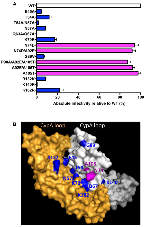
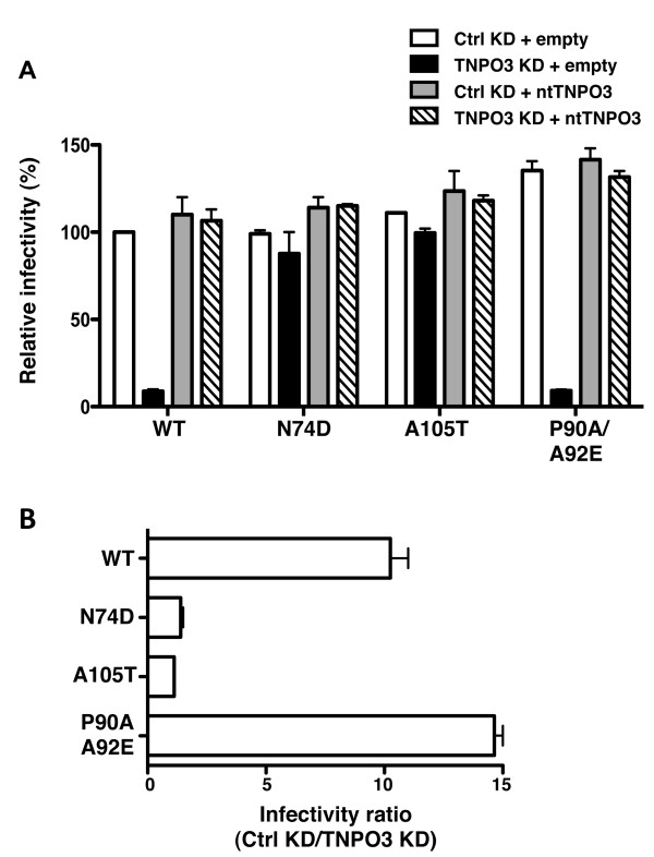
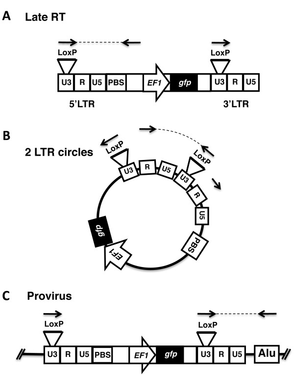
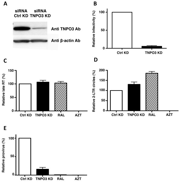
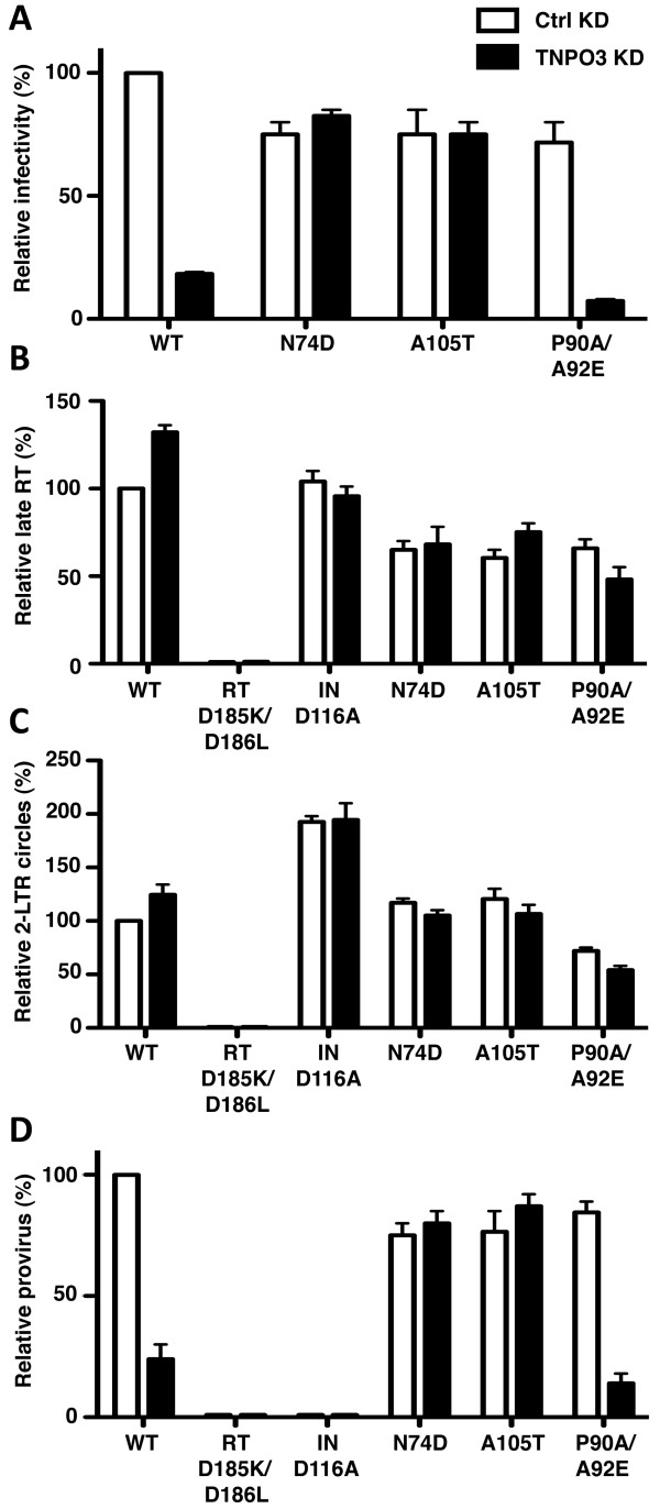
Results: To shed light on the mechanism by which TNPO3 contributes to HIV-1 infection we engineered a panel of twenty-seven single-cycle HIV-1 vectors each bearing a different CA mutation and characterized them for the ability to transduce cells in which TNPO3 had been knocked down (KD). Fourteen CA mutants were relatively TNPO3-independent, as compared to wild-type (WT) HIV-1. Two mutants were more TNPO3-dependent than the WT, and eleven mutants were actually inhibited by TNPO3. The efficiency of the synthesis of viral cDNA, 2-LTR circles, and proviral DNA was then assessed for WT HIV-1 and three select CA mutants. Controls included rescue of TNPO3 KD with non-targetable coding sequence, RT- and IN- mutant viruses, and pharmacologic inhibitors of RT and IN. TNPO3 KD blocked transduction and establishment of proviral DNA by wild-type HIV-1 with no significant effect on the level of 2-LTR circles. PCR results were confirmed by achieving TNPO3 KD using two different methodologies (lentiviral vector and siRNA oligonucleotide transfection); by challenging three different cell types; by using two different challenge viruses, each necessitating different sets of PCR primers; and by pseudotyping virus with VSV G or using HIV-1 Env.

Conclusion: TNPO3 promotes HIV-1 infectivity at a step in the virus life cycle that is detectable after the preintegration complex arrives in the nucleus and CA is the viral determinant for TNPO3 dependence.


Figures

Figure 1
Figure 1
TNPO3 depletion and rescue with non-targetable TNPO3 cDNA. (A) Schematic representation of the lentiviral vectors used to generate TNPO3 knockdown (KD) and rescue cell lines. (B) The sequence of steps used to obtain the four pools of stable cell lines. HeLa cells were transduced with control (Ctrl) KD vector or with TNPO3 KD vector, and selected in pools with 10 μg/ml of puromycin. Each pool of KD cells was then transduced a second time with the rescue vector, either empty or bearing non-targetable TNPO3 cDNA (ntTNPO3), and selected in pools with 10 μg/ml of blasticidin, as well as 1 μg/ml puromycin. (C) Steady-state levels of TNPO3 protein in each of the four pools of doubly-tranduced cells. Cell lysate was probed in western blots with anti-TNPO3 antibody (upper panel) and anti-β-actin antibody (lower panel).
Figure 2
Figure 2
The effect of TNPO3 KD on the infectivity of HIV-1 CA mutants in HeLa cells. HeLa control (ctrl) KD cells and TNPO3 KD cells were challenged with a panel of 27 HIV-1-GFP reporter vectors bearing either WT CA or the indicated CA mutants. At 72 hrs the percent GFP+ cells was determined by flow cytometry as an indication of infectivity. The ratio of HIV-1 infectivity in Ctrl KD vs TNPO3 KD cells is shown. Error bars represent ± SEM (n = 3). Black bars indicate mutants that were significantly less sensitive than the WT to TNPO3 KD (p < 0.01, t-test). Gray bars indicate mutants that were significantly more sensitive than the WT to TNPO3 KD (p < 0.01, t-test).
Figure 3
Figure 3
HIV-1 CA mutants that confer TNPO3-independence localize to the interface between two monomers in the hexameric CA lattice. (A) Absolute infectivity relative to WT virus of TNPO3-independent CA mutants. Data represent one of at least three independent experiments. Error bars represent ± SEM (n = 3). (B) Location of CA amino acid residues important for TNPO3-dependence of HIV-1. Space-fill model of a CA dimer extracted from the hexameric structure (PDB: 3H4E). The location of mutants with absolute infectivity similar WT is indicated in magenta. The location of mutants with an absolute infectivity defect are shown in blue; Q63 is in the dimer interface and not visible in this view.
Figure 4
Figure 4
Infectivity of HIV-1-GFP vectors carrying WT or mutant CA on KD and rescue HeLa cells. Expression of the GFP reporter gene was checked by flow cytometry 72 hrs after challenge with virus. Infectivity relative to WT is shown in (A). The ratio of the infectivity in TNPO3 KD compared to the control KD cells is shown in (B). Data represent one of at least three independent experiments. Error bars represent ± SEM (n = 3).
Figure 5
Figure 5
Strategy for detecting cDNA from HIV-1 reporter virus after challenge of cells that had been previously transduced with HIV-1-based, lentivirus KD and rescue vectors. Schematic diagram showing methods of detection for HIV-1 late RT products (A), 2-LTR circles (B) and provirus (C) in KD cell lines. Identification of nascent cDNA is made possible by the presence of a loxP sequence engineered within a region of the 3'LTR U3 that is dispensable for retrotransposition.
Figure 6
Figure 6
Effect of TNPO3 on the de novo synthesis and fate of HIV-1 cDNA after acute infection. WT or CA mutant HIV-1 reporter virus were used to challenge the TNPO3 KD and rescue cells, as indicated. 24 hrs later, late RT (A), 2-LTR circles (B) and provirus (C) were assayed by qPCR. Viruses bearing the enzymatic site mutants RT-D185K/D186L or IN-D116A IN were used as controls for de novo reverse transcription and integration, respectively. Data represent one of at least three independent experiments. Error bars represent ± SEM (n = 3).
Figure 7
Figure 7
Effect of TNPO3 KD by transfection of siRNA oligonucleotides on HIV-1 transduction and cDNA synthesis. HeLa cells were transfected with the indicated siRNAs and challenged 72 hrs later with HIV-1NL4-3GFP reporter virus. (A). Cell lysate was probed in western blots with anti-TNPO3 antibody (upper panel) and anti-β-actin antibody (lower panel). (B) HIV-1NL4-3GFP reporter gene expression was checked by flow cytometry 72 hrs after virus challenge. 24 hrs after challenge with HIV-1NL4-3GFP, cell-associated DNA was harvested and processed by qPCR for late RT (C), 2-LTR circles (D) and provirus (E). Azidothymidine (AZT) and Raltegravir (RAL) were used to block de novo reverse transcription and integration, respectively. Data represent one of at least three independent experiments. Error bars represent ± SEM (n = 3).
Figure 8
Figure 8
Effect of TNPO3 KD on HIV-1 infection of CD4+ T cells. Jurkat T cells, modified with TNPO3 KD or control KD lentiviruses, were challenged with WT or CA mutant HIV-1 viral vectors, as indicated. (A) GFP reporter gene expression was checked by flow cytometry 72 hrs after HIV-1 challenge. Late RT (B), 2-LTR circles (C) and provirus (D) were assayed by qPCR 24 hrs after infection. Viruses bearing the enzymatic site mutants RT-D185K/D186L or IN-D116A IN were used as controls for de novo reverse transcription and integration, respectively. Data represent one of at least three independent experiments. Error bars represent ± SEM (n = 3).

Similar articles

Cited by

References

    1. Hulme AE, Perez O, Hope TJ. Complementary assays reveal a relationship between HIV-1 uncoating and reverse transcription. Proc Natl Acad Sci USA. 2011;108:9975–9980. doi: 10.1073/pnas.1014522108. - DOI - PMC - PubMed
    1. McDonald D, Vodicka MA, Lucero G, Svitkina TM, Borisy GG, Emerman M, Hope TJ. Visualization of the intracellular behavior of HIV in living cells. J Cell Biol. 2002;159:441–452. doi: 10.1083/jcb.200203150. - DOI - PMC - PubMed
    1. Arhel NJ, Souquere-Besse S, Munier S, Souque P, Guadagnini S, Rutherford S, Prevost MC, Allen TD, Charneau P. HIV-1 DNA Flap formation promotes uncoating of the pre-integration complex at the nuclear pore. EMBO J. 2007;26:3025–3037. doi: 10.1038/sj.emboj.7601740. - DOI - PMC - PubMed
    1. Yamashita M, Perez O, Hope TJ, Emerman M. Evidence for direct involvement of the capsid protein in HIV infection of nondividing cells. PLoS Pathog. 2007;3:1502–1510. - PMC - PubMed
    1. Santoni FA, Hartley O, Luban J. Deciphering the code for retroviral integration target site selection. PLoS Comput Biol. 2010;6:e1001008. doi: 10.1371/journal.pcbi.1001008. - DOI - PMC - PubMed

Publication types

MeSH terms