

Calcitonin gene-related peptide promotes cellular changes in trigeminal neurons and glia implicated in peripheral and central sensitization
- PMID: 22145886
- PMCID: PMC3267674
- DOI: 10.1186/1744-8069-7-94
Calcitonin gene-related peptide promotes cellular changes in trigeminal neurons and glia implicated in peripheral and central sensitization
Abstract
Background: Calcitonin gene-related peptide (CGRP), a neuropeptide released from trigeminal nerves, is implicated in the underlying pathology of temporomandibular joint disorder (TMD). Elevated levels of CGRP in the joint capsule correlate with inflammation and pain. CGRP mediates neurogenic inflammation in peripheral tissues by increasing blood flow, recruiting immune cells, and activating sensory neurons. The goal of this study was to investigate the capability of CGRP to promote peripheral and central sensitization in a model of TMD.
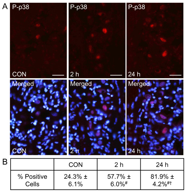
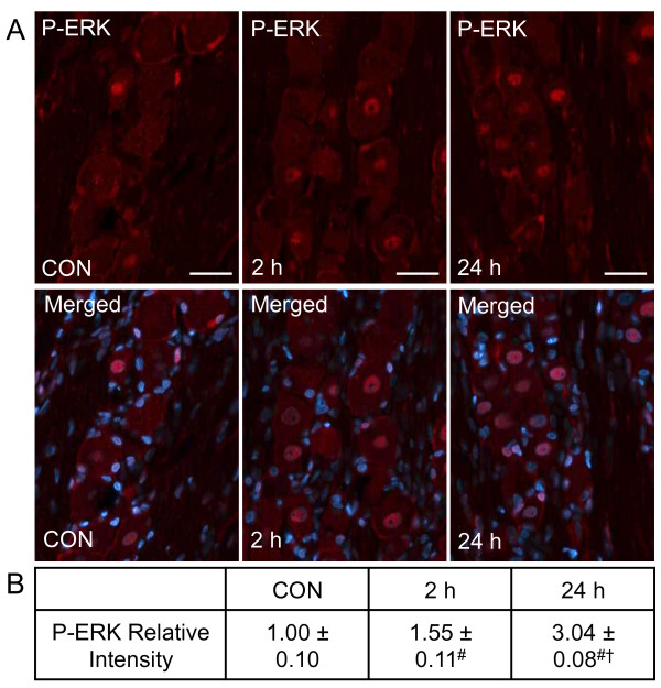
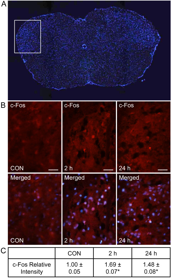
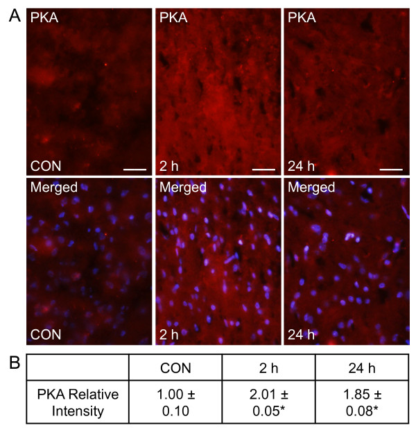
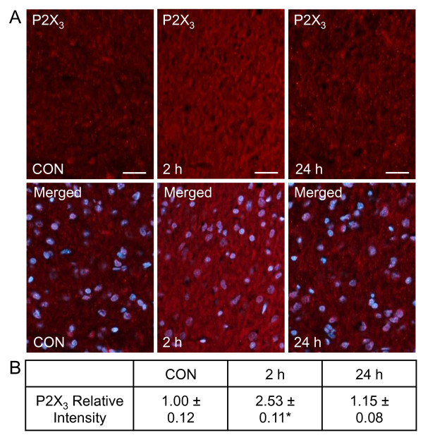
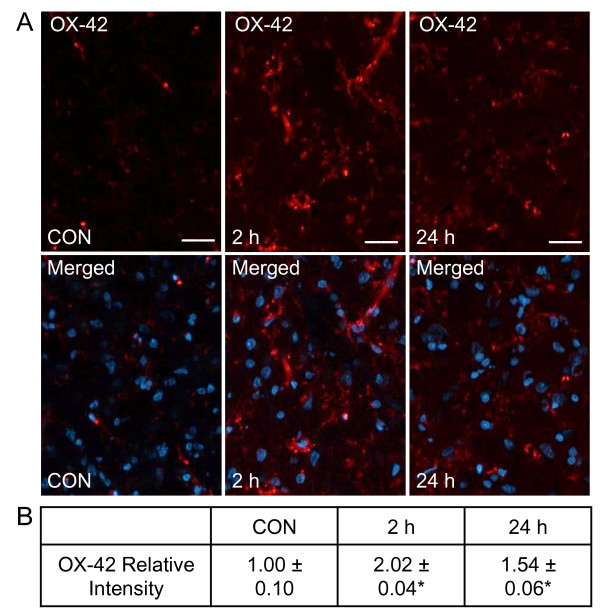
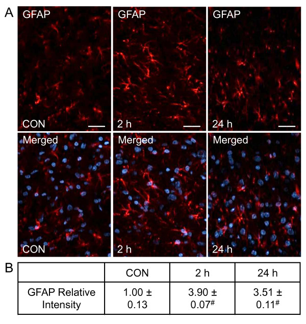
Results: Temporal changes in protein expression in trigeminal ganglia and spinal trigeminal nucleus were determined by immunohistochemistry following injection of CGRP in the temporomandibular joint (TMJ) capsule of male Sprague-Dawley rats. CGRP stimulated expression of the active forms of the MAP kinases p38 and ERK, and PKA in trigeminal ganglia at 2 and 24 hours. CGRP also caused a sustained increase in the expression of c-Fos neurons in the spinal trigeminal nucleus. In contrast, levels of P2X3 in spinal neurons were only significantly elevated at 2 hours in response to CGRP. In addition, CGRP stimulated expression of GFAP in astrocytes and OX-42 in microglia at 2 and 24 hours post injection.
Conclusions: Our results demonstrate that an elevated level of CGRP in the joint, which is associated with TMD, stimulate neuronal and glial expression of proteins implicated in the development of peripheral and central sensitization. Based on our findings, we propose that inhibition of CGRP-mediated activation of trigeminal neurons and glial cells with selective non-peptide CGRP receptor antagonists would be beneficial in the treatment of TMD.
Figures
Similar articles
-
Elevated levels of calcitonin gene-related peptide in upper spinal cord promotes sensitization of primary trigeminal nociceptive neurons.Neuroscience. 2016 Dec 17;339:491-501. doi: 10.1016/j.neuroscience.2016.10.013. Epub 2016 Oct 13. Neuroscience. 2016. PMID: 27746346 Free PMC article.
-
Neuron-glia signaling in trigeminal ganglion: implications for migraine pathology.Headache. 2007 Jul-Aug;47(7):1008-23; discussion 24-5. doi: 10.1111/j.1526-4610.2007.00854.x. Headache. 2007. PMID: 17635592 Free PMC article.
-
Reactive oxygen species induce procalcitonin expression in trigeminal ganglia glia.Headache. 2014 Mar;54(3):472-84. doi: 10.1111/head.12301. Epub 2014 Feb 11. Headache. 2014. PMID: 24512072 Free PMC article.
-
Sensory Neuron-TRPV4 Modulates Temporomandibular Disorder Pain Via CGRP in Mice.J Pain. 2023 May;24(5):782-795. doi: 10.1016/j.jpain.2022.12.001. Epub 2022 Dec 9. J Pain. 2023. PMID: 36509176 Free PMC article. Review.
-
Current understanding of trigeminal ganglion structure and function in headache.Cephalalgia. 2019 Nov;39(13):1661-1674. doi: 10.1177/0333102418786261. Epub 2018 Jul 10. Cephalalgia. 2019. PMID: 29989427 Free PMC article. Review.
Cited by
-
Antagonism of CGRP Receptor: Central and Peripheral Mechanisms and Mediators in an Animal Model of Chronic Migraine.Cells. 2022 Sep 30;11(19):3092. doi: 10.3390/cells11193092. Cells. 2022. PMID: 36231054 Free PMC article.
-
Calcitonin gene-related peptide: An intra-articular therapeutic target for TMJ disorders.Clin Exp Dent Res. 2022 Oct;8(5):1158-1166. doi: 10.1002/cre2.606. Epub 2022 Jun 14. Clin Exp Dent Res. 2022. PMID: 35700066 Free PMC article.
-
Transcriptional Alterations in the Trigeminal Ganglia, Nucleus and Peripheral Blood Mononuclear Cells in a Rat Orofacial Pain Model.Front Mol Neurosci. 2018 Jun 26;11:219. doi: 10.3389/fnmol.2018.00219. eCollection 2018. Front Mol Neurosci. 2018. PMID: 29997476 Free PMC article.
-
The role of calcitonin gene-related peptide in peripheral and central pain mechanisms including migraine.Pain. 2017 Apr;158(4):543-559. doi: 10.1097/j.pain.0000000000000831. Pain. 2017. PMID: 28301400 Free PMC article. Review.
-
Migraine: Calcium Channels and Glia.Int J Mol Sci. 2021 Mar 7;22(5):2688. doi: 10.3390/ijms22052688. Int J Mol Sci. 2021. PMID: 33799975 Free PMC article. Review.
References
-
- Sessle BJ. Neural mechanisms and pathways in craniofacial pain. Can J Neurol Sci. 1999;26(Suppl 3):S7–11. - PubMed
-
- Sessle BJ. Peripheral and central mechanisms of orofacial pain and their clinical correlates. Minerva Anestesiol. 2005;71:117–136. - PubMed
-
- Okeson J. Management of Temporomandibular Disorders and Occlusion. 6. St. Louis, MO; 2008.
-
- Bonjardim LR, Lopes-Filho RJ, Amado G, Albuquerque RL, Goncalves SR. Association between symptoms of temporomandibular disorders and gender, morphological occlusion, and psychological factors in a group of university students. Indian J Dent Res. 2009;20:190–194. - PubMed
-
- Shankland W. The trigeminal nerve. Part IV: the mandibular division. Journal of Craniomandibular Practice. 2001;19:153–161. - PubMed
Publication types
MeSH terms
Substances
LinkOut - more resources
Full Text Sources
Medical
Research Materials
Miscellaneous
