

Erythropoietin attenuates the sequels of ischaemic spinal cord injury with enhanced recruitment of CD34+ cells in mice
- PMID: 22145921
- PMCID: PMC3822692
- DOI: 10.1111/j.1582-4934.2011.01489.x
Erythropoietin attenuates the sequels of ischaemic spinal cord injury with enhanced recruitment of CD34+ cells in mice
Abstract
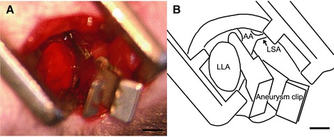
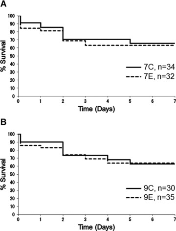
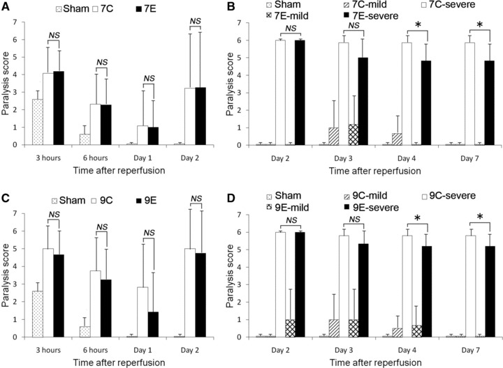
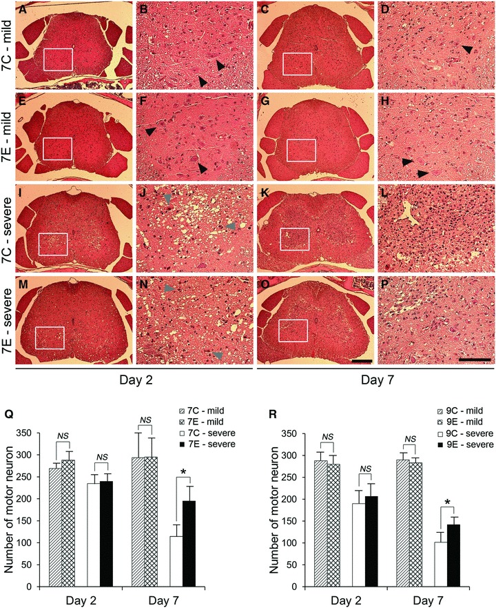
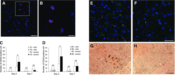
Erythropoietin has been shown to promote tissue regeneration after ischaemic injury in various organs. Here, we investigated whether Erythropoietin could ameliorate ischaemic spinal cord injury in the mouse and sought an underlying mechanism. Spinal cord ischaemia was developed by cross-clamping the descending thoracic aorta for 7 or 9 min. in mice. Erythropoietin (5000 IU/kg) or saline was administrated 30 min. before aortic cross-clamping. Neurological function was assessed using the paralysis score for 7 days after the operation. Spinal cords were histologically evaluated 2 and 7 days after the operation. Immunohistochemistry was used to detect CD34(+) cells and the expression of brain-derived neurotrophic factor and vascular endothelial growth factor. Each mouse exhibited either mildly impaired function or complete paralysis at day 2. Erythropoietin-treated mice with complete paralysis demonstrated significant improvement of neurological function between day 2 and 7, compared to saline-treated mice with complete paralysis. Motor neurons in erythropoietin-treated mice were more preserved at day 7 than those in saline-treated mice with complete paralysis. CD34(+) cells in the lumbar spinal cord of erythropoietin-treated mice were more abundant at day 2 than those of saline-treated mice. Brain-derived neurotrophic factor and vascular endothelial growth factor were markedly expressed in lumbar spinal cords in erythropoietin-treated mice at day 7. Erythropoietin demonstrated neuroprotective effects in the ischaemic spinal cord, improving neurological function and attenuating motor neuron loss. These effects may have been mediated by recruited CD34(+) cells, and enhanced expression of brain-derived neurotrophic factor and vascular endothelial growth factor.
© 2012 The Authors Journal of Cellular and Molecular Medicine © 2012 Foundation for Cellular and Molecular Medicine/Blackwell Publishing Ltd.
Figures
Similar articles
-
Erythropoietin activates the phosporylated cAMP [adenosine 3'5' cyclic monophosphate] response element-binding protein pathway and attenuates delayed paraplegia after ischemia-reperfusion injury.J Thorac Cardiovasc Surg. 2015 Mar;149(3):920-4. doi: 10.1016/j.jtcvs.2014.11.011. Epub 2014 Nov 13. J Thorac Cardiovasc Surg. 2015. PMID: 25500291
-
Optimized induction of beta common receptor enhances the neuroprotective function of erythropoietin in spinal cord ischemic injury.J Thorac Cardiovasc Surg. 2018 Jun;155(6):2505-2516. doi: 10.1016/j.jtcvs.2017.12.132. Epub 2018 Feb 9. J Thorac Cardiovasc Surg. 2018. PMID: 29523405
-
Erythropoietin preconditioning improves clinical and histologic outcome in an acute spinal cord ischemia and reperfusion rabbit model.J Vasc Surg. 2016 Dec;64(6):1797-1804. doi: 10.1016/j.jvs.2015.10.011. Epub 2015 Nov 21. J Vasc Surg. 2016. PMID: 26610640
-
Dexmedetomidine, an α-2a adrenergic agonist, promotes ischemic tolerance in a murine model of spinal cord ischemia-reperfusion.J Thorac Cardiovasc Surg. 2014 Jan;147(1):500-6. doi: 10.1016/j.jtcvs.2013.07.043. Epub 2013 Sep 13. J Thorac Cardiovasc Surg. 2014. PMID: 24035379
-
Synergetic Induction of NGF With Diazoxide and Erythropoietin Attenuates Spinal Cord Ischemic Injury.J Surg Res. 2019 Jan;233:124-131. doi: 10.1016/j.jss.2018.07.021. Epub 2018 Aug 20. J Surg Res. 2019. PMID: 30502238
Cited by
-
Value of micro-CT for monitoring spinal microvascular changes after chronic spinal cord compression.Int J Mol Sci. 2014 Jul 7;15(7):12061-73. doi: 10.3390/ijms150712061. Int J Mol Sci. 2014. PMID: 25003643 Free PMC article.
-
Transplantation of Hematopoietic Stem Cells Promotes Functional Improvement Associated with NT-3-MEK-1 Activation in Spinal Cord-Transected Rats.Front Cell Neurosci. 2017 Jul 19;11:213. doi: 10.3389/fncel.2017.00213. eCollection 2017. Front Cell Neurosci. 2017. PMID: 28769769 Free PMC article.
-
Spinal Stroke: Outcome Attenuation by Erythropoietin and Carbamylated Erythropoietin and Its Prediction by Sphingosine-1-Phosphate Serum Levels in Mice.Int J Mol Sci. 2022 Aug 23;23(17):9558. doi: 10.3390/ijms23179558. Int J Mol Sci. 2022. PMID: 36076955 Free PMC article.
-
Postnatal Erythropoietin Mitigates Impaired Cerebral Cortical Development Following Subplate Loss from Prenatal Hypoxia-Ischemia.Cereb Cortex. 2015 Sep;25(9):2683-95. doi: 10.1093/cercor/bhu066. Epub 2014 Apr 9. Cereb Cortex. 2015. PMID: 24722771 Free PMC article.
-
Regulation of Inflammatory Cytokines for Spinal Cord Injury Repair Through Local Delivery of Therapeutic Agents.Adv Sci (Weinh). 2018 Jul 31;5(11):1800529. doi: 10.1002/advs.201800529. eCollection 2018 Nov. Adv Sci (Weinh). 2018. PMID: 30479916 Free PMC article. Review.
References
-
- Wong DR, Parenti JL, Green SY, et al. Open repair of thoracoabdominal aortic aneurysm in the modern surgical era: contemporary outcomes in 509 patients. J Am Coll Surg. 2011;212:569–79. - PubMed
-
- Conrad MF, Crawford RS, Davison JK, et al. Thoracoabdominal aneurysm repair: a 20-year perspective. Ann Thorac Surg. 2007;83:856–61. - PubMed
-
- Wong DR, Coselli JS, Amerman K, et al. Delayed spinal cord deficits after thoracoabdominal aortic aneurysm repair. Ann Thorac Surg. 2007;83:1345–55. - PubMed
-
- Bavaria JE, Appoo JJ, Makaroun MS, et al. Endovascular stent grafting versus open surgical repair of descending thoracic aortic aneurysms in low-risk patients: a multicenter comparative trial. J Thorac Cardiovasc Surg. 2007;133:369–77. - PubMed
Publication types
MeSH terms
Substances
LinkOut - more resources
Full Text Sources
Medical
