

Low doses of selenium specifically stimulate the repair of oxidative DNA damage in LNCaP prostate cancer cells
- PMID: 22145923
- PMCID: PMC3332102
- DOI: 10.3109/10715762.2011.647009
Low doses of selenium specifically stimulate the repair of oxidative DNA damage in LNCaP prostate cancer cells
Abstract
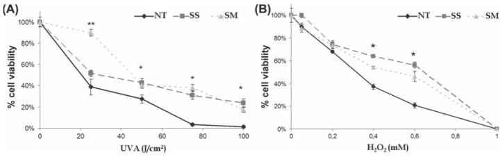
Epidemiological studies have demonstrated an inverse relationship between selenium (Se) intake and cancer incidence and/or mortality. However, the molecular mechanisms underlying the cancer chemopreventive activity of Se compounds remain largely unknown. The objective of this study was to investigate the effect of low doses of Se on the stimulation of DNA repair systems in response to four different qualities of DNA damage. P53-proficient LNCaP human prostate adenocarcinoma cells were grown either untreated or in the presence of low concentrations of two Se compounds (30° nM sodium selenite, or 10 μM selenomethionine) and exposed to UVA, H2O2, methylmethane sulfonate (MMS) or UVC. Cell viability as well as DNA damage induction and repair were evaluated by the alkaline Comet assay. Overall, Se was shown to be a very potent protector against cell toxicity and genotoxicity induced by oxidative stress (UVA or H2O2) but not from the agents that induce other types of deleterious lesions (MMS or UVC). Furthermore, Se-treated cells exhibited increased oxidative DNA repair activity, indicating a novel mechanism of Se action. Therefore, the benefits of Se could be explained by a combination of antioxidant activity, the reduction in DNA damage and the enhancement of oxidative DNA repair capacity.
Conflict of interest statement
This work was supported by University Joseph Fourier and the CEA. Some methods have been developed thought LODORA project which is funded by the National research Agency. Also, the work was supported in part by a grant from the National Cancer Institute (RO1 CA127943) to AMD. The authors report no conflict of interest. The authors alone are responsible for the content and writing of the paper.
Figures


Similar articles
-
Aqueous extracts of selenium-fertilized broccoli increase selenoprotein activity and inhibit DNA single-strand breaks, but decrease the activity of quinone reductase in Hepa 1c1c7 cells.Food Chem Toxicol. 2006 May;44(5):695-703. doi: 10.1016/j.fct.2005.10.002. Epub 2005 Dec 22. Food Chem Toxicol. 2006. PMID: 16377050
-
Effects of selenium compounds on induction of DNA damage by broadband ultraviolet radiation in human keratinocytes.Br J Dermatol. 2003 May;148(5):1001-9. doi: 10.1046/j.1365-2133.2003.05267.x. Br J Dermatol. 2003. PMID: 12786833
-
Interactions of selenium compounds with other antioxidants in DNA damage and apoptosis in human normal keratinocytes.Cancer Epidemiol Biomarkers Prev. 2001 Apr;10(4):385-90. Cancer Epidemiol Biomarkers Prev. 2001. PMID: 11319180
-
Selenium: a double-edged sword for defense and offence in cancer.Arch Toxicol. 2010 Dec;84(12):919-38. doi: 10.1007/s00204-010-0595-8. Epub 2010 Sep 25. Arch Toxicol. 2010. PMID: 20871980 Review.
-
Selenium metabolism, selenoproteins and mechanisms of cancer prevention: complexities with thioredoxin reductase.Carcinogenesis. 1999 Sep;20(9):1657-66. doi: 10.1093/carcin/20.9.1657. Carcinogenesis. 1999. PMID: 10469608 Review.
Cited by
-
The effects of selenium and the GPx-1 selenoprotein on the phosphorylation of H2AX.Biochim Biophys Acta. 2013 Jun;1830(6):3399-406. doi: 10.1016/j.bbagen.2013.03.010. Epub 2013 Mar 18. Biochim Biophys Acta. 2013. PMID: 23518201 Free PMC article.
-
Selenium as a Bioactive Micronutrient in the Human Diet and Its Cancer Chemopreventive Activity.Nutrients. 2021 May 13;13(5):1649. doi: 10.3390/nu13051649. Nutrients. 2021. PMID: 34068374 Free PMC article. Review.
-
Selenium supplementation and prostate cancer mortality.J Natl Cancer Inst. 2014 Dec 12;107(1):360. doi: 10.1093/jnci/dju360. Print 2015 Jan. J Natl Cancer Inst. 2014. PMID: 25505227 Free PMC article.
-
Nutrition Can Help DNA Repair in the Case of Aging.Nutrients. 2020 Nov 1;12(11):3364. doi: 10.3390/nu12113364. Nutrients. 2020. PMID: 33139613 Free PMC article. Review.
-
Does a role for selenium in DNA damage repair explain apparent controversies in its use in chemoprevention?Mutagenesis. 2013 Mar;28(2):127-34. doi: 10.1093/mutage/ges064. Epub 2012 Nov 30. Mutagenesis. 2013. PMID: 23204505 Free PMC article. Review.
References
-
- Meuillet E, Stratton S, Prasad Cherukuri D, Goulet AC, Kagey J, Porterfield B, Nelson MA. Chemoprevention of prostate cancer with selenium: an update on current clinical trials and preclinical findings. J Cell Biochem. 2004;91(3):443–458. - PubMed
-
- Lu J, Holmgren A. Selenoproteins. J Biol Chem. 2009;284(2):723–727. - PubMed
-
- Rayman MP. The use of high-selenium yeast to raise selenium status: how does it measure up? Br J Nutr. 2004;92:557–573. - PubMed
-
- Letavayova L, Vlckova V, Brozmanova J. Selenium: from cancer prevention to DNA damage. Toxicology. 2006;227:1–14. - PubMed
-
- Arthur JR, McKenzie RC, Beckett GJ. Selenium in the immune system. J Nutr. 2003;133(5 Suppl 1):1457S–1459S. - PubMed
Publication types
MeSH terms
Substances
Grants and funding
LinkOut - more resources
Full Text Sources
Other Literature Sources
Medical
Research Materials
Miscellaneous