

Isoflurane does not cause neuroapoptosis but reduces astroglial processes in young adult mice
- PMID: 22146123
- PMCID: PMC3253045
- DOI: 10.1186/2045-9912-1-27
Isoflurane does not cause neuroapoptosis but reduces astroglial processes in young adult mice
Abstract
Background: Isoflurane, a volatile anesthetic widely used clinically, has been implicated to be both neuroprotective and neurotoxic. The claim about isoflurane causing neural apoptosis remains controversial. In this study, we investigated the effects of isoflurane exposures on apoptotic and anti-apoptotic signals, cell proliferation and neurogenesis, and astroglial processes in young adult mouse brains.
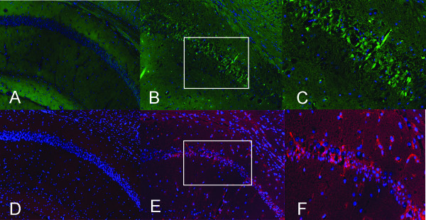
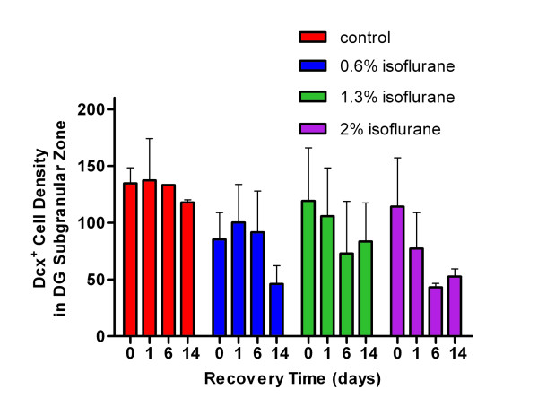
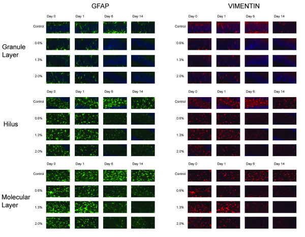
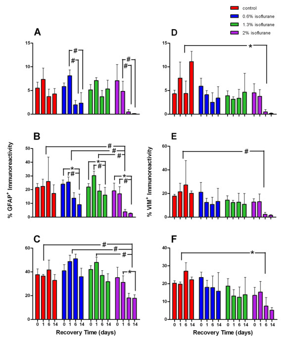
Methods: Sixty 6-week-old mice were randomly assigned to four anesthetic concentration groups (0 as control and 0.6%, 1.3%, and 2%) with four recovery times (2 h and 1, 6, and 14 d) after 2-h isoflurane exposure. Immunohistochemistry measurements of activated caspase-3 and Bcl-xl for apoptotic and anti-apoptotic signals, respectively, glial fibrillary acidic protein (GFAP) and vimentin for reactive astrocytosis, doublecortin (Dcx) for neurogenesis, and BrdU for cell proliferation were performed.
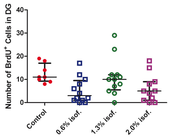
Results: Contrary to the previous conclusion derived from studies with neonatal rodents, we found no evidence of isoflurane-induced apoptosis in the adult mouse brain. Neurogenesis in the subgranule zone of the dentate gyrus was not affected by isoflurane. However, there is a tendency of reduced cell proliferation after 2% isoflurane exposure. VIM and GFAP staining showed that isoflurane exposure caused a delayed reduction of astroglial processes in the hippocampus and dentate gyrus.
Conclusion: Two-hour exposure to isoflurane did not cause neuroapoptosis in adult brains. The delayed reduction in astroglial processes after isoflurane exposure may explain why some volatile anesthetics can confer neuroprotection after experimental stroke because reduced glial scarring facilitates better long-term neuronal recoveries.
Figures
Similar articles
-
The neurotoxic effect of isoflurane on age-defined neurons generated from tertiary dentate matrix in mice.Brain Behav. 2021 Jan;11(1):e01949. doi: 10.1002/brb3.1949. Epub 2020 Nov 17. Brain Behav. 2021. PMID: 33201600 Free PMC article.
-
Characterization and quantification of isoflurane-induced developmental apoptotic cell death in mouse cerebral cortex.Anesth Analg. 2013 Apr;116(4):845-54. doi: 10.1213/ANE.0b013e318281e988. Epub 2013 Mar 4. Anesth Analg. 2013. PMID: 23460572
-
From the Cover: Volatile Anesthetics Transiently Disrupt Neuronal Development in Neonatal Rats.Toxicol Sci. 2016 Dec;154(2):309-319. doi: 10.1093/toxsci/kfw164. Epub 2016 Aug 25. Toxicol Sci. 2016. PMID: 27562558
-
Beyond anesthetic properties: the effects of isoflurane on brain cell death, neurogenesis, and long-term neurocognitive function.Anesth Analg. 2010 Feb;110(2):431-7. Anesth Analg. 2010. PMID: 25508825 Review.
-
Beyond anesthetic properties: the effects of isoflurane on brain cell death, neurogenesis, and long-term neurocognitive function.Anesth Analg. 2010 Feb;110(2):431-7. doi: 10.1213/ANE.0b013e3181af8015. Anesth Analg. 2010. PMID: 19917621 Review.
Cited by
-
Progress of Research on Diffuse Axonal Injury after Traumatic Brain Injury.Neural Plast. 2016;2016:9746313. doi: 10.1155/2016/9746313. Epub 2016 Dec 19. Neural Plast. 2016. PMID: 28078144 Free PMC article. Review.
-
New progress of isoflurane, sevoflurane and propofol in hypoxic-ischemic brain injury and related molecular mechanisms based on p75 neurotrophic factor receptor.Ibrain. 2021 Jun 28;7(2):132-140. doi: 10.1002/j.2769-2795.2021.tb00075.x. eCollection 2021 Jun. Ibrain. 2021. PMID: 37786902 Free PMC article. Review.
-
Perinatal supplementation with omega-3 polyunsaturated fatty acids improves sevoflurane-induced neurodegeneration and memory impairment in neonatal rats.PLoS One. 2013 Aug 13;8(8):e70645. doi: 10.1371/journal.pone.0070645. eCollection 2013. PLoS One. 2013. PMID: 23967080 Free PMC article.
-
Adverse effects of vapocoolant and topical anesthesia for tail biopsy of preweanling mice.J Am Assoc Lab Anim Sci. 2015 May;54(3):291-8. J Am Assoc Lab Anim Sci. 2015. PMID: 26045455 Free PMC article.
-
Mechanistic insights into neurotoxicity induced by anesthetics in the developing brain.Int J Mol Sci. 2012;13(6):6772-6799. doi: 10.3390/ijms13066772. Epub 2012 Jun 4. Int J Mol Sci. 2012. PMID: 22837663 Free PMC article. Review.
References
-
- Stratmann G, Sall JW, Bell JS, Alvi RS, May LV, Ku B, Dowlatshahi M, Dai R, Bickler PE, Russell I. et al.Isoflurane does not affect brain cell death, hippocampal neurogenesis, or long-term neurocognitive outcome in aged rats. Anesthesiology. 2010;112:305–315. doi: 10.1097/ALN.0b013e3181ca33a1. - DOI - PMC - PubMed
Grants and funding
LinkOut - more resources
Full Text Sources
Research Materials
Miscellaneous
