

Voxel-wise meta-analysis of fMRI studies in patients at clinical high risk for psychosis
- PMID: 22146150
- PMCID: PMC3297070
- DOI: 10.1503/jpn.110021
Voxel-wise meta-analysis of fMRI studies in patients at clinical high risk for psychosis
Abstract
Background: Reliable neurofunctional markers of increased vulnerability to psychosis are needed to improve the predictive value of psychosis risk syndrome and inform preventive interventions.
Methods: I performed a signed differential mapping (SDM) voxel-wise meta-analysis of functional magnetic resonance imaging (fMRI) studies of patients at clinical high risk for psychosis.
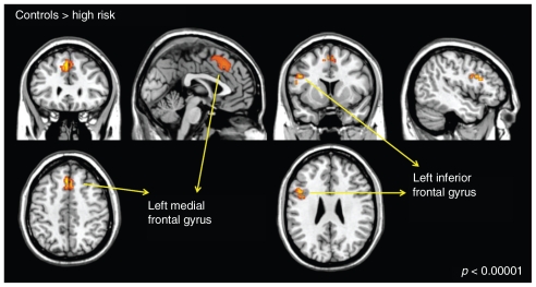
Results: Ten studies were included in the analysis. Compared with controls, high-risk patients showed reduced neural activation in the left inferior frontal gyrus (Brodmann area [BA] 9) and in a cluster spanning the bilateral medial frontal gyrus (BA 8,6), bilateral superior frontal gyrus (BA 8,6)and the left anterior cingulate (BA 32). There was no publication bias. Heterogeneity across studies was low. Sensitivity analysis confirmed the robustness of the findings.
Limitations: The cross-sectional nature of the included studies prevented the comparison of high-risk patients who later experienced a psychotic episode with those who did not. Other caveats are reflected in methodologic heterogeneity across tasks employed by different individual imaging studies.
Conclusion: Reduced neurofunctional activation in prefrontal regions may represent a neurophysiologic correlate of increased vulnerability to psychosis.
Figures
Similar articles
-
Neuroanatomical markers of genetic liability to psychosis and first episode psychosis: a voxelwise meta-analytical comparison.World J Biol Psychiatry. 2014 Apr;15(3):219-28. doi: 10.3109/15622975.2011.630408. Epub 2012 Jan 27. World J Biol Psychiatry. 2014. PMID: 22283467
-
Neuroanatomical maps of psychosis onset: voxel-wise meta-analysis of antipsychotic-naive VBM studies.Schizophr Bull. 2012 Nov;38(6):1297-307. doi: 10.1093/schbul/sbr134. Epub 2011 Nov 10. Schizophr Bull. 2012. PMID: 22080494 Free PMC article.
-
Multimodal voxel-based meta-analysis of structural and functional magnetic resonance imaging studies in those at elevated genetic risk of developing schizophrenia.Psychiatry Res. 2014 Jan 30;221(1):69-77. doi: 10.1016/j.pscychresns.2013.07.008. Epub 2013 Nov 14. Psychiatry Res. 2014. PMID: 24239093
-
Brain structural abnormalities as potential markers for detecting individuals with ultra-high risk for psychosis: A systematic review and meta-analysis.Schizophr Res. 2019 Jul;209:22-31. doi: 10.1016/j.schres.2019.05.015. Epub 2019 May 16. Schizophr Res. 2019. PMID: 31104914
-
Neuroanatomy of vulnerability to psychosis: a voxel-based meta-analysis.Neurosci Biobehav Rev. 2011 Apr;35(5):1175-85. doi: 10.1016/j.neubiorev.2010.12.005. Epub 2010 Dec 17. Neurosci Biobehav Rev. 2011. PMID: 21168439 Review.
Cited by
-
Meta-analysis of structural MRI studies in anorexia nervosa and the role of recovery: a systematic review protocol.Syst Rev. 2021 Sep 13;10(1):247. doi: 10.1186/s13643-021-01799-y. Syst Rev. 2021. PMID: 34517926 Free PMC article.
-
Disrupted functional connectivity between visual and emotional networks in psychosis risk syndromes through representational similarity analysis.Front Psychiatry. 2025 Apr 17;16:1533675. doi: 10.3389/fpsyt.2025.1533675. eCollection 2025. Front Psychiatry. 2025. PMID: 40313238 Free PMC article.
-
Altered relationships between age and functional brain activation in adolescents at clinical high risk for psychosis.Psychiatry Res. 2014 Jan 30;221(1):21-9. doi: 10.1016/j.pscychresns.2013.08.004. Epub 2013 Oct 19. Psychiatry Res. 2014. PMID: 24144510 Free PMC article.
-
Neuropsychiatric symptoms and regional neocortical atrophy in mild cognitive impairment and Alzheimer's disease.Am J Alzheimers Dis Other Demen. 2014 Mar;29(2):159-65. doi: 10.1177/1533317513507373. Epub 2013 Oct 27. Am J Alzheimers Dis Other Demen. 2014. PMID: 24164929 Free PMC article.
-
Altered functional connectivity of the hippocampus in cortico-subcortical networks in early-stage and emerging psychosis.Eur Arch Psychiatry Clin Neurosci. 2025 Aug 19. doi: 10.1007/s00406-025-02079-9. Online ahead of print. Eur Arch Psychiatry Clin Neurosci. 2025. PMID: 40828422
References
-
- Ruhrmann S, Schultze-Lutter F, Bechdolf A, et al. Intervention in at-risk states for developing psychosis. Eur Arch Psychiatry Clin Neurosci. 2010;260(Suppl 2):S90–4. - PubMed
-
- Ruhrmann S, Schultze-Lutter F, Klosterkotter J. Probably at-risk, but certainly ill — advocating the introduction of a psychosis spectrum disorder in DSM-V. Schizophr Res. 2010;120:23–37. - PubMed
-
- Nelson B, Yung AR. Should a risk syndrome for first episode psychosis be included in the DSM-V? Curr Opin Psychiatry. 2011;24:128–33. - PubMed
-
- McGuire P, Howes OD, Stone J, et al. Functional neuroimaging in schizophrenia: diagnosis and drug discovery. Trends Pharmacol Sci. 2008;29:91–8. - PubMed
Publication types
MeSH terms
LinkOut - more resources
Full Text Sources
Medical