. 2012 Apr;32(4):720-30.
doi: 10.1038/jcbfm.2011.176. Epub 2011 Dec 7.

Release of mitochondrial apoptogenic factors and cell death are mediated by CK2 and NADPH oxidase

Affiliations

Release of mitochondrial apoptogenic factors and cell death are mediated by CK2 and NADPH oxidase

Gab Seok Kim et al. J Cereb Blood Flow Metab. 2012 Apr.

Abstract

Activation of the NADPH oxidase subunit, NOX2, and increased oxidative stress are associated with neuronal death after cerebral ischemia and reperfusion. Inhibition of NOX2 by casein kinase 2 (CK2) leads to neuronal survival, but the mechanism is unknown. In this study, we show that in copper/zinc-superoxide dismutase transgenic (SOD1 Tg) mice, degradation of CK2α and CK2α' and dephosphorylation of CK2β against oxidative stress were markedly reduced compared with wild-type (WT) mice that underwent middle cerebral artery occlusion. Inhibition of CK2 pharmacologically or by ischemic reperfusion facilitated accumulation of poly(ADP-ribose) polymers, the translocation of apoptosis-inducing factor (AIF), and cytochrome c release from mitochondria after ischemic injury. The eventual enhancement of CK2 inhibition under ischemic injury strongly increased 8-hydroxy-2'-deoxyguanosine and phosphorylation of H2A.X. Furthermore, CK2 inhibition by tetrabromocinnamic acid (TBCA) in SOD1 Tg and gp91 knockout (KO) mice after ischemia reperfusion induced less release of AIF and cytochrome c than in TBCA-treated WT mice. Inhibition of CK2 in gp91 KO mice subjected to ischemia reperfusion did not increase brain infarction compared with TBCA-treated WT mice. These results strongly suggest that NOX2 activation releases reactive oxygen species after CK2 inhibition, triggering release of apoptogenic factors from mitochondria and inducing DNA damage after ischemic brain injury.


Figures

Figure 1
Figure 1
Reactive oxygen species (ROS) mediate casein kinase 2 (CK2) downregulation in brain injury. (A) Protein samples from the ipsilateral and contralateral hemispheres of WT mice or mice that overexpress SOD1 that were harvested 24 hours after 45 minutes of transient focal cerebral ischemia. The samples were separated by SDS-PAGE and immunoblotted with anti-CK2α, CK2α′, phosphorylated CK2β (Ser209), and CK2β antibodies. β-Tubulin was used as a loading control. Quantitative data show that expression of CK2α (B), CK2α′ (C), and phosphorylated CK2β (D) was markedly reduced at 24 hours in the ipsilateral hemispheres compared with the contralateral hemispheres in the WT mice, but not in the ipsilateral hemispheres of the superoxide dismutase transgenic (SOD1 Tg) mice after transient focal cerebral ischemia (n=5). One-way analysis of variance (ANOVA) followed by Bonferroni correction. *P<0.05 compared with the ipsilateral hemispheres of the WT mice. I/R, ischemic reperfusion; C, contralateral; I, ipsilateral; OD, optical density; WT, wild type.
Figure 2
Figure 2
PARP-1 activation is more intense in tetrabromocinnamic acid (TBCA)-treated mice (CD1) after ischemic injury. Brain sections from sham mice and mice (CD1) that underwent middle cerebral artery occlusion (MCAO) after vehicle or TBCA treatment were incubated with an anti-PAR polymer antibody (green), followed by incubation with a secondary antibody conjugated with Alexa-488. The sections were then mounted with medium containing DAPI (blue) (n=6). Scale bar=50 μmol/L. I/R, ischemic reperfusion; DAPI, 4′,6 diamidino-2-phenylindole; PAR, poly(ADP-ribose); PARP-1, PAR polymerase-1.
Figure 3
Figure 3
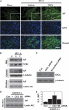
Reactive oxygen species (ROS) generation through inhibition of casein kinase 2 (CK2) leads to facilitation of apoptosis-inducing factor (AIF) translocation to the nucleus. (A) AIF-positive cells were detected by an immunofluorescent technique with an anti-AIF antibody using slices from brains of vehicle- and tetrabromocinnamic acid (TBCA)-injected mice (CD1) 3 hours after ischemia and reperfusion. 4′,6 diamidino-2-phenylindole (DAPI) (blue) was used to counterstain the nucleus (n=6). Scale bar=50 μmol/L. The insets are magnification images showing AIF translocation to the nucleus. (B) Western blotting was performed with an anti-AIF antibody using fractionated samples (mitochondrial, cytosolic, and nuclear) from sham, vehicle- or TBCA-treated mice (CD1) 3 hours after ischemia and reperfusion injury. Cyclooxygenase 4 (COX4), Lamin A/C, and β-tubulin antibodies were used as mitochondrial, nuclear, and cytoplasmic markers, respectively (n=6). (C) Western blotting was performed using samples from control neuronal cells and CK2α small-interfering RNA (siRNA)-transfected and scrambled siRNA-transfected primary neuronal cells to confirm the effect of CK2α siRNA on downregulation of the CK2α protein. (D, E) Primary cortical neurons were transfected with CK2α siRNA or scrambled siRNA for 48 hours. These cells were then subjected to 4 hours of OGD and 3 hours of reoxygenation. AIF translocation to the nucleus was tested by western blotting using the nuclear fraction samples (n=4). One-way analysis of variance (ANOVA) followed by Tukey's test. *P<0.05 compared with OGD/3 hours of reoxygenation only or scrambled siRNA plus OGD and reoxygenation. I/R, ischemic reperfusion; S, sham; V, vehicle; T, TBCA; OGD/R, oxygen-glucose deprivation/reoxygenation; Con, control; —, OGD only (no siRNA transfection); SS, scrambled siRNA; OD, optical density.
Figure 4
Figure 4
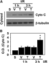
Tetrabromocinnamic acid (TBCA) treatment promotes the release of cytochrome c to the cytoplasm after ischemic-reperfusion injury. (A) Mouse (CD1) brains were isolated 1 and 3 hours after ischemic reperfusion. Cytochrome c release was detected by western blotting using cytoplasmic protein samples from the sham and damaged brains, which were injected with the vehicle or TBCA. (B) Summary graph shows that 3 hours after ischemic reperfusion, cytochrome c release was increased in the TBCA-injected brains (n=4). One-way analysis of variance (ANOVA) followed by Bonferroni correction. *P<0.05 compared with vehicle-treated brains 3 hours after middle cerebral artery occlusion (MCAO). I/R, ischemic reperfusion; S, sham; V, vehicle; T, TBCA; OD, optical density.
Figure 5
Figure 5
Inhibition of casein kinase 2 (CK2) by tetrabromocinnamic acid (TBCA) increases DNA damage after brain injury. (A) Twelve hours after ischemic reperfusion, mouse (CD1) brains were isolated and sectioned. DNA damage was revealed by immunohistochemistry using an anti-8-OHdG antibody on undamaged or damaged brain sections from mice injected intracerebroventricularly with TBCA or the vehicle (n=4). Scale bar=50 μm. (B) Phosphorylation of H2A.X was assessed 3 hours after ischemic reperfusion by western blotting with an anti-phospho-H2A.X antibody using nuclear extracts from sham or damaged brains, which were injected with the vehicle or TBCA. Lamin A/C was used as an internal control and nuclear marker. (C) The graph shows that TBCA increased phosphorylation of H2A.X 3 hours after ischemic injury (n=4). One-way analysis of variance (ANOVA) followed by Tukey's test. *P<0.05 compared with 3 hours of ischemic reperfusion with vehicle treatment. I/R, ischemic reperfusion; NC, negative control; S, sham; V, vehicle; T, TBCA; OD, optical density; 8-OHdG, 8-hydroxy-2′-deoxyguanosine.
Figure 6
Figure 6
Reactive oxygen species (ROS) generated by NADPH oxidase induced by casein kinase 2 (CK2) inhibition are responsible for the increase in mitochondria-mediated brain damage. (A) Apoptosis-inducing factor (AIF) translocation to the nucleus by CK2 inhibition after ischemic injury was measured by western blotting with an anti-AIF antibody using the nuclear fraction from WT (CD1) and superoxide dismutase transgenic (SOD1 Tg) (CD1) mouse brains injected with the vehicle or tetrabromocinnamic acid (TBCA) 3 hours after ischemic reperfusion. Lamin A/C was used as an internal control and nuclear marker (n=6). One-way analysis of variance (ANOVA) followed by Bonferroni correction. *P<0.05 compared with the WT vehicle-injected group. (B) Cytochrome c release from mitochondria to the cytoplasm induced by CK2 inhibition after ischemic injury was assessed by western blotting with an anti-cytochrome c antibody using cytoplasmic samples from WT mouse brains and SOD1 Tg mouse brains injected with the vehicle or TBCA 3 hours after ischemic reperfusion. β-Tubulin was used as an internal control (n=6). One-way ANOVA followed by Bonferroni correction. *P<0.05 compared with the WT vehicle-injected group. (C) AIF translocation was measured by western blotting using samples from WT (C57BL/6J) or gp91phox knockout (KO) (C57BL/6J) mouse brains injected with the vehicle or TBCA (n=4). *P<0.05 compared with the WT vehicle-injected group. (D) The vehicle and TBCA were injected intracerebroventricularly in WT (C57BL/6J) or gp91 KO (C57BL/6J) mice 1 hour before onset of middle cerebral artery occlusion (MCAO). Twenty-four hours after ischemic reperfusion, brains were isolated and brain slices were analyzed to measure infarction volume using 2,3,5-triphenyltetrazolium chloride staining. (E) A summary graph shows comparative data on infarction volumes after vehicle or TBCA treatment in WT and gp91 KO mouse brains 24 hours after ischemic injury (n=5 to 6 in each group). One-way ANOVA followed by Bonferroni correction. *P<0.05 compared with the WT/vehicle group. I/R, ischemic reperfusion; V, vehicle; T, TBCA; NS, not significant; WT, wild type; OD, optical density.

Similar articles

Cited by

References

    1. Ames BN. Endogenous oxidative DNA damage, aging, and cancer. Free Radic Res Commun. 1989;7:121–128. - PubMed
    1. Andrabi SA, Kim NS, Yu S-W, Wang H, Koh DW, Sasaki M, Klaus JA, Otsuka T, Zhang Z, Koehler RC, Hurn PD, Poirier GG, Dawson VL, Dawson TM. Poly(ADP-ribose) (PAR) polymer is a death signal. Proc Natl Acad Sci USA. 2006;103:18308–18313. - PMC - PubMed
    1. Breusing N, Grune T. Regulation of proteasome-mediated protein degradation during oxidative stress and aging. Biol Chem. 2008;389:203–209. - PubMed
    1. Chan PH. Mitochondrial dysfunction and oxidative stress as determinants of cell death/survival in stroke. Ann NY Acad Sci. 2005;1042:203–209. - PubMed
    1. Chan PH, Epstein CJ, Kinouchi H, Kamii H, Chen SF, Carlson E, Gafni J, Yang G, Reola L. Neuroprotective role of CuZn–superoxide dismutase in ischemic brain damage. Adv Neurol. 1996;71:271–280. - PubMed

Publication types

MeSH terms