. 2012 Feb;32(2):203-12.
doi: 10.1038/jcbfm.2011.169. Epub 2011 Dec 7.

Delayed cerebral ischemia and spreading depolarization in absence of angiographic vasospasm after subarachnoid hemorrhage

Affiliations

Delayed cerebral ischemia and spreading depolarization in absence of angiographic vasospasm after subarachnoid hemorrhage

Johannes Woitzik et al. J Cereb Blood Flow Metab. 2012 Feb.

Abstract

It has been hypothesized that vasospasm is the prime mechanism of delayed cerebral ischemia (DCI) after aneurysmal subarachnoid hemorrhage (aSAH). Recently, it was found that clusters of spreading depolarizations (SDs) are associated with DCI. Surgical placement of nicardipine prolonged-release implants (NPRIs) was shown to strongly attenuate vasospasm. In the present study, we tested whether SDs and DCI are abolished when vasospasm is reduced or abolished by NPRIs. After aneurysm clipping, 10 NPRIs were placed next to the proximal intracranial vessels. The SDs were recorded using a subdural electrode strip. Proximal vasospasm was assessed by digital subtraction angiography (DSA). 534 SDs were recorded in 10 of 13 patients (77%). Digital subtraction angiography revealed no vasospasm in 8 of 13 patients (62%) and only mild or moderate vasospasm in the remaining. Five patients developed DCI associated with clusters of SD despite the absence of angiographic vasospasm in three of those patients. The number of SDs correlated significantly with the development of DCI. This may explain why reduction of angiographic vasospasm alone has not been sufficient to improve outcome in some clinical studies.


Figures
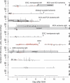
Figure 1
Figure 1
Temporal correlation of neurologic and clinical scores, spreading depolarization (SD) and coregistered responses of the tissue partial pressure of oxygen (ptiO2). The figure summarizes 10 of 13 patients in whom SDs occurred. The length of the black horizontal lines marks the individual monitoring period in each patient. The length of the black vertical lines illustrates the duration of the depression period. Negative black vertical lines denote the occurrence of a slow potential change before recovery of the electrocorticography (ECoG) activity. Red vertical lines illustrate the coregistered increases and decreases of the ptiO2 in response to SD. The gray-shaded area indicates periods with sedation. Arrows indicate the occurrence of delayed neurologic deficits, defined by focal deficits or a decrease of the level of consciousness by at least two Glasgow coma scale points (DOC=decrease of consciousness). In a similar manner, new CT- or MRI-proven infarcts are given (MCA, middle cerebral artery; ACA, anterior cerebral artery; PCA, posterior cerebral artery). Patient no. 7 suffered from an early postoperative frontal MCA ischemia, which is marked by an asterisk. Patient no. 6 developed a long lasting cluster of SDs without recovery of the ECoG activity (striped area). CT, computed tomography; MRI, magnetic resonance imaging; SAH, subarachnoid hemorrhage.
Figure 2
Figure 2
Delayed cerebral ischemia (DCI) is possibly associated with the number of spreading depolarizations (SDs) and total duration of electrocorticography (ECoG) depression periods.
Figure 3
Figure 3
Electrocorticography (ECoG) of a 71-year-old female (patient no. 1) suffering from aneurysmal subarachnoid hemorrhage (aSAH) (World Federation of Neurological Surgeons (WFNS) grade 4/Fisher grade 3). The patient showed several spreading depolarizations (SDs) (arrows) during the first 7 days after aneurysm rupture (compare Figure 1 for the temporal course). On day 7, the patient developed a left-sided hemiparesis and loss of consciousness which necessitated reintubation. Digital subtraction angiography (DSA) (D) excluded proximal vasospasm as a source for the delayed neurologic deficit. Magnetic resonance imaging with diffusion weighted imaging (DWI, E) and fluid-attenuated inversion recovery (Flair) pulse sequences (F) on day 13 revealed a laminar cortical necrosis in the right middle cerebral artery territory and anterior cerebral artery territory (not shown). The ECoG in panel A shows bipolar recordings between electrodes 2 and 3 (channel A), 3 and 4 (channel B), 4 and 5 (channel C), and 5 and 6 (channel D). Recordings from a coimplanted Clark-type oxygen sensor showed monophasic increases (not shown), biphasic changes (not shown), or monophasic decreases (lower trace) of tissue partial pressure of oxygen (ptiO2) in close temporal relationship to SDs. (B) The initial computed tomography (CT) with major aSAH is shown. The CT scan performed on day 6 (C) shows artifacts of the electrode strip implanted over the right frontal cortex. No signs of ischemic stroke are visualized at this time point.
Figure 4
Figure 4
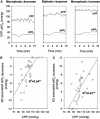
Coregistered responses of tissue partial pressure of oxygen (ptiO2) in temporal relationship to spreading depolarizations (SDs) (A) observed during the first 4 days in patient no. 6. Monophasic increases, decreases, or biphasic responses were detected. A lower baseline level of cerebral perfusion pressure (CPP) was associated with less pronounced increase and more pronounced decrease of the ptiO2 response. Whereas panel B depicts the negative correlation between baseline CPP and the ptiO2 decrease in response to SD (**P<0.001, linear regression correlation), panel C shows the positive correlation between baseline CPP and the ptiO2 increase in response to SD (**P<0.001, linear regression correlation).

Comment in

References

    1. Abdelmoneim SS, Wijdicks EF, Lee VH, Daugherty WP, Bernier M, Oh JK, Pellikka PA, Mulvagh SL. Real-time myocardial perfusion contrast echocardiography and regional wall motion abnormalities after aneurysmal subarachnoid hemorrhage. Clinical article. J Neurosurg. 2009;111:1023–1028. - PubMed
    1. Adams HP, Jr, del Zoppo G, Alberts MJ, Bhatt DL, Brass L, Furlan A, Grubb RL, Higashida RT, Jauch EC, Kidwell C, Lyden PD, Morgenstern LB, Qureshi AI, Rosenwasser RH, Scott PA, Wijdicks EF. Guidelines for the early management of adults with ischemic stroke: a guideline from the American Heart Association/American Stroke Association Stroke Council, Clinical Cardiology Council, Cardiovascular Radiology and Intervention Council, and the Atherosclerotic Peripheral Vascular Disease and Quality of Care Outcomes in Research Interdisciplinary Working Groups: The American Academy of Neurology affirms the value of this guideline as an educational tool for neurologists. Circulation. 2007;115:478–534. - PubMed
    1. Barth M, Capelle HH, Weidauer S, Weiss C, Munch E, Thome C, Luecke T, Schmiedek P, Kasuya H, Vajkoczy P. Effect of nicardipine prolonged-release implants on cerebral vasospasm and clinical outcome after severe aneurysmal subarachnoid hemorrhage: a prospective, randomized, double-blind phase IIa study. Stroke. 2007;38:330–336. - PubMed
    1. Barth M, Woitzik J, Weiss C, Muench E, Diepers M, Schmiedek P, Kasuya H, Vajkoczy P. Correlation of clinical outcome with pressure-, oxygen-, and flow-related indices of cerebrovascular reactivity in patients following aneurysmal SAH. Neurocrit Care. 2010;12:234–243. - PubMed
    1. Bosche B, Graf R, Ernestus RI, Dohmen C, Reithmeier T, Brinker G, Strong AJ, Dreier JP, Woitzik J. Recurrent spreading depolarizations after subarachnoid hemorrhage decreases oxygen availability in human cerebral cortex. Ann Neurol. 2010;67:607–617. - PMC - PubMed

Publication types

MeSH terms