. 2011 Dec 6:7:555.
doi: 10.1038/msb.2011.89.

Non-DNA-binding cofactors enhance DNA-binding specificity of a transcriptional regulatory complex

Affiliations

Non-DNA-binding cofactors enhance DNA-binding specificity of a transcriptional regulatory complex

Trevor Siggers et al. Mol Syst Biol. .

Abstract

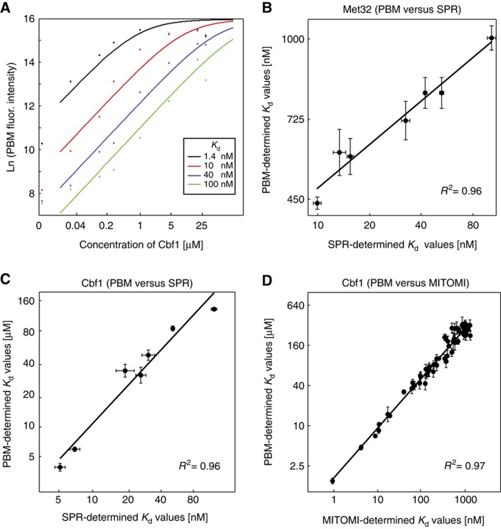
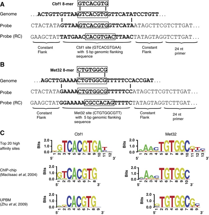
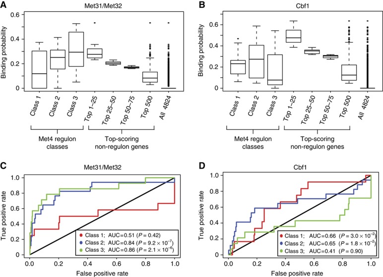
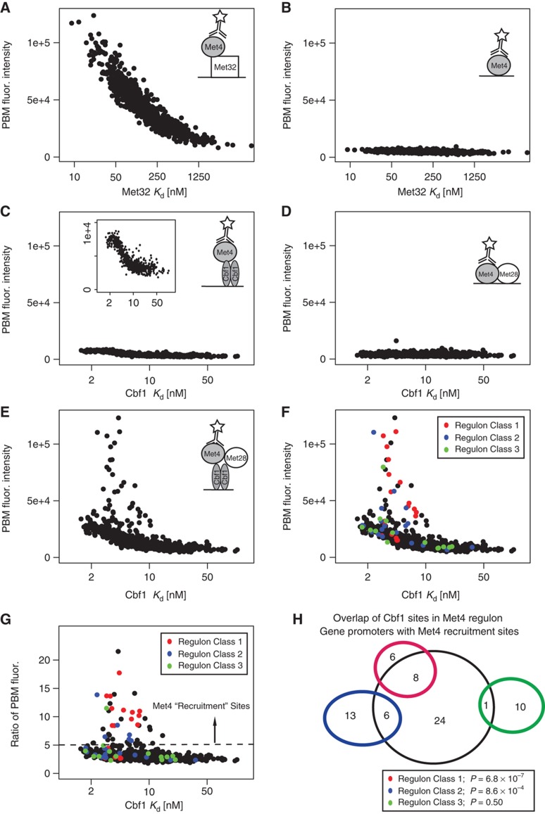
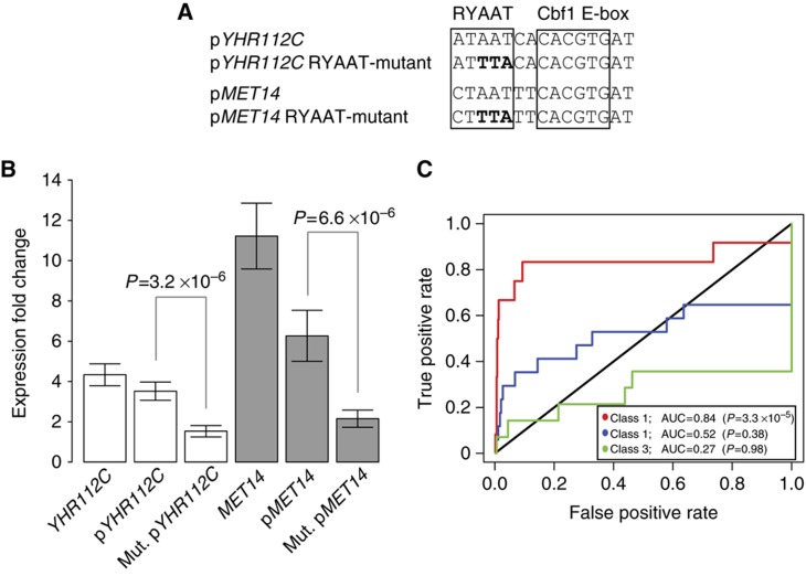
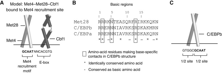
Recruitment of cofactors to specific DNA sites is integral for specificity in gene regulation. As a model system, we examined how targeting and transcriptional control of the sulfur metabolism genes in Saccharomyces cerevisiae is governed by recruitment of the transcriptional co-activator Met4. We developed genome-scale approaches to measure transcription factor (TF) DNA-binding affinities and cofactor recruitment to >1300 genomic binding site sequences. We report that genes responding to the TF Cbf1 and cofactor Met28 contain a novel 'recruitment motif' (RYAAT), adjacent to Cbf1 binding sites, which enhances the binding of a Met4-Met28-Cbf1 regulatory complex, and that abrogation of this motif significantly reduces gene induction under low-sulfur conditions. Furthermore, we show that correct recognition of this composite motif requires both non-DNA-binding cofactors Met4 and Met28. Finally, we demonstrate that the presence of an RYAAT motif next to a Cbf1 site, rather than Cbf1 binding affinity, specifies Cbf1-dependent sulfur metabolism genes. Our results highlight the need to examine TF/cofactor complexes, as novel specificity can result from cofactors that lack intrinsic DNA-binding specificity.


Conflict of interest statement

The authors declare that they have no conflict of interest.

Figures

Figure 1
Figure 1
PBM-determined protein-DNA binding affinities (Kd's) and comparison with SPR- and MITOMI-determined values. (A) Fitted saturation binding curves to PBM fluorescence values at eight concentrations of applied Cbf1 protein are shown for four representative PBM probes (i.e., sequences) with varying SPR-PBM-determined affinities. (B, C) Comparison of PBM- and SPR-determined Kd values for Met32 and Cbf1, respectively. Error bars indicate the standard deviation calculated over replicate measurements (n=4 for PBM, n=2 for SPR). (D) Comparison of PBM- and MITOMI-determined (Maerkl and Quake, 2007) Kd values for Cbf1 to 64 binding site variants of the form GTCACNNN. Plots are shown on a log-log scale. See Supplementary information for linear regression parameters.
Figure 2
Figure 2
Genome-wide binding affinity analysis: PBM probe design and sequence specificity of high-affinity sites. (A, B) Schematic illustrating the design of PBM probe sequences from binding site sequences identified in the yeast genome. Boxed are high-scoring 8-mers described for Cbf1 or Met32 (Zhu et al, 2009), 8-mers for each factor are put in a common register based on binding motifs from Zhu et al and the 20-bp genomic sequence is incorporated into the probe within constant flanking sequence (see Materials and methods for details). All 20 bp binding sites are present on the PBM in duplicate and in their reverse complement (RC) orientation (four probes in total). (C) Logos for Cbf1 and Met32 constructed by aligning the top 20 highest affinity sites (top); determined by MacIsaac et al (2006) by analyzing ChIP-chip data for each factor (middle); and determined by Zhu et al (2009) using a universal PBM approach (bottom). Palindromic Cbf1 sites were randomly oriented in constructing the logo, Met32 sites were oriented with respect to the motif from Zhu et al.
Figure 3
Figure 3
Specificity of Met32- and Cbf1-specific binding models for Met4 regulon genes. (A, B) Probability distribution for Met32 and Cbf1 binding to gene promoters from different gene sets. Met4 regulon genes are grouped according to Met4 regulon class designation of Lee et al (2010) and non-regulon genes are grouped into the top-scoring gene sets of 25 genes (3 sets) and top-scoring 500 genes (1 set). (C, D) ROC curve analysis for the prediction of each regulon gene class using the 500 top-scoring background (i.e., non-regulon) genes as false positives. Genes are scored and ranked according to the Met32-specific and Cbf1-specific binding probabilities calculated for each gene promoter (Materials and methods). Wilcoxon–Mann–Whitney U-test was applied to each regulon gene set to calculate significance of the AUC values.
Figure 4
Figure 4
Sequence dependence of Met4 recruitment. (AE) The median PBM probe fluorescence intensities for GST-tagged Met4 binding to 685 Met32 sites (A, B) and 673 Cbf1 sites (C–E) in the presence of different 6xHis-tagged proteins are shown: (A) Met4 binding to Met32 sites assayed in the presence of Met32; (B) Met4 binding to Met32 sites by itself; (C) Met4 binding to Cbf1 sites assayed in the presence of Cbf1; (D) Met4 binding to Cbf1 sites in the presence of Met28; (E) Met4 binding to Cbf1 sites in the presence of Met28 and Cbf1. X-axis coordinates are the PBM/SPR-determined Kd values for Met32 and Cbf1 binding to the respective sites. Cartoons in each panel represent the hypothesis being tested. (F) The plot from (E) with Cbf1 sites identified in the promoters of Met4 regulon genes highlighted according to Met4 regulon Class designations of Lee et al (2010) is shown. (G) Ratio of PBM fluorescence values for the Met4/Met28/Cbf1 experiment (E) over the Met4/Cbf1 experiment (C). Individual sites are colored as in (F). Met4 ‘recruitment sites’ are indicated as sites having a ratio >5.0. (H) Overlap of Cbf1 sites identified in upstream promoter region of Met4 regulon genes and Met4 recruitment sites in (G). Promoter regions are defined as 1500 bp upstream of TSS or until next coding region. Significance of observed overlap is calculated using Fisher's one-tail exact test (hypergeometric distribution).
Figure 5
Figure 5
Sequence specificity of the Met4 recruitment motif. (A) Logo determined from top 20 Met4 recruitment sites (Supplementary Table S2). These sequences were manually oriented to align the common AAT motif. (B) Ratio of PBM probe fluorescence values (Met4/Met28/Cbf1 PBM experiment over the Met4/Cbf1 PBM experiment) are shown for wild-type and mutant versions of three Met4 recruitment sites (shown in box). Mean and standard deviation are shown for measurements to wild-type or mutant versions of the three sequences. X-axis indicates identity of mutated base (numbering as in (A)). BG indicates measurements over 200 Cbf1 sites with the lowest ratio scores (i.e., background).
Figure 6
Figure 6
RYAAT motif is critical to promoter activity and specification of Class 1 regulon genes. (A) Schematic of wild-type and mutant versions of the composite Met4 recruitment sites from YHR112C and MET14 gene promoters (pYHR112C and pMET14) cloned upstream of LYS2 reporter gene. (B) Expression fold change, under switch to low-sulfur growth conditions, for endogenous genes (YHR112C and MET14) and the LYS2 reporter gene driven by wild-type (pYHR112C and pMET14) and RYAAT-mutant (Mut. pYHR112C and Mut. pMET14) gene promoters. (C) ROC analysis for the prediction (identification) of the Met4 regulon genes using the Met4 recruitment ‘strength’ of the 673 Cbf1 sites used in our genome-wide affinity analysis. Met4 recruitment strength is the ratio of PBM fluorescence intensities shown in Figure 3G (i.e., ratio of PBM fluorescence intensities for the Met4/Met28/Cbf1 and Met4/Cbf1 PBM experiments). ROC analysis was performed using 500 top-scoring non-regulon genes as false positives. Wilcoxon–Mann–Whitney U-test was applied to each regulon gene set to calculate significance of the AUC value. Source data is available for this figure in the Supplementary information.
Figure 7
Figure 7
Proposed model of Met4 recruitment complexes. (A) Diagram of proposed orientation for DNA-binding domains of Met4 (bZIP domain), Met28 (bZIP domain), and Cbf1 (bHLH domain) in the Met4–Met28–Cbf1 complex bound to a Met4 recruitment site. Met4 recruitment motif is in bold. Direct physical contacts that exist between the Met4 and/or Met28 subunits and the Cbf1 homodimer are not explicitly indicated. (B) Sequence alignment of basic region from Met28, human C/EBPb, and human C/EBPa. (C) Diagram of C/EBPb bZIP region homodimer bound to DNA. Binding site sequence for C/EBPb complex is from X-ray crystal structure of C/EBPb complex (PDB code 1H8A) (Tahirov et al, 2002). GCAAT half-site motif is in bold.

Comment in

References

    1. Babb R, Huang CC, Aufiero DJ, Herr W (2001) DNA recognition by the herpes simplex virus transactivator VP16: a novel DNA-binding structure. Mol Cell Biol 21: 4700–4712 - PMC - PubMed
    1. Badis G, Berger MF, Philippakis AA, Talukder S, Gehrke AR, Jaeger SA, Chan ET, Metzler G, Vedenko A, Chen X, Kuznetsov H, Wang CF, Coburn D, Newburger DE, Morris Q, Hughes TR, Bulyk ML (2009) Diversity and complexity in DNA recognition by transcription factors. Science 324: 1720–1723 - PMC - PubMed
    1. Badis G, Chan ET, van Bakel H, Pena-Castillo L, Tillo D, Tsui K, Carlson CD, Gossett AJ, Hasinoff MJ, Warren CL, Gebbia M, Talukder S, Yang A, Mnaimneh S, Terterov D, Coburn D, Li Yeo A, Yeo ZX, Clarke ND, Lieb JD et al. (2008) A library of yeast transcription factor motifs reveals a widespread function for Rsc3 in targeting nucleosome exclusion at promoters. Mol Cell 32: 878–887 - PMC - PubMed
    1. Berger MF, Bulyk ML (2006a) Protein binding microarrays (PBMs) for rapid, high-throughput characterization of the sequence specificities of DNA binding proteins. Methods Mol Biol 338: 245–260 - PMC - PubMed
    1. Berger MF, Philippakis AA, Qureshi AM, He FS, Estep PW III, Bulyk ML (2006b) Compact, universal DNA microarrays to comprehensively determine transcription-factor binding site specificities. Nat Biotechnol 24: 1429–1435 - PMC - PubMed

Publication types

MeSH terms

Substances