. 2011 Dec 6:2:576.
doi: 10.1038/ncomms1585.

Activin enhances skin tumourigenesis and malignant progression by inducing a pro-tumourigenic immune cell response

Affiliations
Free PMC article

Activin enhances skin tumourigenesis and malignant progression by inducing a pro-tumourigenic immune cell response

Maria Antsiferova et al. Nat Commun. .
Free PMC article

Abstract

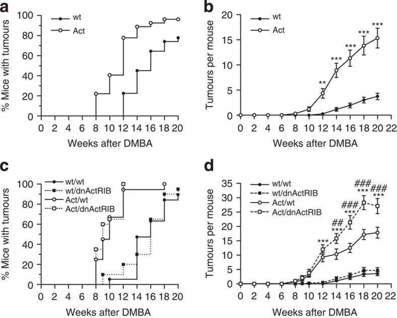
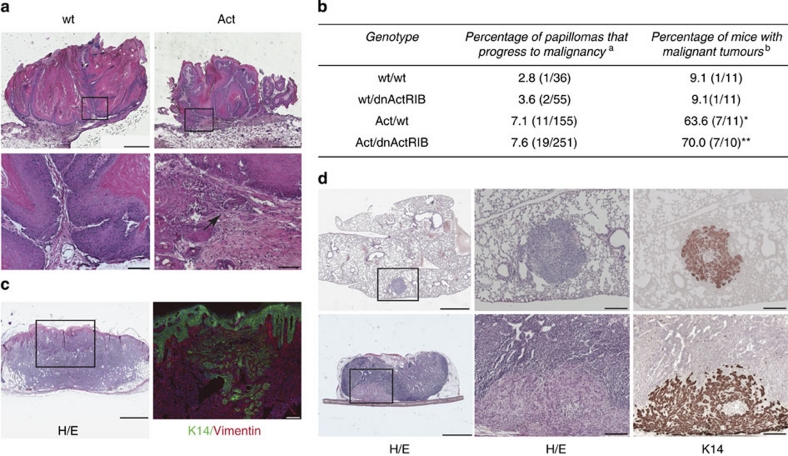
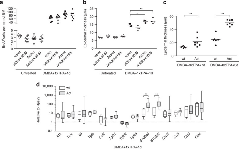
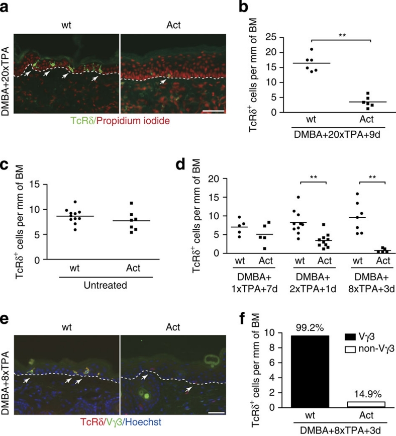
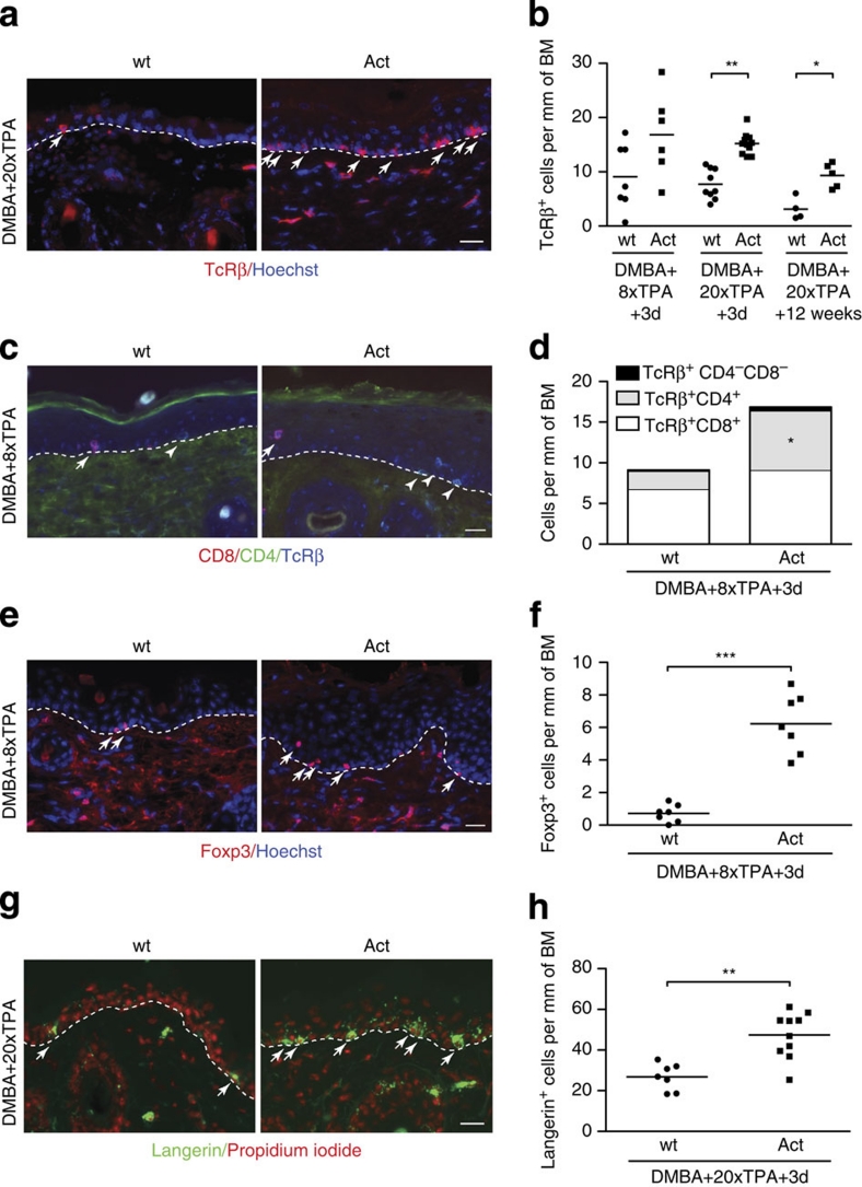
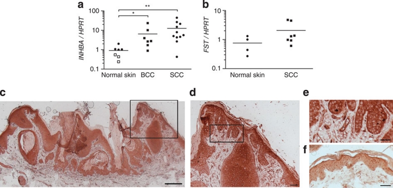
Activin is an important orchestrator of wound repair, but its potential role in skin carcinogenesis has not been addressed. Here we show using different types of genetically modified mice that enhanced levels of activin in the skin promote skin tumour formation and their malignant progression through induction of a pro-tumourigenic microenvironment. This includes accumulation of tumour-promoting Langerhans cells and regulatory T cells in the epidermis. Furthermore, activin inhibits proliferation of tumour-suppressive epidermal γδ T cells, resulting in their progressive loss during tumour promotion. An increase in activin expression was also found in human cutaneous basal and squamous cell carcinomas when compared with control tissue. These findings highlight the parallels between wound healing and cancer, and suggest inhibition of activin action as a promising strategy for the treatment of cancers overexpressing this factor.


Figures

Figure 1
Figure 1. Activin promotes chemically induced skin tumourigenesis in mice via action on stromal cells.
(a,b) Kinetics of tumour incidence (a) or multiplicity (b) observed in experiment I (Table 1). N=31 wild-type (wt) mice; N=29 activin-transgenic (Act) mice. (c,d) Kinetics of tumour incidence (c) and multiplicity (d) in experiment III. N=18 mice per genotype. Kaplan–Meier survival curves are shown in (a) and (c); mean±s.e.m. are shown in (b) and (d); **P<0.01, ***P<0.001 for the comparison between wt/wt and Act/wt mice; ## P<0.01, ### P<0.001 for the comparison between Act/wt and Act/dnActRIB mice (two-way repeated measures ANOVA and Bonferroni post-test).
Figure 2
Figure 2. Activin increases malignancy and metastatic spread of skin tumours.
(a) Hematoxylin/eosin (H/E)-stained sections from papillomas 20 weeks after initiation. Local micro-invasive foci (arrow) were often observed in papillomas of Act mice (lower panel). Bars: 1,000 μm (top), 100 μm (bottom). Areas indicated by the box on the top pictures are shown at higher magnification on the bottom pictures. (b) Malignant conversion rate of tumours 32 weeks after initiation (experiment III). a()=number of carcinomas/total number of tumours. b()=number of mice with carcinomas/total number of mice. N=10 Act/dnActRIB mice, N=11 wt/wt, Act/wt, wt/dnActRIB mice; *P=0.0237 for Act/wt versus wt/wt, **P=0.0075 for Act/dnActRIB versus wt/wt (Fisher's exact test). (c) H/E staining (left) and double immunofluorescence for keratin (K)14 and vimentin (right) of a spindle cell variant of squamous cell carcinoma (SCC) in an Act mouse. Left bar: 1,000 μm; right bar: 200 μm. The area indicated by a box on the left picture is shown at higher magnification on the right picture. (d) Lung (top panel) and lymph node (bottom panel) metastases in carcinoma-bearing Act mice. Left and middle: H/E staining; right: K14 immunohistochemistry. Left bar: 1,000 μm; middle and right bars: 200 μm.
Figure 3
Figure 3. Activin overexpression does not affect DMBA or TPA responsiveness.
(a) Epidermal BrdU+ cells per mm of basement membrane (BM) in untreated back skin or 1 day after the first TPA treatment. Scatter plot and mean values are shown; N=5–8 mice per group; n=6–23 microscopic fields of skin sections per mouse. (b) Epidermal thickness in untreated mice or 1 day after the first TPA treatment. N=3–4; n=6–13; *P≤0.05, **P≤0.01 (one-way ANOVA with Bonferroni post-test). (c) Epidermal thickness 7 days after the third TPA treatment or 3 days after the eighth TPA treatment. N=6–7; n=3–30; **P=0.0047 for 3×TPA, **P=0.0012 for 8×TPA (Mann–Whitney test). (d) RNA samples from back skin 1 day after the first TPA treatment were analysed for expression of pro-inflammatory cytokines and growth factors by quantitative reverse transcription PCR. N=5–10 mice per genotype. Rps29 was used as reference. Box-and-whisker plot is depicted, with boxes showing median and 25th and 75th percentile, whiskers showing minimal and maximal values. **P=0.0047 for S100a8, **P=0.0027 for S100a9 (Mann–Whitney test).
Figure 4
Figure 4. Loss of epidermal γδ T cells in Act mice after TPA treatment.
(a,b) Sections from non-tumourigenic skin of wt and Act mice after 20 TPA applications were stained with antibodies against TcRδ. Nuclei were counterstained with propidium iodide. (b) Quantification of epidermal TcRδ+ cells per mm of basement membrane (BM). N=6 mice per genotype; n=6–16 microscopic fields of skin sections per mouse; **P=0.0022 (Mann–Whitney test). (c,d) Epidermal TcRδ+ cells per mm of BM in untreated adult mice (c) and at different time points of TPA treatment (d). +1d, +3d and +7d indicate the number of days after TPA application. N=5–11; n=9–33; **P=0.0011 for DMBA+2×TPA+1d, **P=0.0025 for DMBA+8×TPA+3d (Mann–Whitney test). Scatter plot and mean values are shown in (b), (c) and (d). (e) Sections from back skin of wt and Act mice 3 days after the eighth TPA application were co-stained with antibodies against the pan-γδ TcR (TcRδ) and the Vγ3 variant of γδ TcR. Nuclei were counterstained with Hoechst. Arrows indicate TcRδ+ cells. Representative micrographs are shown in (a) and (e). Arrows indicate TcRδ+ cells. Bar: 50 μm. Dotted lines indicate dermal/epidermal border. (f) Epidermal TcRδ+ cells and Vγ3+ cells per mm of BM. Bar graph shows the mean number of TcRδ+ cells per mm of BM, numbers above the columns indicate the corresponding percentage of Vγ3+ cells among TcRδ+ cells. N=7; n=3–13.
Figure 5
Figure 5. Activin inhibits proliferation of epidermal γδ T cells.
(a) CD3+TcRδ+ (γδ T cells) and remaining cells (rest) were FACS sorted from the epidermis of three wt mice. RNA was analysed by reverse transcription PCR for expression of activin receptors, Gapdh, Krt14, and the γδ T cell receptor (Tcrg). (b,c) Sections from TPA-treated back skin of wt and Act mice stained for TcRδ and Ki67. (b) The dotted line indicates the epidermal–dermal border. Arrows and arrowheads indicate Ki67+TcRδ+ or Ki67TcRδ+ cells, respectively. Bar: 25 μm. (c) Percentage of Ki67+ cells among TcRδ+ cells 7 days after the first or 1 day after the second TPA application. N=4–10 mice per genotype; n=17–22 microscopic fields of skin sections per mouse; **P=0.0019 (Mann–Whitney test). (d) Untreated or DMBA/TPA-treated mice were injected with EdU twice a day for 7 days. Epidermal single-cell suspensions were analysed for EdU incorporated in TcRδ+ cells by flow cytometry. Representative pseudo-colour plots are shown (gated on viable TcRδ+ cells). The percentage of EdU+ cells among the TcRδ+ cells is indicated. Data shown are representative of at least four independent analyses. (e,f) The percentage of EdU+ cells among the TcRδ+ cells was quantified by flow cytometry before and at different time points after TPA treatment. (e) EdU was injected twice a day for 7 days; N=4–6; *P=0.0159 (Mann–Whitney test). (f) EdU was injected twice a day for 2 days; N=5–7; *P≤0.05; **P≤0.01 (one-way ANOVA with Bonferroni post-test). (g,h) RNA samples from trypsin-separated epidermis from untreated mice (g) or from mice after the second TPA application (h) were analysed for expression of Il15 and Il7 by quantitative reverse transcription PCR using Rps29 or Gapdh as reference. Expression in one of the wt mice was arbitrarily set to 1. N=4–5; ***P≤0.001 (one-way ANOVA with Bonferroni post-test). Scatter plot and mean values are shown in (c), (e), (f), (g) and (h). (i) Vγ3+Thy1.2+ cells were FACS sorted and cultured for 48 h with immobilized anti-CD3 antibodies (0.1 μg ml−1) in the absence (ctrl) or presence of 50 ng ml−1 activin A (Act). 3H-thymidine incorporation assay was performed in quadruplicates. Data are representative of two independent experiments; mean values±s.d. are shown; *P=0.0286 (Mann–Whitney test).
Figure 6
Figure 6. Accumulation of αβ T cells and Langerhans cells in the epidermis of activin-overexpressing mice.
(a,b) Sections from back skin at different time points of TPA treatment stained with TcRβ antibodies. (a) Arrows indicate TcRβ+ cells. (b) TcRβ+ cells per mm of basement membrane (BM). N=4–11 mice per group; n=3–25 microscopic fields of skin sections per mouse; *P=0.0159; **P=0.0022 (Mann–Whitney test). (c,d) Sections from back skin after eight TPA applications stained with antibodies against CD8, CD4 and TcRβ. (c) Arrows and arrowheads indicate CD8+ or CD4+ cells, respectively. (d) Different subpopulations of T cells per mm of BM. N=6–7; n=4–24; mean values are shown; *P=0.01 for comparison of TcRβ+CD4+ cells between wt and Act mice (Mann–Whitney test). (e,f) Sections from back skin after eight TPA applications stained with Foxp3 antibodies. (e) Arrows indicate Foxp3+ cells. (f) Epidermal Foxp3+ cells per mm of BM. N=7; n=6–25; ***P=0.0006 (Mann–Whitney test). (g,h) Sections from back skin after 20 TPA applications stained with langerin antibodies. (g) Arrows indicate langerin+ cells. (h) Langerin+ cells per mm of BM. N=7–10; n=3–25; **P=0.0012 (Mann–Whitney test). Representative sections are shown in (a), (c), (e) and (g), and the dermal–epidermal border is indicated by dotted lines. Nuclei were counterstained with Hoechst (a), (c), and (e) or propidium iodide (g). Bars: 25 μm. Scatter plots and mean values are shown in (b), (f) and (h).
Figure 7
Figure 7. Activin is overexpressed in human skin carcinomas.
(a) RNA samples from normal human skin, basal cell carcinomas (BCCs) and SCCs were analysed for expression of activin βA (INHBA) by quantitative reverse transcription PCR. Hypoxanthine-guanine phosphoribosyl-transferase (HPRT) was used as a reference. Expression levels in one of the normal skin samples were set to 1. Closed and open squares represent values from normal skin collected from the edge of tumours or from healthy adult volunteers, respectively. N=7 normal skin samples; N=7 BCC samples; N=11 SCC samples; *P=0.0111, **P=0.0038 (Mann–Whitney test). (b) RNA samples from normal human skin and SCCs were analysed for expression of follistatin (FST) by quantitative reverse transcription PCR. HPRT was used as a reference. N=4 normal skin samples; N=7 SCC samples. Scatter plot and median values are shown in (a) and (b). (cf) Sections of human SCCs (ce) and normal skin (f) were stained with an activin A antibody and counterstained with hematoxylin. The areas indicated by the box in (c) and (d) are shown at high magnification in (d) and (e), respectively. Note the expression of activin in the tumour cells and in blood vessels. Bar: 500 μm (c), 100 μm (d) or 50 μm (e,f).

References

    1. Schäfer M. & Werner S. Cancer as an overhealing wound: an old hypothesis revisited. Nat. Rev. Mol. Cell. Biol. 9, 628–638 (2008). - PubMed
    1. Chang H. Y. et al.. Gene expression signature of fibroblast serum response predicts human cancer progression: similarities between tumors and wounds. PLoS Biol. 2, E7 (2004). - PMC - PubMed
    1. Chen Y. G. et al.. Activin signaling and its role in regulation of cell proliferation, apoptosis, and carcinogenesis. Exp. Biol. Med. (Maywood) 231, 534–544 (2006). - PubMed
    1. Werner S. & Alzheimer C. Roles of activin in tissue repair, fibrosis, and inflammatory disease. Cytokine Growth Factor Rev. 17, 157–171 (2006). - PubMed
    1. Risbridger G. P., Schmitt J. F. & Robertson D. M. Activins and inhibins in endocrine and other tumors. Endocr. Rev. 22, 836–858 (2001). - PubMed

Publication types

MeSH terms