. 2012 Jan 17;106(2):348-57.
doi: 10.1038/bjc.2011.521. Epub 2011 Dec 6.

The proline TP53 variant stimulates likely lymphangiogenesis in an orthotopic mouse model of pancreatic cancer

Affiliations

The proline TP53 variant stimulates likely lymphangiogenesis in an orthotopic mouse model of pancreatic cancer

N Otto et al. Br J Cancer. .

Abstract

Background: Pancreatic cancer is a deadly disease characterised by high incidence of TP53 mutations. Restoration of TP53 function is perceived as a highly attractive therapeutic strategy, whose effects are not well characterised.

Methods: The current work adapted an inducible strategy of stage-specific reexpression of wild-type (wt) TP53 in an in vivo orthotopic mouse model of pancreatic cancer.

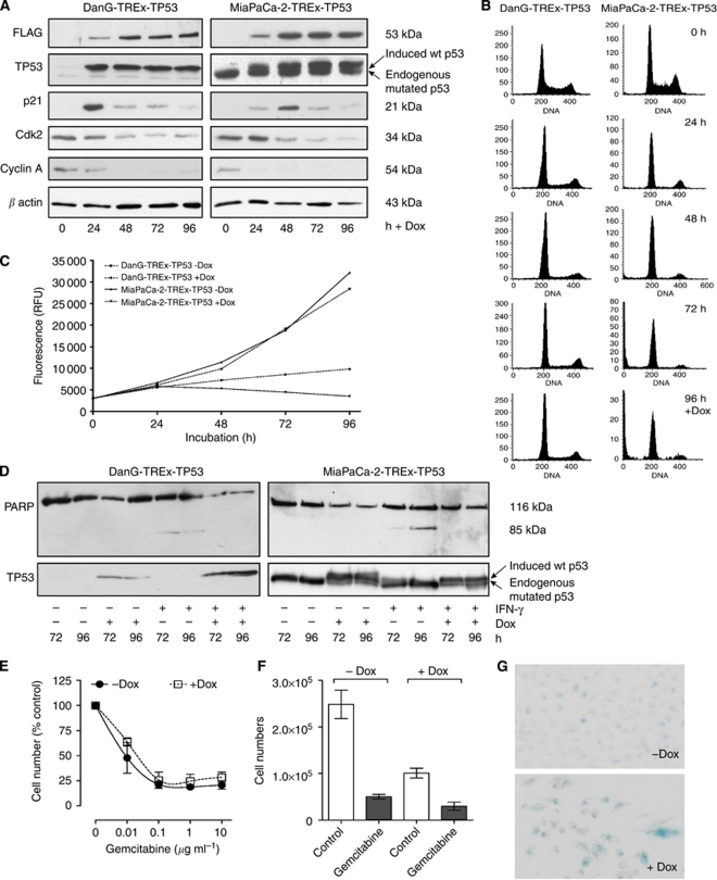
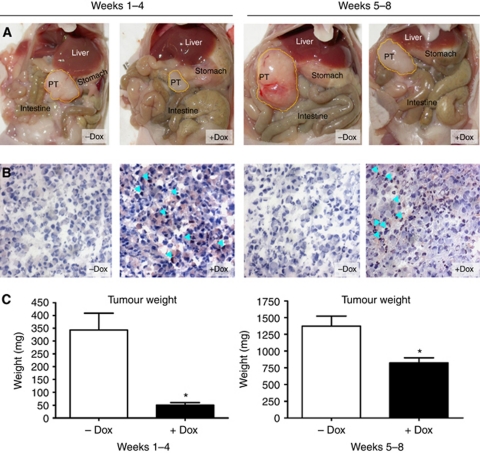
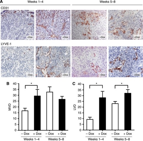
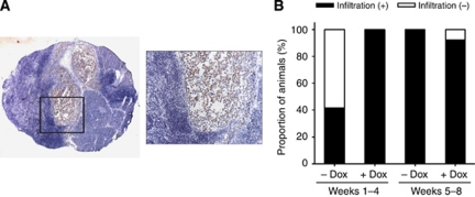
Results: The reconstitution of wt TP53 function in TP53-mutant DanG and MiaPaCa-2 cells caused G1 cell cycle arrest but no evidence of apoptosis induction. Consistent with subcutaneous xenograft models, we found that wt TP53 reduced primary tumour growth. Wt TP53 reexpression during early tumour growth led to significant increase in vascularisation. This correlated with an unexpectedly high rate of micro-metastases in lymph nodes of animals with wt TP53 induction, despite the 90% decrease in median primary tumour weight. Reexpression of wt TP53 later in tumour development did not significantly affect the number of CD31-reactive vessels, but increased lymphatic vessel density.

Conclusion: The increased number of lymphatic vessels and micro-metastases suggests that wt TP53 induction complexly affected the biology of different tumour constituents of pancreatic cancer. Our observation suggests that combination of the inducible system with an orthotopic model can yield important insights into in vivo pancreatic cancer biology.


Figures

Figure 1
Figure 1
Generation of DanG-TREx-TP53 and MiaPaCa-2-TREx-TP53 cells with Dox-inducible expression of functionally active wt TP53. (A) The plasmids pcDNA6/TR and pcDNA4/TO-TP53FLAG were sequentially transfected into DanG and MiaPaCa-2 cells bearing mutant endogenous p53 and clones with inducible expression of wt TP53 were selected (DanG-TREx-TP53 and MiaPaCa-2-TREx-TP53). Cells were cultured for 96 h in the presence or absence of Dox and lysates were prepared for western blot analysis of FLAG (upper panel) and TP53 (second panel) at the indicated time points. Equal amounts of protein (10 μg) were separated on SDS polyacrylamide gel electrophoresis (PAGE). Furthermore, effects of Dox-induced wt TP53 expression on downstream targets were investigated by western blot analysis: p21WAF1/CIP1, cyclin-dependent kinase-2, cyclin A and β-actin (lower panels). Equal amounts of protein (30 μg) were separated on SDS–PAGE. (B) Effects on cell cycle after wt TP53 expression in DanG-TREx-TP53 and MiaPaCa-2-TREx-TP53 were analysed by FACS over a time course of 96 h. At every 24 h, cells were fixed with 70% ethanol and stained with propidium iodide. (C) Time-dependent proliferation of DanG-TREx-TP53 and MiaPaCa-2-TREx-TP53 cells in the presence or absence of Dox as determined by alamar blue assay. At every 24 h, alamar blue dye was applied to the media of cells and fluorescence was determined as an indirect measurement of cell numbers. (D) Immunoblot demonstrating expression of poly(ADP-ribose)polymerase (p116) and/or its apoptosis-related cleavage product p85 in untreated controls and cells that had been treated with Dox, IFN-γ or a combination of both for 72 or 96 h. Whole-cell lysates were separated by 7.5% SDS–PAGE. (E) The diagram depicts the concentration dependence of the growth inhibition by gemcitabine with data presented as a percentage of the respective controls (vehicle and +Dox, respectively). (F) The response of the MiaPaCa-2-TREx-TP53 cells to gemcitabine was evaluated by determination of cell numbers. The cells were treated with −/+Dox and −/+gemcitabine, as indicated, for 48 h. The diagram presents cell numbers after treatment with or without 10 μg ml−1 gemcitabine. (G) Cellular senescence (β-Gal activity) in MiaPaCa-2-TREx-TP53 cells was detected by a cell senescence assay after −/+Dox treatment. Senescence-positive cells appeared blue ( × 200 magnification). A representative experiment out of two is shown.
Figure 2
Figure 2
Induction of wt TP53 expression reduced primary tumour growth in orthotopic MiaPaCa-2 pancreatic carcinomas at both time points. MiaPaCa-2-TREx-TP53 cells were grown orthotopically in the pancreas of mice treated with or without Dox as indicated. At the end of treatment (either 4 weeks or 8 weeks) tumour weights were determined. (A) Open situs of representative mice from each control (−Dox) and treatment group (+Dox). (B) Conformation of wt TP53 induction in vivo. Primary tumours were analysed for TP53 expression by immunohistochemistry using an antibody against the FLAG-tag. Examples of wt TP53 expression at 4 and 8 weeks of tumour growth, with detection of the wt TP53-FLAG protein in the nucleus of tumour cells from Dox-treated mice indicated by the light blue arrows. In contrast, no signal was obtained in control tumours (−Dox) (images at × 200 magnification). (C) Summary of primary tumour weights of Dox-treated or -untreated animals receiving Dox either in weeks 1–4 or in weeks 5–8. Data represent mean±s.e.m. for each group. *P<0.05.
Figure 3
Figure 3
Effects of wt TP53 restitution on vascularisation in orthotopic MiaPaCa-2-TREx-TP53 pancreatic carcinomas. Cryosection of the tumours from control (−Dox) and treatment groups (+Dox) were stained with endothelial marker CD31 orlymphatic endothelial marker LYVE-1. To determine vessel densities, the number of CD31- and LYVE-1-expressing vessels were quantitated from hotspot areas, as described in Materials and Methods. (A) Shown are representative CD31 (upper panel) and LYVE-1 stainings (lower panel) from −Dox- and +Dox-treated tumours of both groups (B and C). Summary of the quantitative evaluation of the results is given as MVD or LVD, *P<0.05 (images at × 100 magnification).
Figure 4
Figure 4
(A) Effects of wt TP53 induction on metastatic spread of MiaPaCa-2 cells to the liver hilus lymph node. Micro-metastatic spread of MiaPaCa-2-TREx-TP53 cells was evaluated based on detection of human cytokeratin in available mouse lymph nodes (n=7). Inspection of lymph node metastasis, which was stained by pan-cytokeratin antibody. Representative picture of a lymph node stained with an anti-human pan-cytokeratin antibody, revealing positive human tumour cells (stained red) surrounded by lymphocytes (image at × 25 magnification and × 200 magnification (image inset)). (B) Quantitative analysis of lymph node metastasis. Data represent the proportion of animals with presence of lymph node metastasis in the liver hilus lymph node, compared between the treatment (+Dox) and control group (−Dox) of different time periods with wt TP53 reexpression.

Similar articles

Cited by

References

    1. Alves F, Contag S, Missbach M, Kaspareit J, Nebendahl K, Borchers U, Heidrich B, Streich R, Hiddemann W (2001) An orthotopic model of ductal adenocarcinoma of the pancreas in severe combined immunodeficient mice representing all steps of the metastatic cascade. Pancreas 23: 227–235 - PubMed
    1. American Cancer Society (2007) Cancer Facts & Figures 2007. American Cancer Society: Atlanta, GA, pp 1–52
    1. Bardeesy N, DePinho RA (2002) Pancreatic cancer biology and genetics. Nat Rev Cancer 2: 897–909 - PubMed
    1. Blouw B, Song H, Tihan T, Bosze J, Ferrara N, Gerber HP, Johnson RS, Bergers G (2003) The hypoxic response of tumors is dependent on their microenvironment. Cancer Cell 4: 133–146 - PubMed
    1. Braithwaite AW, Prives CL (2006) p53: more research and more questions. Cell Death Differ 13: 877–880 - PubMed

Publication types