. 2011 Aug 16;1(1):20.
doi: 10.1186/2045-9912-1-20.

ACS6, a Hydrogen sulfide-donating derivative of sildenafil, inhibits homocysteine-induced apoptosis by preservation of mitochondrial function

Affiliations

ACS6, a Hydrogen sulfide-donating derivative of sildenafil, inhibits homocysteine-induced apoptosis by preservation of mitochondrial function

Xiao-Qing Tang et al. Med Gas Res. .

Abstract

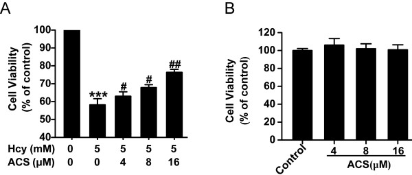
Background: The hydrogen sulfide-releasing sildenafil, ACS6, has been demonstrated to inhibit superoxide formation through donating hydrogen sulfide (H2S). We have found that H2S antagonizes homocysteine-induced oxidative stress and neurotoxicity. The aim of the present study is to explore the protection of ACS6 against homocysteine-triggered cytotoxicity and apoptosis and the molecular mechanisms underlying in PC12 cells.

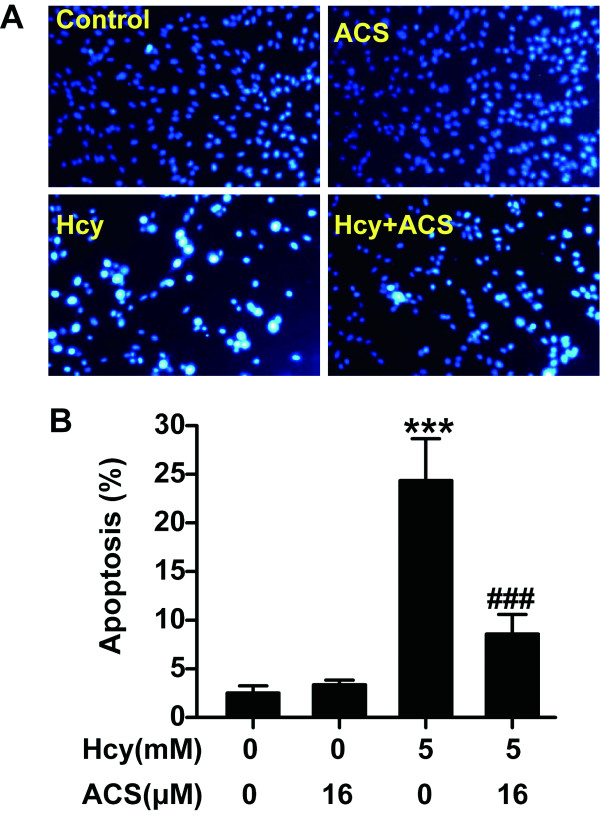
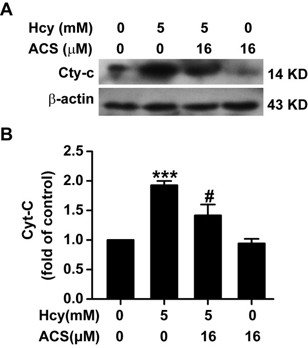
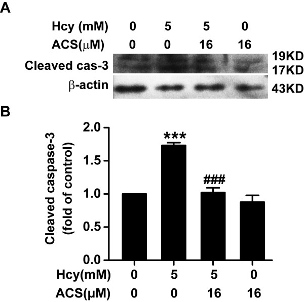
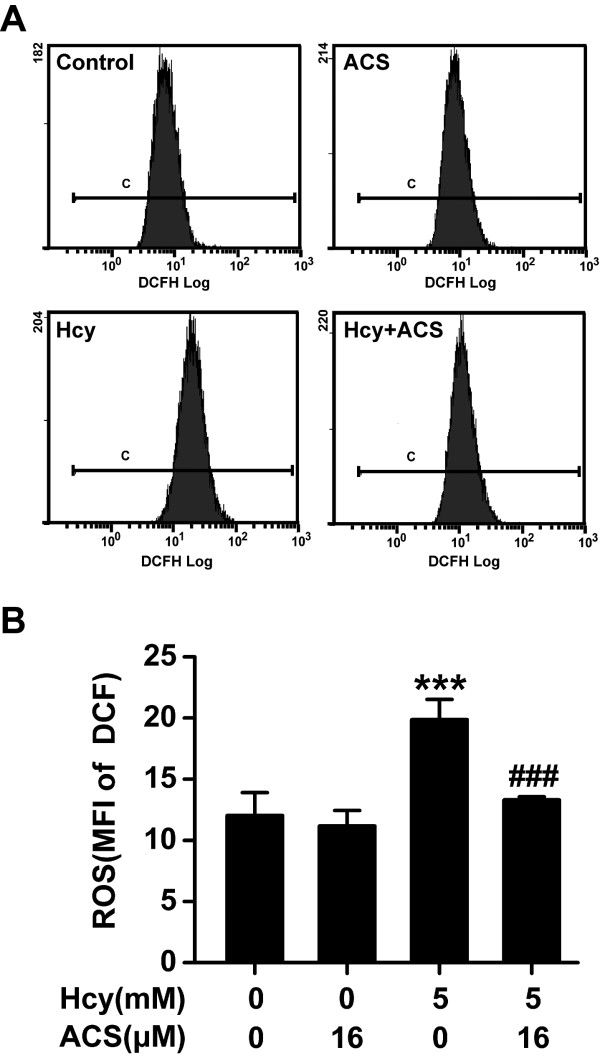
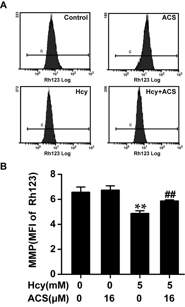
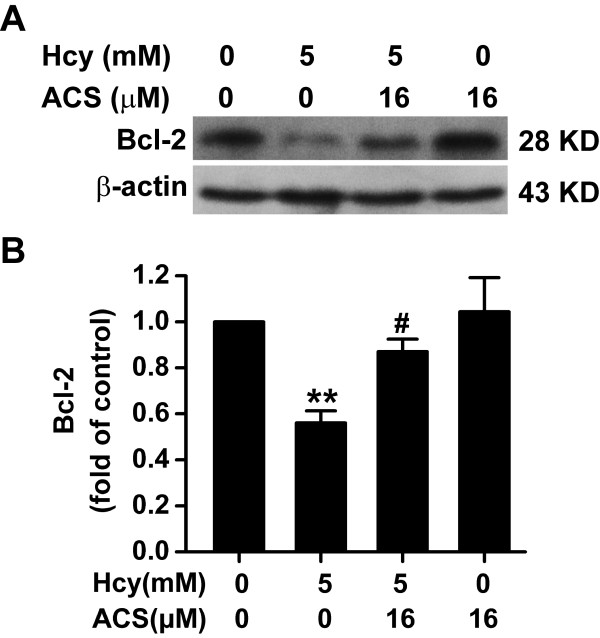
Methods: Cell viability was determined by Cell Counting Kit-8 assay. Cell apoptosis was observed using the chromatin dye Hoechst 33258 and analyzed by Flow Cytometry after propidium iodide staining. Mitochondrial membrane potential was monitored using the fluorescent dye Rh123. Intracellular reactive oxygen species were determined by oxidative conversion of cell permeable 2',7'-dichlorfluorescein-diacetate to fluorescent 2',7'-dichlorfluorescein. The expression of cleaved caspase-3 and bcl-2 and the accumulation of cytosolic cytochrome c were analyzed by Western blot.

Results: We show that ACS6 protects PC12 cells against cytotoxicity and apoptosis induced by homocysteine and blocks homocysteine-triggered cytochrome c release and caspase-3 activation. ACS6 treatment results in not only prevention of homocysteine-caused mitochondrial membrane potential (Δψ) loss and reactive oxygen species (ROS) overproduction but also reversal of Bcl-2 down-expression.

Conclusions: These results indicate that ACS6 protects PC12 cells against homocysteine-induced cytotoxicity and apoptosis by preservation of mitochondrial function though inhibiting both loss of Δψ and accumulation of ROS as well as modulating the expression of Bcl-2. Our study provides evidence both for a neuroprotective effect of ACS6 and for further evaluation of ACS6 as novel neuroprotectants for Alzheimer's disease associated with homocysteine.


Figures

Figure 1
Figure 1
The chemical structure of ACS6.
Figure 2
Figure 2
ACS6 protects PC12 cells against homocysteine-induced cytotoxicity. (A) PC12 cells were pretreated with ACS6 (4, 8, or 16 μmol/L) for 30 min and then exposed to homocysteine (Hcy, 5 mmol/L) for 24 h. (B) PC12 cells were treated with 4, 8, and 16 μmol/L ACS6 for 24 h. Cell viability was determined by CCK-8 assay. Values are the mean ± SEM (n = 3). ***P < 0.001, versus control group; #P < 0.05, ##P < 0.01, versus 5 mmol/L homocysteine-treated alone group.
Figure 3
Figure 3
Nuclear staining to evaluate the anti-apoptotic effect of ACS6. After pretreated with 16 μmol/L ACS6 for 30 min, PC12 cells were exposed to 5 mmol/L homocysteine (Hcy) for 24 h and incubated with 5 mg/L Hoechst 33258 for 30 min. (A) Representative morphology visualized under a fluorescence microscope (10 × objective, BX50-FLA, Olympus). Cells with brightly fluorescent and fragmented nuclei were apoptotic. (B) Quantitative analysis of the percentage of apoptotic cells. Values are the mean ± SEM (n = 5). ***P < 0.001, versus control group; ###P < 0.001, versus 5 mmol/L homocysteine-treated alone group.
Figure 4
Figure 4
Effects of ACS6 on the release of Cyt-c in PC12 cells. After pretreated with 16 μmol/L ACS6 for 30 min, PC12 cells were exposed to 5 mmol/L homocysteine (Hcy) for 24 h. A, representative immunoblots for Cyt-c release from three independent experiments. Cytosolic fractions of the extract were subjected to Western blot analysis using an anti-Cyt-c antibody. In all blots, staining for β-actin was used as a loading control. B, quantification of cytosolic Cyt-c accumulation as a percent of the control. Values are the mean ± SEM (n = 3). ***P <0.001, versus control; #P < 0.05, versus 5 mmol/L homocysteine-treated alone group.
Figure 5
Figure 5
Effects of ACS6 on the activation of caspase-3 in PC12 cells. After pretreated with 16 μmol/L ACS6 for 30 min, PC12 cells were exposed to 5 mmol/L homocysteine (Hcy) for 24 h. The activation of caspase-3 in PC12 cells was analyzed by Western blot using an anti-cleaved caspase-3 antibody. Western blot images show representative results from three independent experiments. In all blots, staining for β-actin was used as a loading control. The level of cleaved caspase-3 expression obtained in each experimental condition was calculated as a fold of the control. Values are the mean ± SEM (n = 3). ***P <0.001, versus control; ###P < 0.001, versus 5 mmol/L homocysteine-treated alone group.
Figure 6
Figure 6
Effects of ACS6 on homocysteine-exerted accumulation of intracellular ROS in PC12 cells. After pretreated with 16 μmol/L ACS6 for 30 min, PC12 cells were exposed to 5 mmol/L homocysteine (Hcy) for 24 h and stained with DCFH-DA for 20 min. The changes of ROS in different treatment groups were quantified by fluorescent sorting FCM analysis (A, B). (A) Representative histogram of DCF-derived fluorescence in PC12 cells exposed to different treatments measured by FCM. (B) Quantitative analysis of the mean fluorescence intensity (MFI) of DCF measured by FCM. Values are the mean ± SEM (n = 3). ***P <0.001, versus control; ###P < 0.001, versus 5 mmol/L homocysteine- treated alone group.
Figure 7
Figure 7
Effects of ACS6 on homocysteine-induced loss of mitochondrial membrane potential (Δψ) in PC12 cells. After pretreated with 16 μmol/L ACS6 for 30 min, PC12 cells were exposed to 5 mmol/L homocysteine (Hcy) for 24 h and stained with Rh123 for 20 min. The changes of Δψ in different treatment groups were quantified by fluorescent sorting FCM analysis. (A) Representative histogram of Rh123-derived fluorescence in PC12 cells exposed to different treatments measured by FCM. (B) Quantitative analysis of the mean fluorescence intensity (MFI) of Rh123 measured by FCM. Values are the mean ± SEM (n = 3). **P <0.01, versus control; ##P < 0.01, versus 5 mmol/L homocysteine-treated alone group.
Figure 8
Figure 8
Effects of ACS6 on the expression of Bcl-2 in PC12 cells. After pretreated with 16 μmol/L ACS6 for 30 min, PC12 cells were exposed to 5 mmol/L homocysteine (Hcy) for 24 h. The levels of Bcl-2 expression in PC12 cells were determined by Western blot using an anti-Bcl-2 antibody. Western blot images show representative results from three independent experiments. In all blots, staining for β-actin was used as a loading control. The level of Bcl-2 expression obtained in each experimental condition was calculated as a fold of the control. Values are the mean ± SEM (n = 3). **P <0.01, versus control; #P < 0.05, versus 5 mmol/L homocysteine-treated alone group.

Similar articles

Cited by

References

    1. Prudova A, Bauman Z, Braun A, Vitvitsky V, Lu SC, Banerjee R. S-adenosylmethionine stabilizes cystathionine beta-synthase and modulates redox capacity. Proc Natl Acad Sci USA. 2006;103(17):6489–94. doi: 10.1073/pnas.0509531103. - DOI - PMC - PubMed
    1. Selhub J. Homocysteine metabolism. Annu Rev Nutr. 1999;19:217–46. doi: 10.1146/annurev.nutr.19.1.217. - DOI - PubMed
    1. Lipton SA, Kim WK, Choi YB, Kumar S, D'Emilia DM, Rayudu PV. et al.Neurotoxicity associated with dual actions of homocysteine at the N-methyl-D-aspartate receptor. Proc Natl Acad Sci USA. 1997;94(11):5923–8. doi: 10.1073/pnas.94.11.5923. - DOI - PMC - PubMed
    1. Parsons RB, Waring RH, Ramsden DB, Williams AC. In vitro effect of the cysteine metabolites homocysteic acid, homocysteine and cysteic acid upon human neuronal cell lines. Neurotoxicology. 1998;19(4-5):599–603. - PubMed
    1. Kim JP, Koh JY, Choi DW. L-homocysteate is a potent neurotoxin on cultured cortical neurons. Brain Res. 1987;437(1):103–10. doi: 10.1016/0006-8993(87)91531-9. - DOI - PubMed