. 2012 Feb 1;362(1):57-64.
doi: 10.1016/j.ydbio.2011.11.010. Epub 2011 Nov 29.

Ongoing Notch signaling maintains phenotypic fidelity in the adult exocrine pancreas

Affiliations

Ongoing Notch signaling maintains phenotypic fidelity in the adult exocrine pancreas

Daniel Kopinke et al. Dev Biol. .

Abstract

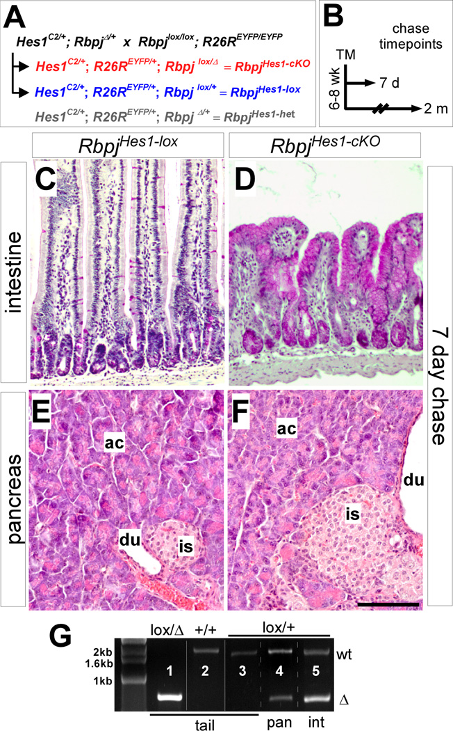
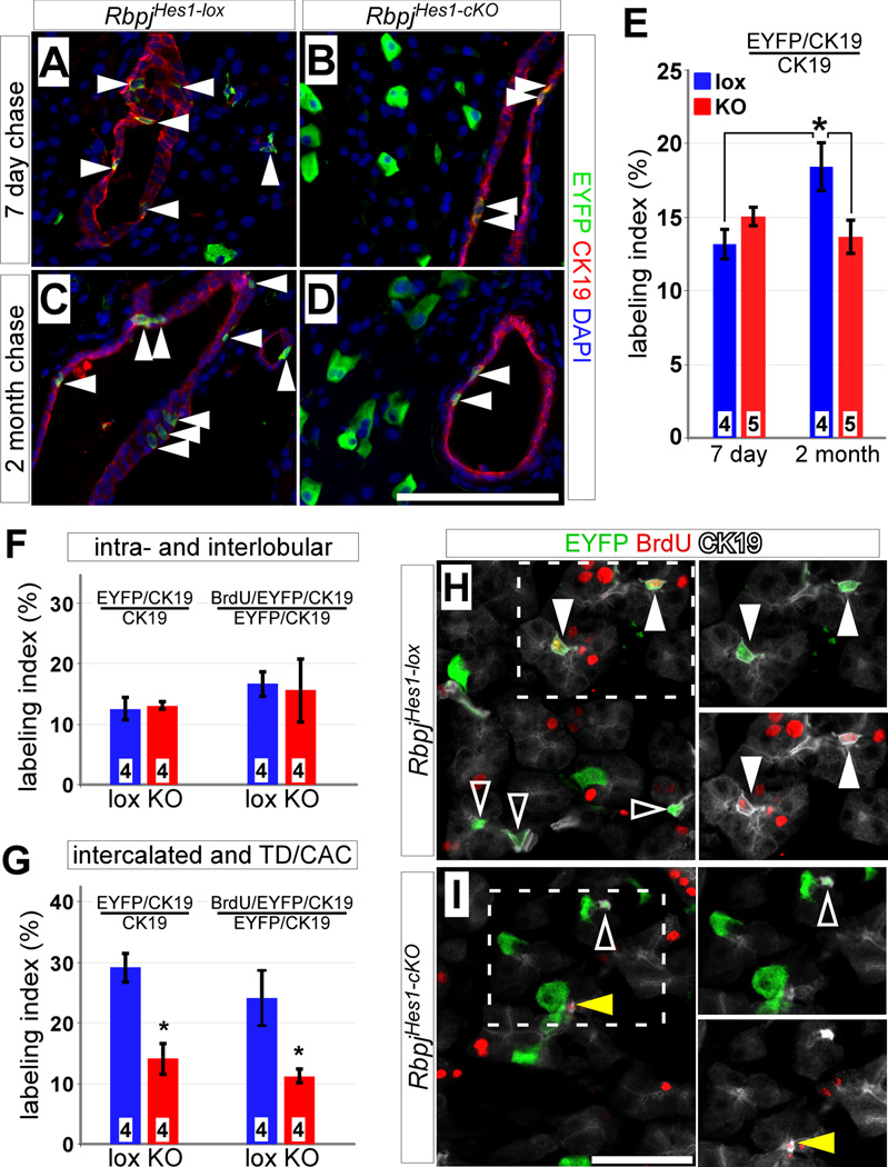
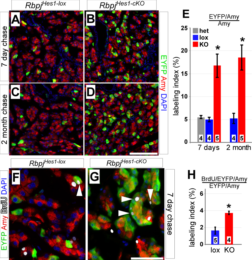
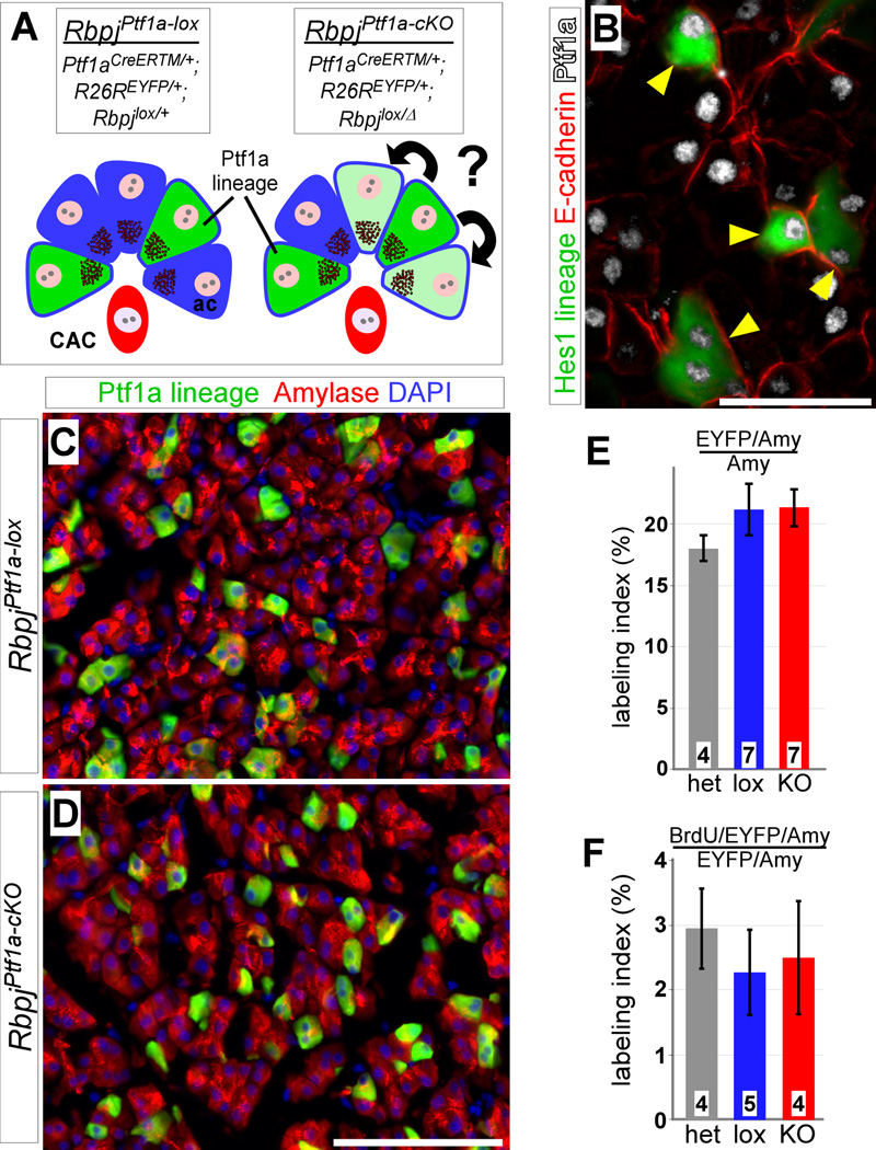
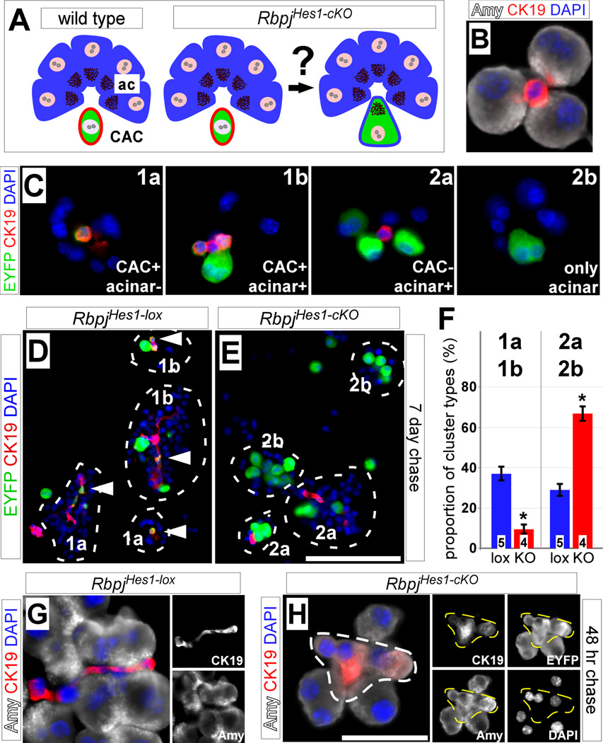
The Notch signaling pathway regulates embryonic development of the pancreas, inhibiting progenitor differentiation into exocrine acinar and endocrine islet cells. The adult pancreas appears to lack progenitor cells, and its mature cell types are maintained by the proliferation of pre-existing differentiated cells. Nonetheless, Notch remains active in adult duct and terminal duct/centroacinar cells (CACs), in which its function is unknown. We previously developed mice in which cells expressing the Notch target gene Hes1 can be labeled and manipulated, by expression of Cre recombinase, and demonstrated that Hes1(+) CACs do not behave as acinar or islet progenitors in the uninjured pancreas, or as islet progenitors after pancreatic duct ligation. In the current study, we assessed the function of Notch signaling in the adult pancreas by deleting the transcription factor partner of Notch, Rbpj, specifically in Hes1(+) cells. We find that loss of Rbpj depletes the pancreas of Hes1-expressing CACs, abrogating their ongoing contribution to growth and homeostasis of more proximal duct structures. Upon Rbpj deletion, CACs undergo a rapid transformation into acinar cells, suggesting that constitutive Notch activity suppresses the acinar differentiation potential of CACs. Together, our data provide direct evidence of an endogenous genetic program to control interconversion of cell fates in the adult pancreas.


Figures

Figure 1
Figure 1. Hes1-specific deletion of Rbpj in the pancreas and intestine
(A) Breeding strategy for conditional knockout of Rbpj. Animals heterozygous for a null (Δ) and a floxed (lox) allele of Rbpj (Rbpj lox/Δ) are abbreviated RbpjHes1-cKO, while Rbpjlox/+ mice serve as controls (RbpjHes1-lox). In some experiments, RbpjΔ/+ mice were included as an additional control (RbpjHes1-het). All mice also carry a R26REYFP reporter allele, allowing for lineage tracing of recombined cells. (B) Pulse-chase strategy. Recombination was induced by TM administration in 6–8 week old adults and animals were chased for 7 days (short term) or 2 months (long term) before analyzing. (C–F) Comparison of PAS-stained intestine (C–D) and H&E-stained pancreata (E–F) between RbpjHes1-lox and RbpjHes1-cKO mice after a 7 day chase. Intestinal KO of Rbpj leads to widespread transformation of the gut epithelium into PAS+ goblet cells. In contrast, no morphological differences were detected between RbpjHes1-lox and RbpjHes1-cKO pancreata. (G) PCR to detect recombination of floxed Rbpj. While the deletion-specific Rbpj amplification product (Δ) can be detected in tail DNA from Rbpjlox/Δ (lane l) but not Rbpj+/+ or Rbpjlox/+ (lanes 2–3) mice, the Δ band can be amplified from the pancreas or duodenum of an Rbpjlox/+ mouse after TM treatment (lanes 4–5). Abbreviations: ac, acinar; is, islet; du, duct. Scale bar: 100 µm.
Figure 2
Figure 2. Deletion of Rbpj inhibits expansion of Hes1C2-labeled ducts
(A–E) Adult RbpjHes1-lox and RbpjHes1-cKO mice were treated with tamoxifen and stained for EYFP (green) and the duct marker CK19 (red) after 7 days (A–B) and 2 months (C–D). While there is no difference in the fraction of EYFP-labeled duct cells (arrowheads) between 7 day chased RbpjHes1-lox and RbpjHes1-cKO animals, an increase is detected between 7 days and 2 month in RbpjHes1-lox mice, which is inhibited in RbpjHes1-cKO animals (E). * P < 0.05. (F) Quantifications of EYFP and BrdU labeling indices of intra- and interlobular ducts indicate no differences between RbpjHes1-lox and RbpjHes1-cKO mice (P=0.75). (G) Quantifications of EYFP and BrdU labeling indices of intercalated ducts, terminal ducts (TD) and centroacinar cells (CACs) reveal a 2-fold decrease in EYFP labeling in RbpjHes1-cKO mice, as well as a 2-fold reduction in the fraction of cycling (BrdU+) EYFP-labeled intercalated ducts, TDs and CACs. * P < 0.05. (H–I) EYFP (green) and BrdU (red) labeling of CK19+ CACs (white), 7 days post-TM and following a 10 day BrdU pulse. CACs expressing EYFP only (open arrowheads) or positive for both EYFP and BrdU (closed arrowheads) can be found in RbpjHes1-lox mice. In contrast, RbpjHes1-cKO animals have fewer EYFP+ CACs, and most of the BrdU+ CACs are EYFP-negative (yellow arrowhead). Numbers in bars (E–G) indicate mice analyzed per group. Scale bars: A–D and J, 100 µm; F–G, 50 µm.
Figure 3
Figure 3. Loss of Rbpj in Hes1+ cells results in dramatic increase of labeled acinar cells
(A–E) Adult RbpjHes1-lox and RbpjHes1-cKO pancreata were analyzed for co-expression of EYFP (green) with the acinar marker amylase (red), 7 days (A–B) and 2 months (C–D) post-TM. The fraction of EYFP+ acini in RbpjHes1-lox animals remains constant between 7 days and 2 months. In contrast, a drastic increase (~3.5-fold) in labeled acinar cells is seen both 7 days and 2 months after loss of Rbpj (E). * P < 0.005. (F–H) BrdU/EYFP labeling at 7 days post-TM. RbpjHes1-cKO animals display a 2-fold increase over RbpjHes1-lox in the fraction of EYFP+ acinar cells that have incorporated BrdU (white arrowheads). * P < 0.005. Numbers in bars (E, H) indicate mice analyzed per group. Scale bars: A–D, 100 µm; F–G, 50 µm.
Figure 4
Figure 4. No increased proliferation after acinar-specific loss of Rbpj
(A) To determine whether Notch generally represses expansion of acinar cells, Rbpj was deleted using an inducible Ptf1aCre-ERTM Cre driver, which induces mosaic recombination in acinar cells but not ducts or CACs. Blue, acinar cells; red, CAC; green, Ptf1a lineage-labeling. (B) Short term lineage tracing of Hes1+ cells (green) demonstrates that all EYFP+ acinar cells (arrowheads) also express Ptf1a (white). (C–D) At 7 days post-TM, no difference is detected, between RbpjPtf1a-lox and RbpjPtf1a-cKO, in EYFP labeling (green) of amylase+ acinar cells (red). (E) There is no change in the fraction of EYFP+ acinar cells after a 7 day chase (P=0.52). (F) Quantification of BrdU labeling analysis 7 days post-TM. The EYFP/BrdU labeling index of acinar cells remains the same between control (lox and het) and Ptf1a1-KO animals (P=0.81). Numbers in bars (E–F) indicate mice analyzed per group. Scale bars: B, 50 µm; C–D, 100 µm.
Figure 5
Figure 5. Hes1+ CACs contribute to acinar cells upon Rbpj deletion
(A) We hypothesize that the increase in acinar labeling after loss of Rbpj is due to CACs adopting an acinar fate. Blue, acinar cells; red, CAC; green, Hes1 lineage-labeling. (B) Dissociation of whole pancreata generates small clusters containing amylase+ acinar cells (white) and CK19+ CACs (red). (C) Clusters from Hes1 lineage traced (green) pancreata were divided into two major categories, based on the presence (class 1) or absence (class 2) of labeled CACs as indicated. Class 1 clusters contain EYFP+ CACs with acinar cells unlabeled (1a) or EYFP+ CACs and acinar cells together (1b), while class 2 clusters contain EYFP+ acinar cells with unlabeled CACs (2a) or EYFP+ acinar cells with no CACs present (2b). Inserts represent schematic representations of cluster types. (D–E) Immunofluorescence for EYFP (green) and CK19 (red) of clusters from dissociated pancreata of RbpjHes1-lox and RbpjHes1-cKO animals at 7 days post-TM. Only clusters containing 3 or more closely-attached acinar cells were scored (circle). Most of the RbpjHes1-lox clusters contain labeled CAC (arrowhead) and belong to class 1, while CACs are either unlabeled or absent altogether in RbpjHes1-cKO clusters (classes 2a and 2b). (F) Scoring of cluster types from two independent experiments demonstrates a shift in the proportion of clusters from type 1a and 1b to 2a and 2b between RbpjHes1-lox and RbpjHes1-cKO mice (* P<0.0005). Note that the denominator represents the number of total clusters present per field, including entirely EYFP-negative ones. (G–H) Immunofluorescence for amylase (white) and CK19 (red) of cell clusters from RbpjHes1-lox (J) and RbpjHes1-cKO (K) pancreata 48 hrs after TM administration. In RbpjHes1-lox mice (J), CK19 and amylase expression are restricted to CAC and acinar cells, respectively. After TM induction (K), some amylase+ acinar cells are also positive for CK19 (dotted outline). Co-expression of duct and acinar markers is seen only in EYFP+ cells (right). Right, single-channel amylase, CK19, EYFP and DAPI staining as indicated. Numbers in bars (I) indicate mice analyzed per group. Scale bar: D–E, 50 µm; G–H, 25 µm.

Similar articles

Cited by

References

    1. Apelqvist A, Li H, Sommer L, Beatus P, Anderson DJ, Honjo T, Hrabe de Angelis M, Lendahl U, Edlund H. Notch signalling controls pancreatic cell differentiation. Nature. 1999;400:877–881. - PubMed
    1. Brennand K, Huangfu D, Melton D. All beta Cells Contribute Equally to Islet Growth and Maintenance. PLoS Biol. 2007;5:e163. - PMC - PubMed
    1. Burlison JS, Long Q, Fujitani Y, Wright CV, Magnuson MA. Pdx-1 and Ptf1a concurrently determine fate specification of pancreatic multipotent progenitor cells. Dev. Biol. 2008;316:74–86. - PMC - PubMed
    1. Chiba S. Notch signaling in stem cell systems. Stem Cells. 2006;24:2437–2447. - PubMed
    1. Collombat P, Xu X, Ravassard P, Sosa-Pineda B, Dussaud S, Billestrup N, Madsen OD, Serup P, Heimberg H, Mansouri A. The ectopic expression of Pax4 in the mouse pancreas converts progenitor cells into alpha and subsequently beta cells. Cell. 2009;138:449–462. - PMC - PubMed

Publication types

MeSH terms

Substances