Review
. 2012 Jun;69(12):1917-29.
doi: 10.1007/s00018-011-0886-0. Epub 2011 Dec 7.

Deciphering the structure and function of FcεRI/mast cell axis in the regulation of allergy and anaphylaxis: a functional genomics paradigm

Affiliations
Review

Deciphering the structure and function of FcεRI/mast cell axis in the regulation of allergy and anaphylaxis: a functional genomics paradigm

Jayapal Manikandan et al. Cell Mol Life Sci. 2012 Jun.

Abstract

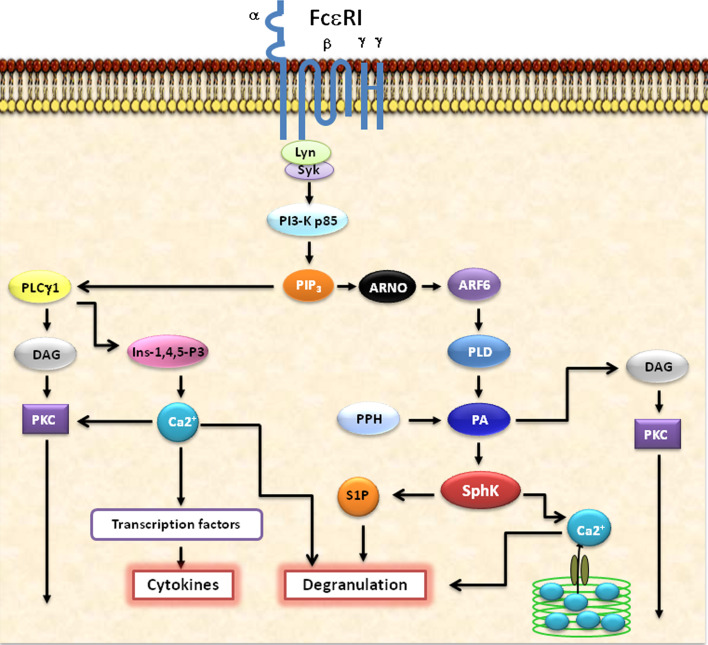
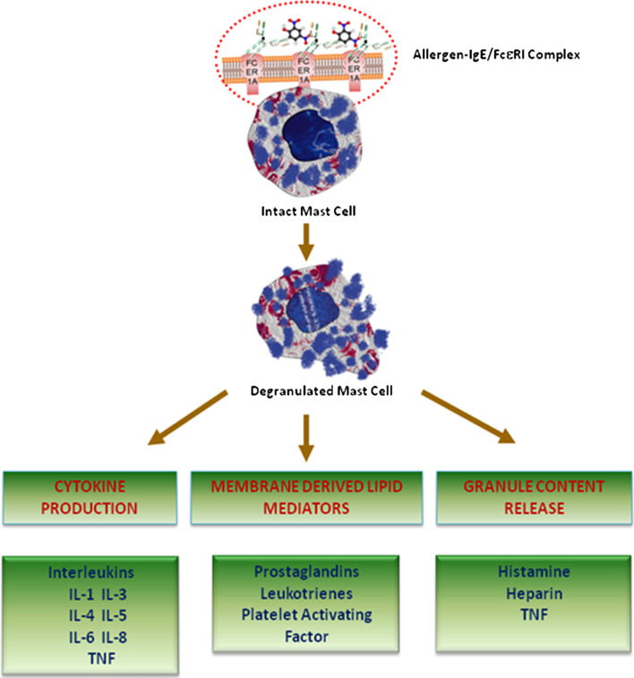
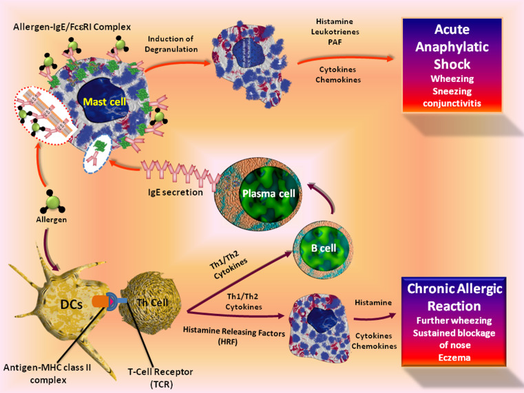
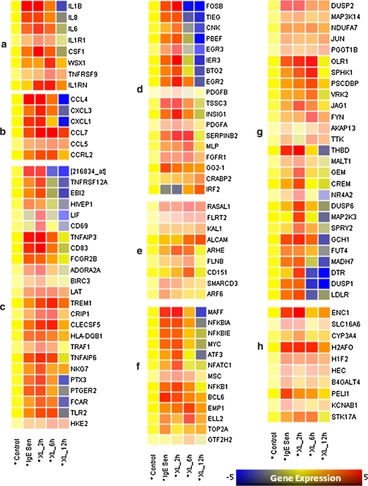
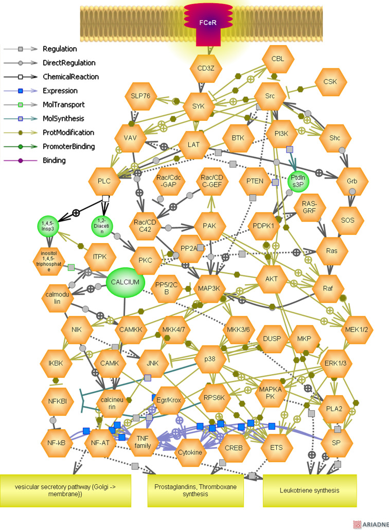
Allergy and anaphylaxis are inflammatory disorders caused by immune reactions mainly induced by immunoglobulin-E that signal through the high-affinity FcεRI receptor to release the inflammatory mediators from innate immune cells. The FcεRI/mast cell axis is potently involved in triggering various intracellular signaling molecules to induce calcium release from the internal stores, induction of transcription factors such as NF-kB, secretion of various cytokines as well as lipid mediators, and degranulation, resulting in the induction of allergy and anaphylaxis. In this review, we discuss various cellular and molecular mechanisms triggered through FcεRI/mast cell axis in allergy and anaphylaxis with a special emphasis on the functional genomics paradigm.


Figures

Fig. 1
Fig. 1
Schematic representation of FcεRI-mediated intracellular signaling events in mast cells and basophils. The allergen cross linking of FcεRI receptors initiate key cellular signaling events such as the activation of PLCγ, PKCs, SPHKs, PI3Ks, etc. An important role for these major pathways in immune cell function is predicted based on their ability to regulate the transcriptional activity of cytokine genes, calcium release from the internal stores which is essential for the degranulation and further intracellular mediation of signaling processes, and the production of lipid mediators such as prostaglandins and leukotrienes. The specific blocking of these key intracellular signaling molecules such as PLCγ, PKCs, SPHKs, S1P, PI3Ks, etc., by specific chemical inhibitors or siRNAs, or therapeutic Intrabodies would certainly be of immense use in the effective management of allergy and anaphylaxis in the near future
Fig. 2
Fig. 2
The key mediators of FcεRI/Mast cell axis in allergy and anaphylaxis. The schematic representation illustrates some of the key mediators released by the mast cells upon stimulation during allergy and anaphylaxis. The mediators released by the mast cells can be categorized into three main groups: preformed granule content, membrane-derived lipid mediators, and cytokines
Fig. 3
Fig. 3
The cellular pathways leading to acute and chronic allergic reactions. The binding of allergen with IgE/FcεRI complex on mast cells triggers the release of pre-formed mediators, production of lipid mediators, and cytokines leading to acute allergic conditions. Allergic conditions broadly consist of two phases: sensitization and elicitation phase. In the self, sensitization occurs upon the initial exposure to an allergen or antigen. Dendritic cells and macrophages are the first line of defense against antigens through phagocytosis and cell-mediated uptake of antigens. The antigen is subsequently degraded as a component of the MHC class II complexes. Then the mature dendritic cells stimulate naive T cells to differentiate into either TH1- or TH2-type CD4+ T cells, which aids in the maturation of B-cells to IgE-secreting plasma cells. Chronic allergic reactions, including the late-phase reaction, may rely on a combination of events and the release of mast-cell products by histamine-releasing factors from T-helper cells
Fig. 4
Fig. 4
Gene expression pattern in human mast cells following IgE sensitization and FcεRI aggregation. The raw Affymetrix GeneChip data was downloaded from gene expression omnibus (GSE1933) and normalized with parametric test based on cross gene error model (PCGEM) and subjected to one-way ANOVA and Bonferroni-Hochberg FDR (p < 0.05) using Genespring 7.3 The differentially expressed genes by the IgE sensitization and FcεRI cross-linking for different time points (2, 6, and 12 h) were then classified and clustered based on gene ontology (GO). Analysis to decode the differentially expressed genes implicated in biological processes such as a cytokines and cytokine receptors, b chemokines and chemokine receptors, c other immunoregulatory genes, d cell proliferation and apoptosis, e adhesion and cytoskeleton remodeling, f transcription factors and regulators, g signal transduction, and h other genes [115]
Fig. 5
Fig. 5
Molecular pathways triggered through FcεRI/mast cell axis. Representation of specific inflammatory and immunoregulatory pathways among the differentially expressed genes triggered through FcεRI/mast cell axis was analyzed using Pathway Studio® software (Ariadne Genomics, Rockville, MD) version 6.0. The software uses information available in the current literature to identify common pathways, targets, or regulators that are associated with the altered genes to generate biological interaction networks. Genes were linked to each other based on the published literature

Similar articles

Cited by

References

    1. MacDonald SM, Rafnar T, Langdon J, Lichtenstein LM. Molecular identification of an IgE-dependent histamine-releasing factor. Science. 1995;269(5224):688–690. - PubMed
    1. Portier P, Richet C. De l’action anaphylatique de certain venins. C R Soc Biol. 1902;54:170.
    1. Theoharides TC, Kalogeromitros D. The critical role of mast cells in allergy and inflammation. Ann NY Acad Sci. 2006;1088:78–99. - PubMed
    1. Theoharides TC, Alysandratos KD, Angelidou A, Delivanis DA, Sismanopoulos N, Zhang B, Asadi S, Vasiadi M, Weng Z, Miniati A, Kalogeromitros D (2010) Mast cells and inflammation. Biochim Biophys Acta. doi:10.1016/j.bbadis.2010.12.014 - PMC - PubMed
    1. Metcalfe DD, Baram D, Mekori YA. Mast cells. Physiol Rev. 1997;77(4):1033–1079. - PubMed