. 2012 Oct;16(5):1451-61.
doi: 10.1007/s00784-011-0650-8. Epub 2011 Dec 8.

Long-term bonding to eroded dentin requires superficial bur preparation

Affiliations

Long-term bonding to eroded dentin requires superficial bur preparation

Brigitte Zimmerli et al. Clin Oral Investig. 2012 Oct.

Abstract

Objectives: This study aims to evaluate the influence of different surface preparation techniques on long-term bonding effectiveness to eroded dentin.

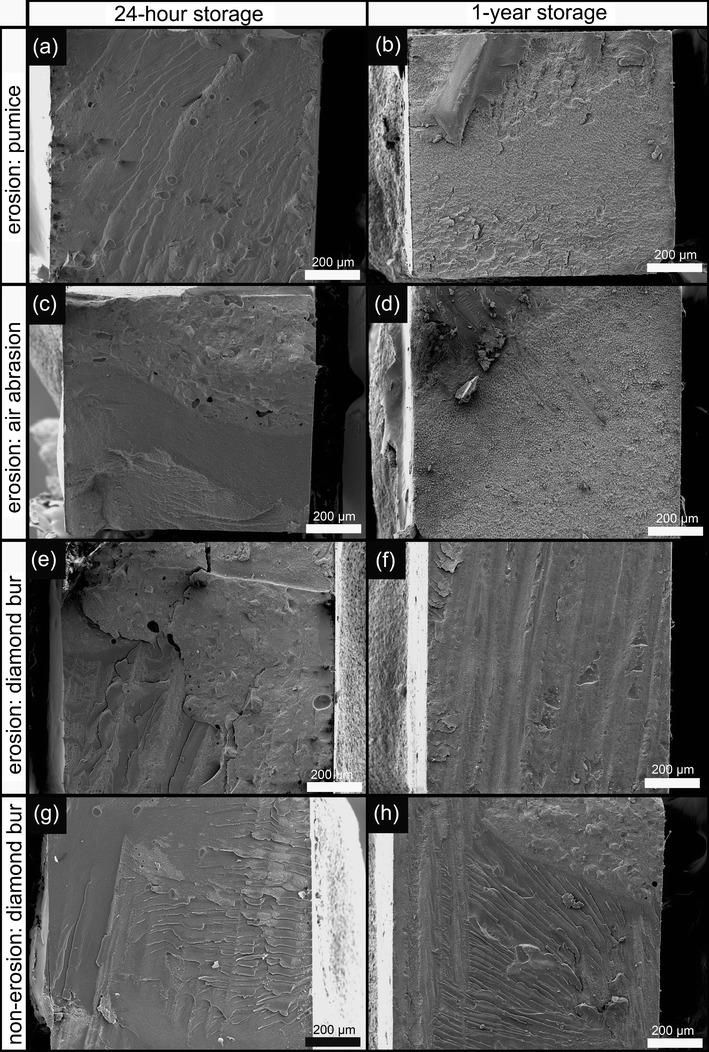
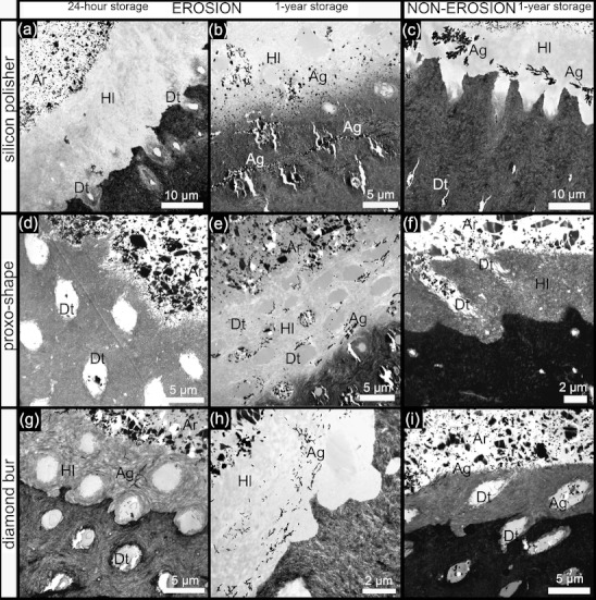
Materials and methods: Dentin specimens were eroded by pH cycling or were left untreated as control, respectively. Five different "preparation" techniques were applied: (1) cleaning with pumice, (2) air abrasion, (3) silicon polisher, (4) proxo-shape, and (5) diamond bur. The three-step etch-and-rinse adhesive OptiBond FL (O-FL; Kerr) and the mild two-step self-etch adhesive Clearfil SE Bond (C-SE; Kuraray) were evaluated. Micro-tensile bond strength was measured after water storage for 24 h and 1 year. Fracture analysis was performed by stereomicroscopy and SEM. Interfaces were characterized by TEM. Differences were statistically analyzed with a linear mixed effects model (α = 0.05).

Results: Erosion reduced bond strength in all groups, but this effect was less prominent when eroded dentin was prepared by diamond bur. Storage lowered bond strength in almost all groups significantly, but this ageing effect was more prominent for the eroded surfaces than for non-eroded controls. Whereas after 1-year control specimens revealed superior bond strength with the three-step etch-and-rinse adhesive (O-FL), the mild two-step self-etch adhesive (C-SE) revealed a better 1-year bond strength to eroded dentin. The interface at eroded dentin appeared very prone to degradation as was shown by the increased amount of adhesive failures and by the silver infiltration detected by TEM.

Conclusions and clinical relevance: Although a minimally invasive approach should clinically always be strived for, superficial preparation (or minimal roughening) with a diamond bur is recommendable for long-term bonding to eroded dentin.


Figures

Fig. 1
Fig. 1
SEM photomicrographs of μTBS-fractured surfaces of Optibond FL (Kerr) specimens. a “Mixed” failure with major part having failed “cohesively” after 24-h water storage. b One-year storage changed clearly the fracture pattern into an ‘adhesive’ failure mode. Resin penetration into the demineralized (eroded) dentin was poor as the failure occurred within the hybrid layer. c Proxo-shape (Intensiv) preparation resulted in a “cohesive” failure pattern after 24-h storage, involving failure within the adhesive resin as well as restorative composite. d Failure pattern after 1-year water storage again turned into an “adhesive” failure mode as in b. Failure within the hybrid layer. e Diamond bur preparation resulted in a “cohesive” failure type after 24 h. However, this specimen included a “cohesive” failure within dentin as well as a “cohesive” failure within resin. f One-year storage turned the “cohesive” failure pattern into an “adhesive” one. However, compared to b and d, this failure mostly occurred at the top of the hybrid layer. Furthermore, some resin remnants that failed “cohesively” are detectable as well. g Only one edge of the stick failed “adhesively” for this non-eroded specimen. Most of the surface remained covered by composite. h The surface percentage “adhesive” failure increased after 1-year storage. However, hybridization seemed to hold out better for the non-eroded specimens as the fracture pattern occurred predominantly near the top of the hybrid layer
Fig. 2
Fig. 2
SEM photomicrographs of μTBS-fractured surfaces of Clearfil SE Bond (Kuraray) specimens. a Pumice cleaning of eroded dentin resulted in an overall “cohesive” failure pattern after 24-h water storage. b One-year storage turned the failure type into an “adhesive” one. The adhesive failure occurred at different levels of the hybrid layer, indicating that the by-erosion exposed collagen fibrils were poorly enveloped by resin. c Air abrasion revealed a “mixed” failure type composed of all types of failure, such as “cohesive in resin,” “cohesive in dentin,” and “adhesive.” d Similar change in failure mode after 1-year storage as in b: “adhesive” failure within the hybrid layer. e Diamond bur preparation resulted in a “cohesive” failure type within the adhesive as well as composite. f Although the incidence of “adhesive” failure was increased after 1-year storage, the surface seemed still to be covered by adhesive resin and the hybrid layer seemed to be less prone to fracture than in the case where other surface preparation methods were used. g Non-eroded diamond bur specimen that failed “cohesively.” h One-year storage of non-eroded specimens was clearly less affected by ageing of the interface than in the case of eroded specimens
Fig. 3
Fig. 3
TEM photomicrographs of OptiBond FL (Kerr) specimens. a The use of a silicon polisher did not sufficiently reduce the depth of demineralized (eroded) dentin. Consequently, the hybrid layer thickness was still larger than that typically observed for the non-eroded control in c. b The poor hybridization made the interface at eroded dentin very sensitive to degradation, as was confirmed by the relatively heavy silver deposition. Silver uptake was most prominent in the hybrid layer at the transition zone from the completely demineralised to the more densely mineralized dentin. c The non-eroded (control) specimen showed localized silver deposition after 1-year storage, while the hybrid layer thickness was clearly smaller than that in a. d Collagen fibrils were largely exposed after preparation with Proxo-shape (Intensiv), as was also confirmed by the positively stained specimens. e The central and bottom part of the hybrid layer appeared most susceptible for silver deposition after 1-year storage. f The positively stained non-eroded (control) specimen resulted in a densely organized collagen–fibril structure with some exposed fibrils d. g Diamond bur preparation of eroded dentin resulted in an almost similar interfacial ultrastructure as that observed for the non-eroded control i. h Removal of the most demineralized outer dentin zone resulted in a better long-term bonding performance, as was revealed by the less prominent silver deposition after 1-year water storage. i Nevertheless, the non-eroded specimen still revealed less silver deposition (1-year storage). Ag AgNO3 deposition, Ar adhesive resin, Dt dentin tubule, Hl hybrid layer
Fig. 4
Fig. 4
TEM photomicrographs of Clearfil SE Bond (Kuraray) specimens. a Scrubbing the surface with pumice appeared to have been quite effective to remove the most outer zone of the demineralized (eroded) dentin. The remaining, more poorly mineralized dentin clearly disclosed residual hydroxyapatite crystals. b One-year storage highly affected the interface, as was evidenced by severely affected collagen fibrils. This became especially clear as the exposed collagen zone appeared much less stainable with heavy metals. The bond degradation also made specimen processing more difficult as interface detachment was more often experienced. c After 1-year storage, the non-eroded (control) specimen revealed only few and localized areas of silver deposition. d Air abrasion almost did not change the micro-structure of the eroded specimen as also a wide zone of demineralized (eroded) dentin remained. The many areas where silver was deposited, around the tubules and within the hybrid layer, indicate that resin had not infiltrated the eroded dentin surface sufficiently. e One-year storage resulted in increased interface detachment and collagen fibrils that appeared severely affected by water storage ageing. f After 24-h water storage, much less nano-leakage was detected in the hybrid layer, as what appeared from the rather limited silver deposition. g Diamond bur preparation again reduced the hybrid layer thickness to that typically observed for the control (non-eroded) specimens. Some localized silver deposition however remained detectable. h One-year storage showed good performance in TEM regarding silver deposition. Even the underlying dentin zone seemed normally mineralized, though some silver-infiltrated areas could be detected as well. i At non-eroded dentin, 1-year storage revealed rather limited silver deposition, mainly appearing as small spot-like infiltrations in the hybrid layer. Ag AgNO3 deposition, Ar adhesive resin, Dt, dentin tubule, Er embedding resin, Hl hybrid layer

Similar articles

Cited by

References

    1. Myklebust S, Espelid I, Svalestad S, Tveit AB. Dental health behavior, gastroesophageal disorders and dietary habits among Norwegian recruits in 1990 and 1999. Acta Odontol Scand. 2003;61:100–104. doi: 10.1080/00016350310001433. - DOI - PubMed
    1. Karamanolis G, Sifrim D. Developments in pathogenesis and diagnosis of gastroesophageal reflux disease. Curr Opin Gastroenterol. 2007;23:428–433. doi: 10.1097/MOG.0b013e328133f56a. - DOI - PubMed
    1. Waterhouse PJ, Auad SM, Nunn JH, Stehen IN, Moynihan PJ. Diet and dental erosion in young people in south-east Brazil. Int J Paediatr Dent. 2008;18:353–360. doi: 10.1111/j.1365-263X.2008.00919.x. - DOI - PubMed
    1. Brunton PA, Kalsi KS, Watts DC, Wilson NH. Resistance of two dentin-bonding agents and a dentin desensitizer to acid erosion in vitro. Dent Mater. 2000;16:351–355. doi: 10.1016/S0109-5641(00)00027-0. - DOI - PubMed
    1. Azzopardi A, Bartlett SW, Watson TF, Sheriff M. The measurement and prevention of erosion and abrasion. J Dent. 2001;29:295–400. doi: 10.1016/S0300-5712(01)00031-8. - DOI - PubMed

Publication types