. 2012 Feb 15;590(4):777-92.
doi: 10.1113/jphysiol.2011.220236. Epub 2011 Dec 6.

Phosphatase and tensin homologue (PTEN) regulates synaptic plasticity independently of its effect on neuronal morphology and migration

Affiliations

Phosphatase and tensin homologue (PTEN) regulates synaptic plasticity independently of its effect on neuronal morphology and migration

Margaret Sperow et al. J Physiol. .

Abstract

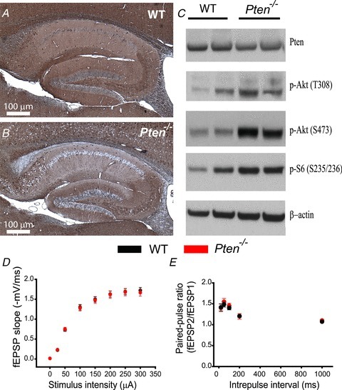
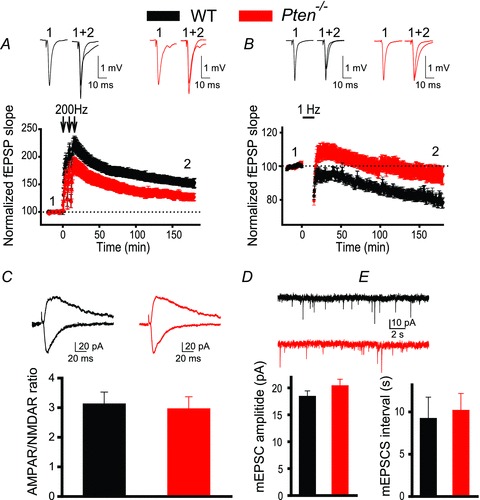
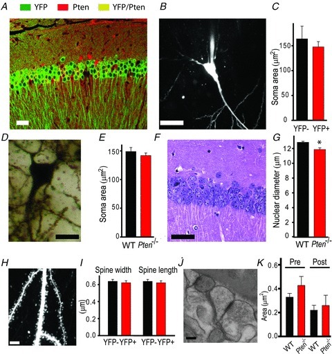
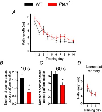
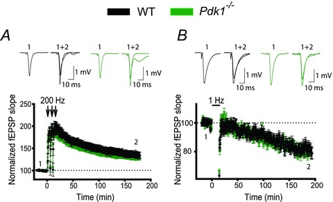
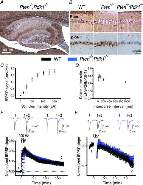
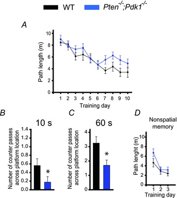
The tumour suppressor PTEN is the central negative regulator of the phosphatidylinositol 3-kinase (PI3K) signalling pathway, which mediates diverse processes in various tissues. In the nervous system, the PI3K pathway modulates proliferation, migration, cellular size, synaptic transmission and plasticity. In humans, neurological abnormalities such as autism, seizures and ataxia are associated with inherited PTEN mutations. In rodents, Pten loss during early development is associated with extensive deficits in neuronal migration and substantial hypertrophy of neurons and synaptic densities; however, whether its effect on synaptic transmission and plasticity is direct or mediated by structural abnormalities remains unknown. Here we analysed neuronal and synaptic structures and function in Pten-conditional knockout mice in which the gene was deleted from excitatory neurons postnatally. Using two-photon imaging, Golgi staining, immunohistochemistry, electron microscopy, and electrophysiological tools, we determined that Pten loss does not affect hippocampus development, neuronal or synaptic structures, or basal excitatory synaptic transmission. However, it does cause deficits in both major forms of synaptic plasticity, long-term potentiation and long-term depression, of excitatory synaptic transmission. These deficits coincided with impaired spatial memory, as measured in water maze tasks. Deletion of Pdk1, which encodes a positive downstream regulator of the PI3K pathway, rescued Pten-mediated deficits in synaptic plasticity but not in spatial memory. These results suggest that PTEN independently modulates functional and structural properties of hippocampal neurons and is directly involved in mechanisms of synaptic plasticity.


Figures

Figure 1
Figure 1. Postnatal Pten deletion preserves hippocampal architecture and basal synaptic transmission
A and B, immunohistochemistry for PTEN in the hippocampus of 8-week-old WT (A) and Pten−/− (B) mice. Note the normal architecture of all hippocampal areas in Pten−/− mice. C, Western blot analysis of protein lysates from hippocampi of WT and Pten−/− littermates. Pten−/− mice show increased phosphorylation of Akt at T308 and S473 and of the downstream substrate S6. D, mean fEPSP slope as a function of stimulation intensity measured at CA3–CA1 synapses in slices from WT or Pten−/− mice shows that basal synaptic transmission is normal in both genotypes. E, mean fEPSP slopes as a function of interpulse interval between the first and second fEPSPs evoked at CA3–CA1 synapses in slices from WT and Pten−/− mice shows that paired-pulse ratio is normal in Pten−/− mice.
Figure 2
Figure 2. Postnatal deletion of Pten causes deficits in LTP and LTD
A and B, mean fEPSPs as a function of time before and after induction of LTP (A) or LTD (B) in slices from WT (black) or Pten−/− (red) mice. Insets show representative fEPSP traces before and 180 min after induction of synaptic plasticity. C, mean ratios of AMPAR/NMDAR currents recorded at CA3–CA1 synapses in slices from WT or Pten−/− mice are similar. Insets show representative traces of AMPAR-mediated (recorded at –70 mV, downward traces) and NMDAR-mediated (recorded at 40 mV, upward traces) EPSCs recorded in YFP (WT) and YFP+ (Pten−/−) neurons. D and E, mean amplitudes (D) and intervals (E) of spontaneous mEPSCs measured in CA1 neurons of WT or Pten−/− mice are comparable. Insets show representative traces of mEPSCs measured in YFP (WT) and YFP+ (Pten−/−) CA1 neurons.
Figure 3
Figure 3. Postnatal Pten deletion in pyramidal neurons does not induce neuronal hypertrophy in the CA3 and CA1 areas of the hippocampus
A, double-immunofluorescence staining for PTEN (red) and YFP (green) reveals that YFP is a reliable indicator of Pten deletion in Cre;Ptenfl/fl; R26LSL-EYFP mice, with only rare false-positive cells expressing both YFP and PTEN (yellow). B and C, TPLSM image of a neuron filled with Alexa 594 shows the soma clearly. This method was used to measure mean soma areas in WT (YFP) or Pten-null (YFP+) neurons (C). D and E, Golgi staining of somata (D) of CA1 neurons was used to measure soma areas in slices from WT and Pten−/− mice (E). F and G, H&E staining of the CA1 area of the hippocampus shows the nuclei of cells. G, the mean nuclear diameter of neurons in H&E-stained slices from Pten−/− mice was smaller than that in WT mice (*P < 0.05). H, a two-photon image of an apical dendrite of a CA1 neuron. Note that the dendritic spines are clearly visible. These images were used to measure the mean width and length of dendritic spines in slices from WT and Pten−/− mice (I). J and K, scanning electron microscopic images of the hippocampus (J) were used to measure the mean presynaptic (Pre) and postsynaptic (Post) areas of synapses in the CA1 areas of WT and Pten−/− mice (K). Scale bars, 40 μm (A), 20 μm (B), 10 μm (D), 30 μm (F), 5 μm (H), and 100 nm (J).
Figure 4
Figure 4. Pten deletion in pyramidal neurons during postnatal development causes a deficit in spatial memory
A, spatial learning is not impaired in Pten−/− mice, as indicated by the lack of difference in the mean path lengths of WT (black) or Pten−/− (red) mice swimming in the Morris water maze to find a hidden platform as a function of training days. B and C, long-term memory retention is, however, impaired in Pten−/− mice. The mean number of counter passes across the platform location measured during the first 10 s (B) and during the entire 60 s probe trial (C) in WT and Pten−/− mice differed significantly (*P < 0.05). D, non-spatial memory is normal in Pten−/− mice. Mean path lengths to swim to a visible platform, as a function of training days, was comparable in WT and Pten−/− mice.
Figure 5
Figure 5. Postnatal deletion of Pdk1 in pyramidal neurons does not induce deficits in synaptic plasticity
A and B, synaptic plasticity is not perturbed in Pdk1−/− mice. The mean fEPSP slope versus time before and after induction of LTP (A) or LTD (B) in slices from WT and Pdk1−/− mice did not differ. Insets show representative fEPSP traces before and 180 min after induction of synaptic plasticity.
Figure 6
Figure 6. Pdk1 deletion rescues the synaptic plasticity deficit in Pten−/−mice
A, a representative image of immunohistochemistry for PTEN in hippocampal slices from Pten−/−;Pdk1−/− mice. As with Pten−/− mice, Pten is effectively deleted in most of the pyramidal neurons from the double-mutant mice, and their hippocampal architecture is normal. B, representative images of double immunostaining for PTEN and p-S6 in hippocampal slices of WT, Pten−/− and Pten−/−;Pdk1−/− littermates. The CA1 areas of the hippocampi are shown. C and D, basal synaptic transmission is not affected in Pten−/−;Pdk1−/− mice. Mean fEPSP slope as a function of stimulation intensity (C) or interpulse interval (D) measured at CA3–CA1 synapses in slices from WT or Pten−/−;Pdk1−/− mice are comparable. E and F, normal synaptic plasticity occurs in double-mutant mice. The mean fEPSP as a function of time before and after induction of LTP (E) and LTD (F) in slices from WT or Pten−/−;Pdk1−/− mice are similar. Insets show representative fEPSP traces before (1) and 180 min after (2) induction of synaptic plasticity.
Figure 7
Figure 7. Spatial memory is impaired in Pten−/−;Pdk1−/− mice
A, spatial learning is normal in double-mutant mice. Mean path length as a function of training days in the Morris water maze task in WT and Pten−/−;Pdk1−/− mice was comparable. B and C, spatial memory, however, is impaired in double-mutant mice. The mean number of counter passes across the platform location during the first 10 s (B) and during the entire (60 s) probe trial (C) for WT or Pten−/−;Pdk1−/−mice were significantly different (*P < 0.05). D, non-spatial memory is normal in Pten−/−;Pdk1−/− mice. The mean path lengths in the Morris water maze as a function of training days during the visual platform task did not differ between WT and Pten−/−;Pdk1−/− mice.

Comment in

References

    1. Backman S, Stambolic V, Mak T. PTEN function in mammalian cell size regulation. Curr Opin Neurobiol. 2002;12:516–522. - PubMed
    1. Backman SA, Stambolic V, Suzuki A, Haight J, Elia A, Pretorius J, Tsao MS, Shannon P, Bolon B, Ivy GO, Mak TW. Deletion of Pten in mouse brain causes seizures, ataxia and defects in soma size resembling Lhermitte-Duclos disease. Nat Genet. 2001;29:396–403. - PubMed
    1. Bangash MA, Park JM, Melnikova T, Wang D, Jeon SK, Lee D, Syeda S, Kim J, Kouser M, Schwartz J, Cui Y, Zhao X, Speed HE, Kee SE, Tu JC, Hu JH, Petralia RS, Linden DJ, Powell CM, Savonenko A, Xiao B, Worley PF. Enhanced polyubiquitination of Shank3 and NMDA receptor in a mouse model of autism. Cell. 2011;145:758–772. - PMC - PubMed
    1. Bayascas JR, Leslie NR, Parsons R, Fleming S, Alessi DR. Hypomorphic mutation of PDK1 suppresses tumorigenesis in PTEN(+/–) mice. Curr Biol. 2005;15:1839–1846. - PubMed
    1. Bayazitov IT, Richardson RJ, Fricke RG, Zakharenko SS. Slow presynaptic and fast postsynaptic components of compound long-term potentiation. J Neurosci. 2007;27:11510–11521. - PMC - PubMed

Publication types

Substances