. 2011 Dec;23(12):4446-61.
doi: 10.1105/tpc.111.090142. Epub 2011 Dec 6.

The Arabidopsis tail-anchored protein PEROXISOMAL AND MITOCHONDRIAL DIVISION FACTOR1 is involved in the morphogenesis and proliferation of peroxisomes and mitochondria

Affiliations

The Arabidopsis tail-anchored protein PEROXISOMAL AND MITOCHONDRIAL DIVISION FACTOR1 is involved in the morphogenesis and proliferation of peroxisomes and mitochondria

Kyaw Aung et al. Plant Cell. 2011 Dec.

Abstract

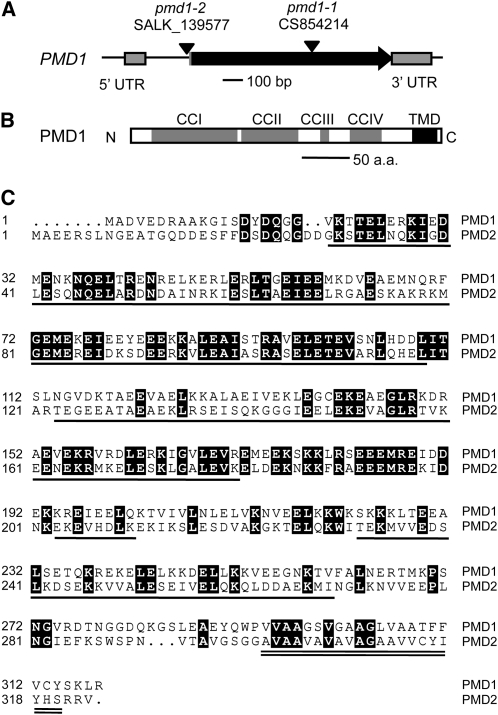
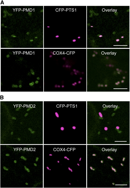
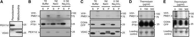
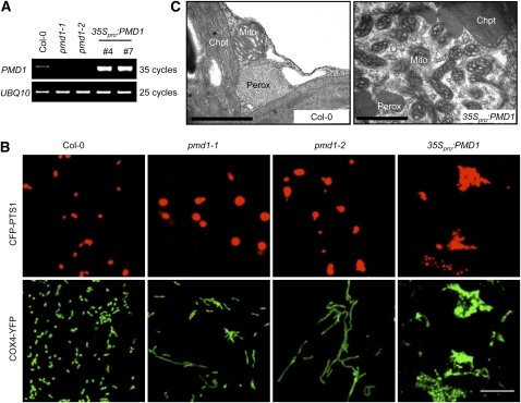
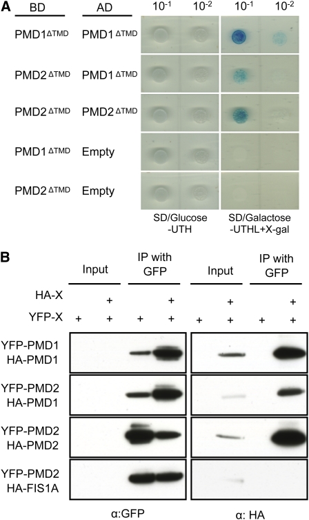
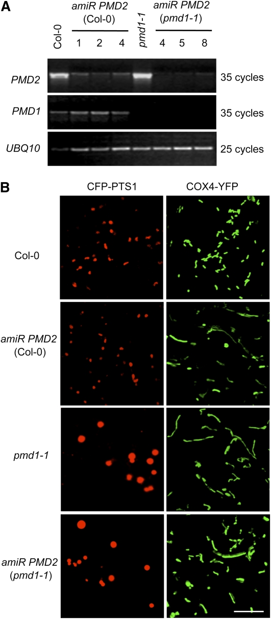
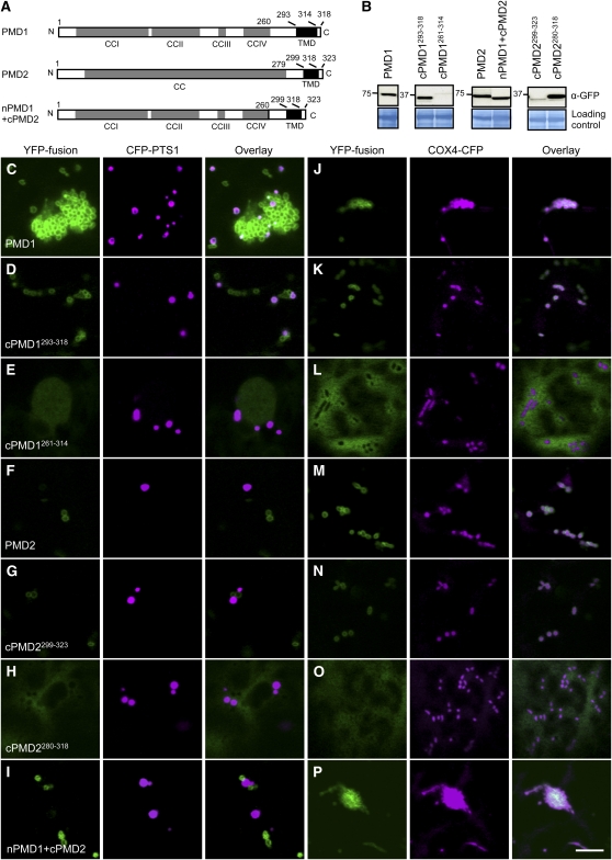
Peroxisomes and mitochondria are multifunctional eukaryotic organelles that are not only interconnected metabolically but also share proteins in division. Two evolutionarily conserved division factors, dynamin-related protein (DRP) and its organelle anchor FISSION1 (FIS1), mediate the fission of both peroxisomes and mitochondria. Here, we identified and characterized a plant-specific protein shared by these two types of organelles. The Arabidopsis thaliana PEROXISOMAL and MITOCHONDRIAL DIVISION FACTOR1 (PMD1) is a coiled-coil protein tethered to the membranes of peroxisomes and mitochondria by its C terminus. Null mutants of PMD1 contain enlarged peroxisomes and elongated mitochondria, and plants overexpressing PMD1 have an increased number of these organelles that are smaller in size and often aggregated. PMD1 lacks physical interaction with the known division proteins DRP3 and FIS1; it is also not required for DRP3's organelle targeting. Affinity purifications pulled down PMD1's homolog, PMD2, which exclusively targets to mitochondria and plays a specific role in mitochondrial morphogenesis. PMD1 and PMD2 can form homo- and heterocomplexes. Organelle targeting signals reside in the C termini of these proteins. Our results suggest that PMD1 facilitates peroxisomal and mitochondrial proliferation in a FIS1/DRP3-independent manner and that the homologous proteins PMD1 and PMD2 perform nonredundant functions in organelle morphogenesis.


Figures

Figure 1.
Figure 1.
Structure and Sequence Analysis of PMD1. (A) Genomic structure of PMD1. The T-DNA insertion sites in pmd1-1 and pmd1-2 are indicated. UTR, untranslated region. (B) Putative protein structure of PMD1. (C) Sequence alignment of PMD1 and PMD2. CC domains are underlined, and the TMD is double underlined. Identical sequences are shaded in black. All domain assignments are based on analysis of PMD1.
Figure 2.
Figure 2.
Subcellular Localization of YFP-PMD1 and YFP-PMD2. Confocal images are from leaf epidermal cells from transgenic plants expressing PMD1pro:YFP-PMD1 (A) or 35Spro:YFP-PMD2 (B) along with the peroxisomal marker CFP-PTS1 or the mitochondrial marker COX4-CFP. YFP signals are in green, and CFP signals are in magenta. Merged images show the colocalization of the YFP fusion protein to peroxisomes or mitochondria. Bars = 5 μm.
Figure 3.
Figure 3.
PMD1 Is Anchored to the Organelle Membranes by the C Terminus. (A) Immunoblotting analysis of peroxisomal and mitochondrial proteins purified from Arabidopsis plants expressing YFP-PMD1 using antibodies against the peroxisomal specific protein PEX11d and the mitochondrial-specific protein VDAC. (B) and (C) Immunoblot analyses of purified Arabidopsis leaf peroxisomes (B) and mitochondria (C) after the proteins were treated with sodium chloride (NaCl) or sodium carbonate (Na2CO3) and fractionated into soluble (S) and pellet (P) fractions. The GFP antibody was used to detect the expression of CFP and YFP fusion proteins. CFP-PTS1 and COX4-CFP served as controls for matrix proteins, and PEX11d and VDAC were organelle-specific membrane protein controls. Asterisk in (C) marks cross-hybridized signals of COX4-CFP, as the same membrane had been probed by the GFP antibody in the previous immunoblot. (D) and (E) Immunoblot analyses of peroxisomes (D) and mitochondria (E) that were treated with various concentrations of thermolysin. The levels of CFP and YFP fusion proteins were detected with the GFP antibody. A nonspecific band was used as loading control. Numbers to the right of each panel indicate molecular mass in kilodaltons.
Figure 4.
Figure 4.
pmd1 Mutants Exhibit Abnormal Peroxisomal and Mitochondrial Morphologies. (A) RT-PCR analyses showing the levels of PMD1 mRNA in the wild type (Col-0), loss-of-function mutants (pmd1-1 and pmd1-2), and gain-of-function mutants (35Spro:PMD1 line numbers 4 and 7). UBQ10 transcripts are used as loading controls. (B) Confocal images from leaf epidermal cells of the indicated plants showing morphologies of peroxisomes (labeled by CFP-PTS1) and mitochondria (marked by COX4-YFP). Bar = 10 μm. (C) TEM images from leaf mesophyll cells illustrating the ultrastructure of peroxisomes (Perox), mitochondria (Mito), and chloroplasts (Chpt) in Col-0 and 35Spro:PMD1 plants. Bars = 1 μm. [See online article for color version of this figure.]
Figure 5.
Figure 5.
PMD1 and PMD2 Are Able to Form Homo- and Heterocomplexes. (A) Y2H analyses. SD/Glc-UTH selects for transformants and SD/Gal-UTHL + X-Gal selects for protein–protein interactions. Empty, pB42AD-GW vector only. (B) Co-IP analyses. Various combinations of the fusions as indicated were transiently expressed in tobacco leaf epidermal cells, followed by immunoprecipitation using the GFP antibody. A GFP or HA antibody was used to detect the proteins. [See online article for color version of this figure.]
Figure 6.
Figure 6.
PMD2 Is Involved in Mitochondrial Morphogenesis. (A) RT-PCR analyses showing the transcripts of PMD1, PMD2, and UBQ10 in Col-0, amiR PMD2, pmd1-1, and pmd1-1 amiR PMD2 plants. Three independent lines that contain amiR PMD2 in the Col-0 (lines 1, 2, and 4) or pmd1-1 (lines 4, 5, and 8) background are shown. (B) Confocal images from leaf epidermal cells of plants coexpressing the organelle markers. Peroxisomes were labeled by CFP-PTS1, and mitochondria were marked by COX4-YFP. Bar = 10 μm. [See online article for color version of this figure.]
Figure 7.
Figure 7.
Organelle Targeting Signals Reside in the C Terminus of PMD1 and PMD2. (A) Schematics of PMD1, PMD2, and nPMD1+cPMD2 with the CC domains, TMD, and amino acids indicated. Despite sequence similarities between PMD1 and PMD2, PMD2 was annotated as having a single and long CC domain. (B) Immunoblot analysis to detect the YFP-PMD variants expressed in tobacco cells. The large subunit of ribulose-1,5-bis-phosphate carboxylase/oxygenase was used as the loading control. Numbers to the left of each panel indicate molecular mass in kilodaltons. (C) to (P) Subcellular targeting of the YFP-PMD1/PMD2 fusions. YFP signals are in green, and CFP signals are in magenta. To better illustrate colocalization between the YFP fusion and CFP-PTS1, images in (C) were taken from a region where peroxisome proliferation/aggregation was not so strong. Bar = 10 μm.

Similar articles

Cited by

References

    1. Abell B.M., Mullen R.T. (2011). Tail-anchored membrane proteins: exploring the complex diversity of tail-anchored-protein targeting in plant cells. Plant Cell Rep. 30: 137–151 - PubMed
    1. Andrade-Navarro M.A., Sanchez-Pulido L., McBride H.M. (2009). Mitochondrial vesicles: An ancient process providing new links to peroxisomes. Curr. Opin. Cell Biol. 21: 560–567 - PubMed
    1. Arimura S., Aida G.P., Fujimoto M., Nakazono M., Tsutsumi N. (2004). Arabidopsis dynamin-like protein 2a (ADL2a), like ADL2b, is involved in plant mitochondrial division. Plant Cell Physiol. 45: 236–242 - PubMed
    1. Arimura S., Fujimoto M., Doniwa Y., Kadoya N., Nakazono M., Sakamoto W., Tsutsumi N. (2008). Arabidopsis ELONGATED MITOCHONDRIA1 is required for localization of DYNAMIN-RELATED PROTEIN3A to mitochondrial fission sites. Plant Cell 20: 1555–1566 - PMC - PubMed
    1. Arimura S., Tsutsumi N. (2002). A dynamin-like protein (ADL2b), rather than FtsZ, is involved in Arabidopsis mitochondrial division. Proc. Natl. Acad. Sci. USA 99: 5727–5731 - PMC - PubMed

Publication types

MeSH terms