. 2011 Dec;59(12):1041-59.
doi: 10.1369/0022155411426780.

The skeletal muscle satellite cell: still young and fascinating at 50

Affiliations

The skeletal muscle satellite cell: still young and fascinating at 50

Zipora Yablonka-Reuveni. J Histochem Cytochem. 2011 Dec.

Abstract

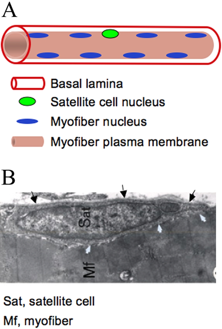
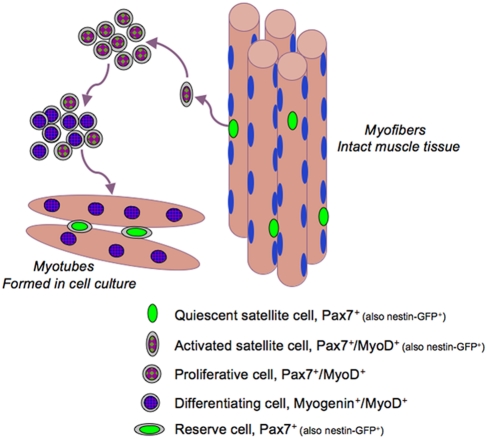
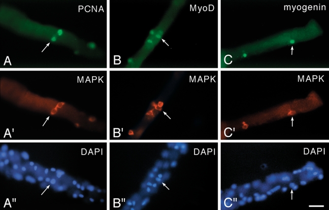
The skeletal muscle satellite cell was first described and named based on its anatomic location between the myofiber plasma and basement membranes. In 1961, two independent studies by Alexander Mauro and Bernard Katz provided the first electron microscopic descriptions of satellite cells in frog and rat muscles. These cells were soon detected in other vertebrates and acquired candidacy as the source of myogenic cells needed for myofiber growth and repair throughout life. Cultures of isolated myofibers and, subsequently, transplantation of single myofibers demonstrated that satellite cells were myogenic progenitors. More recently, satellite cells were redefined as myogenic stem cells given their ability to self-renew in addition to producing differentiated progeny. Identification of distinctively expressed molecular markers, in particular Pax7, has facilitated detection of satellite cells using light microscopy. Notwithstanding the remarkable progress made since the discovery of satellite cells, researchers have looked for alternative cells with myogenic capacity that can potentially be used for whole body cell-based therapy of skeletal muscle. Yet, new studies show that inducible ablation of satellite cells in adult muscle impairs myofiber regeneration. Thus, on the 50th anniversary since its discovery, the satellite cell's indispensable role in muscle repair has been reaffirmed.


Conflict of interest statement

The author(s) declared no potential conflicts of interest with respect to the authorship and publication of this article.
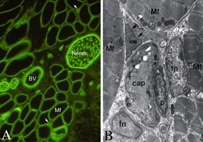
Figures

Figure 1.
Figure 1.
A schematic (A) and electron microscopy image (B) of the satellite cell location. In panel A, nuclei depicted at the myofiber periphery represent the state of healthy adult myofibers; immature myofibers present in regenerating muscles and in muscular dystrophy display centralized myofiber nuclei (not shown) (Yablonka-Reuveni and Day 2011, with kind permission of Springer Science+Business Media). In panel B, black arrows depict the basal lamina, and white arrows depict apposing satellite cell and myofiber membranes; note the sarcomeric organization within the myofiber (Yablonka-Reuveni 1995). The myofiber basement and plasma membranes have been routinely detected by immunostaining with antibodies against laminin and dystrophin, respectively.
Figure 2.
Figure 2.
Immunohistochemical labeling of serial cross-sections of anterior latissimus dorsi muscle from a 2-month-old chicken, depicting Pax7+ satellite cells in intrafusal and extrafusal myofibers. The small intrafusal fibers (clustered within a spindle capsule) are near the center of each parallel image. A, Myosin (in green) highlights myofiber cross-sectional area; laminin (in red) highlights the myofiber basal lamina and the spindle capsule (identified with an asterisk). B and C, laminin (deep blue) and nuclei (light blue). C, Pax7+ nuclei (in red). e, extrafusal fiber; open/large arrow, intrafusal fiber; small arrow, Pax7+ cell associated with intrafusal fiber; arrowhead, Pax7+ cell associated with an extrafusal fiber. Scale bars = 30 µm. This figure is adapted from Kirkpatrick et al. (2009) with kind permission from Dr. Benjamin Rosser. Immunostaining regents and protocols are detailed in Kirkpatrick et al. (2009).
Figure 3.
Figure 3.
The molecular signature of satellite cells and their progeny upon activation, proliferation, differentiation, and self-renewal. The model is based primarily on cell culture studies (Yablonka-Reuveni and Day 2011, with kind permission of Springer Science+Business Media; edited for nestin-GFP information). Such cell cultures have contributed to the identification of autocrine/paracrine factors that can regulate satellite cell activation and proliferation, demonstrating that hepatocyte growth factors and fibroblast growth factors are crucial growth factors involved in the process (e.g., Shefer et al. 2006; Wozniak et al. 2005; Yablonka-Reuveni and Rivera 1997; Yablonka-Reuveni, Seger et al. 1999; Yamada et al. 2009; Tatsumi et al 1998; Gal-Levi et al. 1998). The role of many other growth factors and extracellular matrix components has been studied extensively in cell culture as well; for a comprehensive review of these topics that are beyond the scope of this article, see Shefer and Yablonka-Reuveni (2008).
Figure 4.
Figure 4.
Micrographs of fiber cultures isolated from the flexor digitorum brevis muscle of an 8-week-old rat. Cultures were maintained in basal medium and reacted via double immunofluorescence with a rabbit polyclonal antibody against MAPK (anti-ERK1/2, highlights the cytoplasm of satellite cells as detailed in Yablonka-Reuveni, Seger et al. (1999) and mouse monoclonal antibodies against (A, A′) PCNA, which highlights proliferating cells; (B, B′) MyoD; and (C, C′) myogenin. For each antibody combination, the bottom panel (A′′–C′′) shows a parallel DAPI stain, which highlights both myofiber nuclei and satellite cell nuclei. Arrows in each set of adjacent panels point to the location of the same cell. Immunostaining with the anti-PCNA/anti-MAPK and the anti-MyoD/anti-MAPK is shown for Day 2 cultures, and immunostaining with the anti-myogenin/anti-MAPK is shown for Day 3 cultures. Not all positive nuclei or cells on the fibers are in the same focal plane. Bar = 34 µm. This figure was published first in Yablonka-Reuveni, Seger et al. (1999), where additional details regarding immunostaining reagents and protocols are provided. Note that before high-quality antibodies for satellite cell research were available, researchers investigated activation and proliferation of satellite cells on isolated myofibers by tracing triturated thymidine uptake, which required extra time (even several weeks) to obtain autoradiographic images of the cultures to identify the location of S-phase cells (Bischoff 1989; Schultz 1996; Yablonka-Reuveni and Rivera 1997). This approach provided valuable feedback about response of satellite cells to growth factors, regardless of the tedious method that was used to collect the data (Bischoff 1986).
Figure 5.
Figure 5.
Low- and high-resolution images depicting capillaries of neighboring myofibers. A, immunofluorescent image of a cross-section from rat flexor digitorum brevis muscle reacted with an antibody against laminin (green), which highlights the basement membrane of the myofiber (Mf) and other structures in the muscle, including nerve bundles, blood vessels (BV), and capillaries (indicated by arrows). B, electron microscopy micrograph of adult chicken muscle (Yablonka-Reuveni 1995) demonstrating the fine details of a capillary surrounded by four myofibers. Mf, myofiber; fn, myofiber nucleus (myonucleus); cap, capillary; e, endothelial cell; p, pericyte; mc, an uncharacterized “mysterious cell,” for which a higher resolution EM revealed a large nucleus and some rER structures in the cytoplasm.

Similar articles

Cited by

References

    1. Abou-Khalil R, Le Grand F, Pallafacchina G, Valable S, Authier FJ, Rudnicki MA, Gherardi RK, Germain S, Chretien F, Sotiropoulos A, et al. 2009. Autocrine and paracrine angiopoietin 1/Tie-2 signaling promotes muscle satellite cell self-renewal. Cell Stem Cell. 5:298–309 - PMC - PubMed
    1. Aguennouz M, Vita GL, Messina S, Cama A, Lanzano N, Ciranni A, Rodolico C, Di Giorgio RM, Vita G. 2011. Telomere shortening is associated to TRF1 and PARP1 overexpression in Duchenne muscular dystrophy. Neurobiol Aging. 32:2190–2197 - PubMed
    1. Allouh MZ, Yablonka-Reuveni Z, Rosser BW. 2008. Pax7 reveals a greater frequency and concentration of satellite cells at the ends of growing skeletal muscle fibers. J Histochem Cytochem. 56:77–87 - PMC - PubMed
    1. Anderson JE. 2006. The satellite cell as a companion in skeletal muscle plasticity: currency, conveyance, clue, connector and colander. J Exp Biol. 209:2276–2292 - PubMed
    1. Andres V, Walsh K. 1996. Myogenin expression, cell cycle withdrawal, and phenotypic differentiation are temporally separable events that precede cell fusion upon myogenesis. J Cell Biol. 132:657–666 - PMC - PubMed

Publication types