

Effects of long-term storage on the detection of proteins, DNA, and mRNA in tissue microarray slides
- PMID: 22147608
- PMCID: PMC3283084
- DOI: 10.1369/0022155411423779
Effects of long-term storage on the detection of proteins, DNA, and mRNA in tissue microarray slides
Abstract
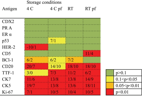
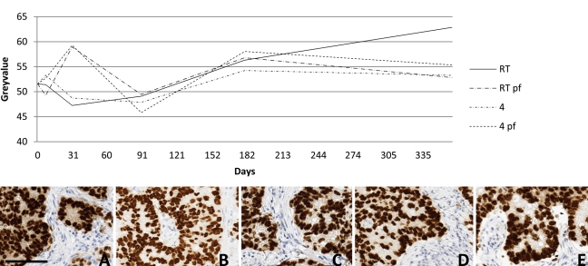
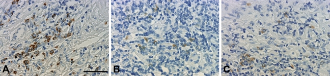
Storage of tissue slides has been claimed to induce dramatically reduced antigen detection particularly for immunohistochemistry (IHC). With tissue microarrays, the necessity to serially cut blocks in order to obtain as much material as possible is obvious. The presumed adverse effect of storage might hamper such an approach. The authors designed an experimental setting consisting of four different storage conditions with storage time of tissue slides of up to 1 year. Detection of proteins, DNA, and mRNA was performed using IHC and in situ hybridization techniques. Slight but significant changes in IHC occurred over time. The most important factor is the primary antibody used: four showed no significant changes, whereas limited decreases in 8 antibodies could be detected by image analysis. Whether the antigen was nuclear or cytoplasmic/membranous did not matter. No major differences between different storage conditions could be shown, but storage at 4C was overall the best procedure. Furthermore, gene copy number aberrations, chromosomal translocations, and the presence of mRNA could be detected on slides stored up to 1 year. In conclusion, in tissues optimally formalin fixed and using modern histological techniques, only minute changes in tissue antigenicity are induced by long-term storage.
Figures
Similar articles
-
Factors affecting immunoreactivity in long-term storage of formalin-fixed paraffin-embedded tissue sections.Histochem Cell Biol. 2015 Jul;144(1):93-9. doi: 10.1007/s00418-015-1316-4. Epub 2015 Mar 11. Histochem Cell Biol. 2015. PMID: 25757745
-
A gene-protein assay for human epidermal growth factor receptor 2 (HER2): brightfield tricolor visualization of HER2 protein, the HER2 gene, and chromosome 17 centromere (CEN17) in formalin-fixed, paraffin-embedded breast cancer tissue sections.Diagn Pathol. 2012 May 30;7:60. doi: 10.1186/1746-1596-7-60. Diagn Pathol. 2012. PMID: 22647525 Free PMC article.
-
Ultrasound-accelerated tissue fixation/processing achieves superior morphology and macromolecule integrity with storage stability.J Histochem Cytochem. 2006 May;54(5):503-13. doi: 10.1369/jhc.5A6802.2005. Epub 2005 Nov 28. J Histochem Cytochem. 2006. PMID: 16314441
-
Excavation of a buried treasure--DNA, mRNA, miRNA and protein analysis in formalin fixed, paraffin embedded tissues.Histol Histopathol. 2011 Jun;26(6):797-810. doi: 10.14670/HH-26.797. Histol Histopathol. 2011. PMID: 21472693 Review.
-
A review of preanalytical factors affecting molecular, protein, and morphological analysis of formalin-fixed, paraffin-embedded (FFPE) tissue: how well do you know your FFPE specimen?Arch Pathol Lab Med. 2014 Nov;138(11):1520-30. doi: 10.5858/arpa.2013-0691-RA. Arch Pathol Lab Med. 2014. PMID: 25357115 Review.
Cited by
-
Spatially resolved analysis of FFPE tissue proteomes by quantitative mass spectrometry.Nat Protoc. 2020 Sep;15(9):2956-2979. doi: 10.1038/s41596-020-0356-y. Epub 2020 Jul 31. Nat Protoc. 2020. PMID: 32737464
-
Factors affecting immunoreactivity in long-term storage of formalin-fixed paraffin-embedded tissue sections.Histochem Cell Biol. 2015 Jul;144(1):93-9. doi: 10.1007/s00418-015-1316-4. Epub 2015 Mar 11. Histochem Cell Biol. 2015. PMID: 25757745
-
Comparison of Different Antibody Clones for Immunohistochemistry Detection of Programmed Cell Death Ligand 1 (PD-L1) on Non-Small Cell Lung Carcinoma.Appl Immunohistochem Mol Morphol. 2018 Feb;26(2):83-93. doi: 10.1097/PAI.0000000000000531. Appl Immunohistochem Mol Morphol. 2018. PMID: 28719380 Free PMC article.
-
Expert opinion on NSCLC small specimen biomarker testing - Part 1: Tissue collection and management.Virchows Arch. 2022 Sep;481(3):335-350. doi: 10.1007/s00428-022-03343-2. Epub 2022 Jul 20. Virchows Arch. 2022. PMID: 35857102 Free PMC article. Review.
-
Understanding preanalytical variables and their effects on clinical biomarkers of oncology and immunotherapy.Semin Cancer Biol. 2018 Oct;52(Pt 2):26-38. doi: 10.1016/j.semcancer.2017.12.008. Epub 2017 Dec 16. Semin Cancer Biol. 2018. PMID: 29258857 Free PMC article. Review.
References
-
- Blind C, Koepenik A, Pacyna-Gengelbach M, Fernahl G, Deutschmann N, Dietel M, Krenn V, Petersen I. 2008. Antigenicity testing by immunohistochemistry after tissue oxidation. J Clin Pathol. 61:79–83 - PubMed
-
- Buesa RJ. 2008. Histology without formalin? Ann Diagn Pathol. 12:387–396 - PubMed
-
- D’Amico F, Skarmoutsou E, Stivala F. 2009. State of the art in antigen retrieval for immunohistochemistry. J Immunol Methods. 341:1–18 - PubMed
-
- De Marzo AM, Fedor HH, Gage WR, Rubin MA. 2002. Inadequate formalin fixation decreases reliability of p27 immunohistochemical staining: probing optimal fixation time using high-density tissue microarrays. Hum Pathol. 33:756–760 - PubMed
MeSH terms
Substances
LinkOut - more resources
Full Text Sources
Research Materials
Miscellaneous
