Comparative Study
. 2012 Feb 10;287(7):4462-9.
doi: 10.1074/jbc.M111.311522. Epub 2011 Dec 6.

Plasma half-life extension of small recombinant antibodies by fusion to immunoglobulin-binding domains

Affiliations
Comparative Study

Plasma half-life extension of small recombinant antibodies by fusion to immunoglobulin-binding domains

Meike Hutt et al. J Biol Chem. .

Abstract

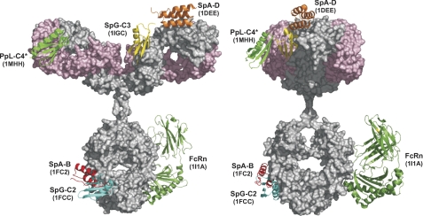
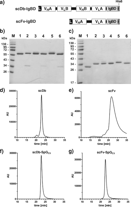
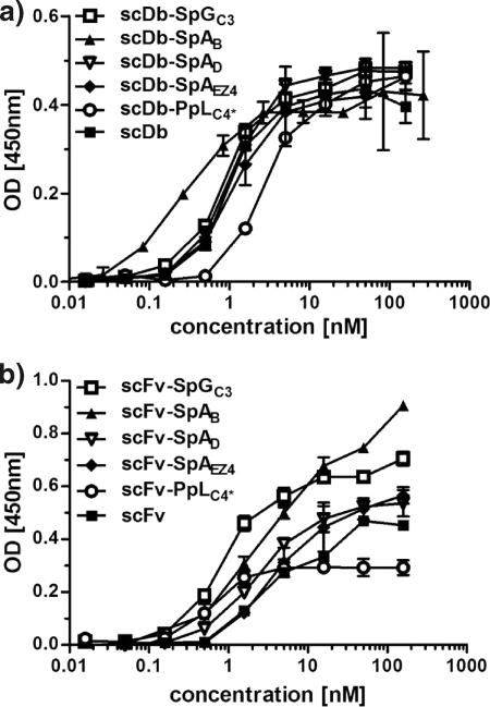
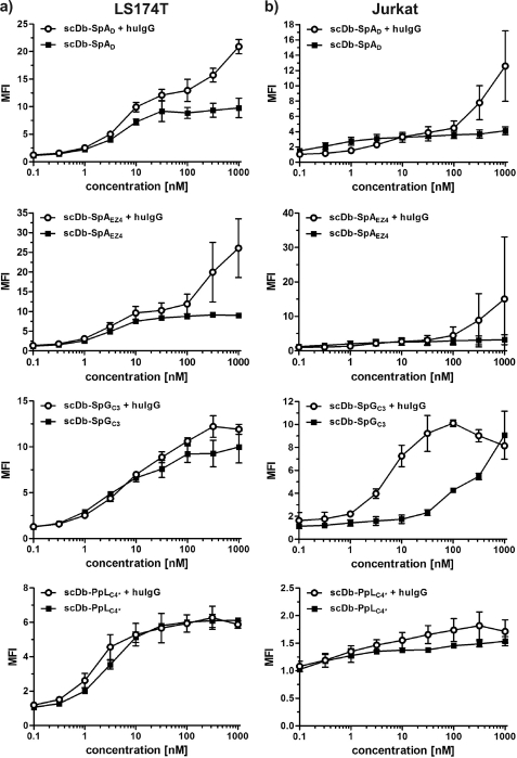
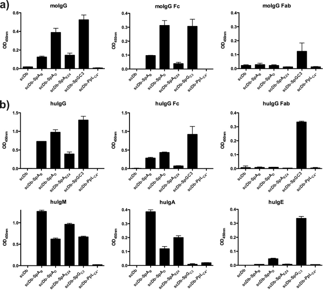
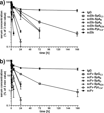
Many therapeutic proteins possessing a small size are rapidly cleared from circulation. Half-life extension strategies have therefore become increasingly important to improve the pharmacokinetic and pharmacodynamic properties of protein therapeutics. Here, we performed a comparative analysis of the half-life extension properties of various bacterial immunoglobulin-binding domains (IgBDs) derived from Staphylococcus protein A (SpA), Streptococcus protein G (SpG), and Finegoldia (formerly Peptostreptococcus) protein L (PpL). These domains, composed of 50-60 amino acid residues, were fused to the C terminus of a single-chain Fv and a bispecific single-chain diabody, respectively. All fusion proteins were produced in mammalian cells and retained their antigen-binding properties. The half-lives of the antibody molecules were prolonged to varying extents for the different IgBDs. The strongest effects in mice were observed for domain C3 of SpG (SpG(C3)) followed by domains B and D of SpA, suggesting that SpG(C3) is particularly useful to extend the plasma half-life of small proteins.


Figures

FIGURE 1.
FIGURE 1.
Summary of IgBD bound to human IgG1. IgBDs from protein A (SpAB, SpAD), protein G (SpGC2, SpGC3), and protein L (PpLC4*) in complex with IgG were visualized on a human IgG1 model (29). In addition, the extracellular region of FcRn bound to the Fc region was included (8). Protein Data Bank entries are indicated for each IgBD and the FcRn. The structures were visualized with PyMOL (30). The IgG1 heavy chain is shown in gray, the light chain in light pink.
FIGURE 2.
FIGURE 2.
Construction of scDb-IgBDs and scFv-IgBDs. a, composition of the scDb-IgBD and scFv-IgBD fusion protein. IgBDs are fused to the C terminus of a bispecific scDb or a scFv. b, SDS-PAGE analysis of purified scDb-CEACD3 (lane 1), scDb-SpAB (lane 2), scDb-SpAD (lane 3), scDb-SpAE4 (lane 4), scDb-SpGC3 (lane 5), and scDb-PpLC4* (lane 6) under reducing conditions. c, SDS-PAGE analysis of purified anti-CEA scFv (lane 1), scFv-SpAB (lane 2), scFv-SpAD (lane 3), scFv-SpAE4 (lane 4), scFv-SpGC3 (lane 5), and scFv-PpLC4* (lane 6) under reducing conditions. Two micrograms of protein were analyzed per lane, and the gel was stained with Coomassie Brilliant Blue G-250. Lane M, molecular mass standards. d–g, purified scDb, scFv as well as the scDb-SpGC3 and scFv-SpGC3 fusion proteins were analyzed by size exclusion chromatography.
FIGURE 3.
FIGURE 3.
Binding of scDb-IgBD and scFv-IgBD fusion proteins to CEA in ELISA. Increasing concentrations of the scDb-IgBD (a) or scFv-IgBD (b) fusion proteins were analyzed for binding to immobilized CEA.
FIGURE 4.
FIGURE 4.
Cell binding of scDb-IgBD fusion proteins. Binding of scDb-IgBD fusion proteins to CD3-positive Jurkat cells and CEA-expressing LS174T cells was analyzed in the absence or presence of human IgG (100 μg/ml IgG), respectively. Binding was detected with a FITC-labeled anti-His tag antibody. Binding is shown as mean fluorescence intensity (MFI) calculated from MFI of cells incubated with fusion protein/MFI of cells alone (background).
FIGURE 5.
FIGURE 5.
Binding of scDb-IgBD to IgG, Fab, and Fc analyzed by ELISA. Human and mouse IgG as well as Fab and Fc fragments thereof were immobilized, and binding of the scDb-IgBD fusion proteins was determined at a concentration of 100 nm. Furthermore, the scDb-IgBD fusion proteins were analyzed for binding to human IgM, human IgA, and human IgE.
FIGURE 6.
FIGURE 6.
Plasma half-life of scDb-IgBD and scFv-IgBD fusion proteins compared with unmodified proteins (scDb, scFv) and a chimeric IgG. scDb-IgBD (a) and scFv-IgBD (b) fusion proteins were injected intraperitoneally into CD1 mice (25 μg/animal), and serum concentrations of the antibody molecules were determined at different time points by ELISA. Data were normalized considering maximal concentration at the first time point (3 min).

Similar articles

Cited by

References

    1. Kontermann R. E. (2009) Strategies to extend plasma half-lives of recombinant antibodies. BioDrugs 23, 93–109 - PubMed
    1. Kontermann R. E. (2011) Strategies for extended serum half-life of protein therapeutics. Curr. Opin. Biotechnol. 22, 868–876 - PubMed
    1. Huang C. (2009) Receptor-Fc fusion therapeutics, traps, and MIMETIBODY technology. Curr. Opin. Biotechnol. 20, 692–699 - PubMed
    1. Chuang V. T., Kragh-Hansen U., Otagiri M. (2002) Pharmaceutical strategies utilizing recombinant human serum albumin. Pharm. Res. 19, 569–577 - PubMed
    1. Unverdorben F., Schwarz A., Richter F., Hutt M., Kontermann R. E. (2012) Half-life extension of a single-chain diabody by fusion to domain B of staphylococcal protein A. Protein Eng. Design Sel., in press - PubMed

Publication types

MeSH terms

Substances