. 2012 Jan 20;287(4):2652-65.
doi: 10.1074/jbc.M111.280867. Epub 2011 Dec 6.

Charge-based interaction conserved within histone H3 lysine 4 (H3K4) methyltransferase complexes is needed for protein stability, histone methylation, and gene expression

Affiliations

Charge-based interaction conserved within histone H3 lysine 4 (H3K4) methyltransferase complexes is needed for protein stability, histone methylation, and gene expression

Douglas P Mersman et al. J Biol Chem. .

Abstract

Histone H3 lysine 4 (H3K4) methyltransferases are conserved from yeast to humans, assemble in multisubunit complexes, and are needed to regulate gene expression. The yeast H3K4 methyltransferase complex, Set1 complex or complex of proteins associated with Set1 (COMPASS), consists of Set1 and conserved Set1-associated proteins: Swd1, Swd2, Swd3, Spp1, Bre2, Sdc1, and Shg1. The removal of the WD40 domain-containing subunits Swd1 and Swd3 leads to a loss of Set1 protein and consequently a complete loss of H3K4 methylation. However, until now, how these WD40 domain-containing proteins interact with Set1 and contribute to the stability of Set1 and H3K4 methylation has not been determined. In this study, we identified small basic and acidic patches that mediate protein interactions between the C terminus of Swd1 and the nSET domain of Set1. Absence of either the basic or acidic patches of Set1 and Swd1, respectively, disrupts the interaction between Set1 and Swd1, diminishes Set1 protein levels, and abolishes H3K4 methylation. Moreover, these basic and acidic patches are also important for cell growth, telomere silencing, and gene expression. We also show that the basic and acidic patches of Set1 and Swd1 are conserved in their human counterparts SET1A/B and RBBP5, respectively, and are needed for the protein interaction between SET1A and RBBP5. Therefore, this charge-based interaction is likely important for maintaining the protein stability of the human SET1A/B methyltransferase complexes so that proper H3K4 methylation, cell growth, and gene expression can also occur in mammals.


Figures

FIGURE 1.
FIGURE 1.
Swd1 is necessary for proper levels of Set1 protein and H3K4 methylation but not Set1 transcript. A, 3XMYC-tagged Set1, expressed from its endogenous chromosomal locus, was immunoprecipitated (IP) from whole-cell lysates prepared from the indicated strains. Protein levels of MYC-tagged Set1 were determined by immunoblotting with an α-MYC antibody. Whole-cell lysates generated from the indicated strains were immunoblotted with H3K4 mono- (me1), di- (me2), and trimethyl (me3) antibodies. Histone H3 immunoblots were used for a loading control. B, transcript levels of 3XMYC-SET1 expressed at its endogenous chromosomal locus were examined in the indicated Set1 complex component deletion strains by quantitative real time PCR. Actin expression was used as an internal loading control. The error bars shown here represent S.D. from six biological repeats with three technical repeats each.
FIGURE 2.
FIGURE 2.
C terminus of Swd1 is important for H3K4 methylation and Set1 protein levels. A, schematic of Swd1 illustrating the positions of the predicted WD40 domains (hash mark boxes). Numbers indicate amino acid positions. B, whole-cell lysates from the indicated strains were immunoblotted with H3K4 mono- (me1), di- (me2), and trimethyl (me3) antibodies. Histone H3 immunoblots were used for a loading control. C, 3XMYC-Set1 was immunoprecipitated (IP) from whole-cell lysates prepared from the indicated strains. Protein levels of 3XMYC-Set1 were determined by immunoblotting with an α-MYC antibody. HA-tagged Swd1 and Swd1 mutants were detected by immunoblotting whole-cell lysates with a rabbit α-HA antibody (the asterisk indicates a nonspecific band). Histone H3 was used as a loading control. D, quantitative real time PCR analysis was used to determine the transcript levels of Swd1 and Swd1 deletion constructs. Actin was used as an internal control. Data represent three biological repeats with three technical repeats each. For B, C, and D, HA-tagged SWD1 and swd1 C-terminal deletion constructs were episomally expressed in an swd1Δ strain with an integrated 3XMYC-SET1 expressed from its endogenous locus. The error bars shown here represent S.D. from three biological samples with three technical repeats each.
FIGURE 3.
FIGURE 3.
Acidic patches in C-terminal tail of Swd1 are important for H3K4 methylation and Set1 protein levels. A, schematic of Swd1 indicating positions of predicted WD40 repeats (hash mark boxes) and C-terminal APs. Numbers indicate amino acid positions. B, SWD1 and swd1 AP deletions were episomally expressed in an swd1Δ strain. Whole-cell lysates from the indicated strains were immunoblotted using H3K4 mono- (me1), di- (me2), and trimethyl (me3) antibodies. Histone H3 was used as a loading control. 3XMYC-Set1, expressed from its endogenous chromosomal locus, was immunoprecipitated (IP) from whole-cell lysates prepared from the indicated strains. 3XMYC-Set1 was detected by immunoblotting with an α-MYC antibody. Whole-cell lysates generated from strains expressing HA-tagged Swd1 and Swd1 acidic patch mutants were immunoblotted with a rabbit α-HA antibody. Immunoblots of glucose-6-phoshate dehydrogenase (G6PDH) were used for a loading control.
FIGURE 4.
FIGURE 4.
Basic patch in nSET domain of Set1 is needed for H3K4 methylation and proper Set1 protein levels. A, schematic of Set1 indicating the positions of the RNA recognition motif (RRM), nSET, SET, and post-SET (Post) domains as well as the position of the nSET domain basic patch (KRKK). Numbers indicate amino acid positions. B, 3XMYC-Set1 was immunoprecipitated (IP) from whole-cell lysates prepared from set1Δ strains expressing the indicated constructs. Set1 protein levels were detected by immunoblotting with an α-MYC antibody. C, 3XMYC-Set1 and 3XMYC-Set1ΔKRKK were immunoprecipitated from whole-cell lysates prepared from the indicated strains. Set1 protein levels were detected by immunoblotting using an α-MYC antibody. D, transcript levels of SET1 and the indicated set1 deletion mutants under the control of the endogenous promoter were examined using quantitative real time PCR. Actin expression was used as an internal control. The error bars shown here represent S.D. from three biological samples with three technical repeats each. E, 3XMYC-Set1 and 3XMYC-Set1ΔHRRR were immunoprecipitated from whole-cell lysates prepared from the indicated strains. Set1 protein levels were detected by immunoblotting using an α-MYC antibody. In B, C and E, whole-cell lysates generated from the indicated strains were immunoblotted with H3K4 mono- (me1), di- (me2), and trimethyl (me3) antibodies. Histone H3 was used for a loading control.
FIGURE 5.
FIGURE 5.
Acidic and basic patches in Swd1 and Set1 are needed for proper cell growth, telomere silencing, and gene expression. A, growth assays. The indicated yeast strains were serially diluted 5-fold and spotted on SC-Ura medium. Plates were photographed after 36 h of incubation at 30 °C. B, silencing assay. The indicated yeast strains were serially diluted 5-fold and spotted on SC-Leu + FOA or SC-Leu medium. The growth control plates (SC-Leu plates) were photographed after 24 h of incubation at 30 °C, and cells on SC-Leu + FOA plates were photographed after 60 h of incubation at 30 °C. Loss of telomere silencing was indicated by reduced or no growth on FOA-containing plates. C, transcript levels of MDH2 were examined using quantitative real time PCR in the indicated strains. Actin expression was used as an internal control. MDH2 transcript levels in Set1 and Swd1 mutant strains were compared with transcript levels in a wild-type strain. The error bars shown here represent S.D. from three biological samples with three technical repeats each.
FIGURE 6.
FIGURE 6.
Acidic patches in C terminus of Swd1 are important for binding to Set1 but not Swd3. A, co-immunoprecipitation assays were used to determine whether Swd1 and Swd1 acidic patch mutants could interact with Set1. 3XMYC-Set1, Swd1-HA, and Swd1-HA acidic patch mutants were either expressed individually or co-expressed in Sf9 cells. 3XMYC-Set1 was immunoprecipitated (IP) using an α-MYC antibody. Immunoprecipitates were analyzed for the presence of HA-tagged Swd1, swd1ΔAP1, swd1ΔAP2, or swd1ΔAP1&2 by immunoblotting with a rabbit α-HA antibody. Immunoblots of whole-cell lysates were performed with α-MYC and α-HA.11 antibodies to determine protein loading. B, co-immunoprecipitation assays were used to determine whether Swd1 and Swd1 acidic patch mutants could interact with Swd3. FLAG-Swd3, Swd1-HA, and Swd1-HA acidic patch mutants were either expressed individually or co-expressed in Sf9 cells. Extracts were prepared, and Swd3 was immunoprecipitated using α-FLAG M2 resin. Immunoprecipitates were analyzed for the presence of HA-tagged Swd1, swd1ΔAP1, swd1ΔAP2, or swd1ΔAP1&2 by immunoblotting with a rabbit α-HA antibody. Immunoblots of whole-cell lysates were performed with rabbit α-FLAG and α-HA.11 antibodies to determine protein loading.
FIGURE 7.
FIGURE 7.
Charge-based interaction mediates association between Swd1 and nSET domain of Set1 in vitro. A and B, GST fusion binding assays were used to determine binding between Swd1 and the nSET domain of Set1. GST-nSET was used to pull down (PD) the indicated HA-tagged Swd1 and Swd1 mutants from Sf9 extracts (A) or to pull down the indicated His-tagged Swd1 and Swd1 mutants from bacterial extracts (B). C, GST fusion binding assays were used to determine binding between Swd1 and the nSET domain of Set1. GST-nSET or GST-nSETΔKRKK was used to pull down (PD) HA-tagged Swd1. D, GST-nSET constructs were engineered in which the basic patch (KRKK) was mutated to conserve the charge (RKRR) or to convert to negatively charged residues (EREK or EEEE). GST fusion binding assays were used to determine binding between Swd1-HA and wild-type nSET domain and nSET domains with conserved charge or opposite charge. In all panels, GST was used as a control for nonspecific binding. Input and bound fractions of Swd1-HA were detected by immunoblotting using an α-HA.11 antibody. Input and bound fraction of His-Swd1 were detected by immunoblotting using an α-His antibody. Input fractions of GST, GST-nSET, or GST-nSET mutants were detected by immunoblotting using an α-GST antibody.
FIGURE 8.
FIGURE 8.
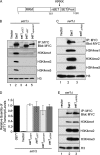
Charge-based interaction is important for association between Swd1 and Set1 and maintaining H3K4 methylation in vivo. A, to determine whether reversing the charge of the nSET domain basic patch affects histone methylation and Set1 protein levels, wild-type 3XMYC-Set1 or the indicated Set1 mutants were expressed in a set1Δ strain. 3XMYC-Set1 was immunoprecipitated (IP) from whole-cell lysates and detected by immunoblotting using an α-MYC antibody. Immunoblots of whole-cell lysates using H3K4 mono- (me1), di- (me2), and trimethyl (me3) antibodies show the H3K4 methylation status from yeast strains expressing the indicated Set1 constructs. An immunoblot probed with an α-H3 antibody was used to determine protein loading. B, co-immunoprecipitation assays were performed to determine whether Swd1 and Swd1 acidic patch mutants could interact with Set1. FLAG-Set1(690–1080), FLAG-Set1 basic patch mutant (FLAG-Set1(690–1080) ΔKRKK), Swd1-HA, and Swd1-HA acidic patch mutants were either expressed individually or co-expressed in yeast. Immunoprecipitates were analyzed for the presence of HA-tagged Swd1, swd1ΔAP1, or swd1ΔAP2 by immunoblotting with a rabbit α-HA antibody. Immunoblots of whole-cell lysates using H3K4 mono-, di-, and trimethyl antibodies show the H3K4 methylation status in yeast. Immunoblots were probed with rabbit α-FLAG, rabbit α-HA, α-H3, or α-glucose-6-phoshate dehydrogenase (G6PDH) antibody to monitor protein loading. The plus (+) and minus (−) symbols represent the expressed construct.
FIGURE 9.
FIGURE 9.
Human SET1A, but not MLL1, interacts with human RBBP5 in vitro. A, sequence alignment of the nSET domain regions of yeast Set1 compared with human orthologs SET1A, SET1B, and MLL1. The conserved basic patch region is bold and boxed. The alignment was generated by ClustalX software using the nSET domain sequences. The y represents budding yeast nSET domain sequence, and h represents human nSET domain sequences. B, sequence alignment of yeast Swd1 compared with other orthologs. The region shown represents the C-terminal region of Swd1 and RBBP5. The conserved acidic patches (AP1 or AP2) and the WDR5 interaction motif are bold and boxed. This result was generated by ClustalX software using the full-length amino acid sequences of Swd1 and RBBP5. Homo represents Homo sapiens, Drosophila represents Drosophila melanogaster, Schizosaccharomyces represents Schizosaccharomyces Pombe, Saccharomyces represents Saccharomyces cerevisiae, and Candida represents Candida albicans. C, GST fusion binding assays were performed to determine whether the nSET domains of SET1A and MLL1 bind to RBBP5. GST-SET1A-nSET (amino acids 1498–1572) or GST-MLL1-nSET (amino acids 3753–3832) was used to pull down bacterially expressed His-tagged RBBP5. GST was used as a control for nonspecific binding. D, GST fusion binding assays were used to determine whether acidic patches in RBBP5 are important for its binding to the nSET domain of SET1A. GST-SET1A-nSET was used to pull down His-tagged RBBP5 and the indicated RBBP5 acidic patch mutants. GST was used as a control for nonspecific binding. The input and bound fractions of His-RBBP5 were detected by immunoblotting with an α-His antibody. The amount of GST or GST fusion protein fragments was analyzed by Coomassie Blue staining.

Similar articles

Cited by

References

    1. Roguev A., Schaft D., Shevchenko A., Pijnappel W. W., Wilm M., Aasland R., Stewart A. F. (2001) The Saccharomyces cerevisiae Set1 complex includes an Ash2 homologue and methylates histone 3 lysine 4. EMBO J. 20, 7137–7148 - PMC - PubMed
    1. Miller T., Krogan N. J., Dover J., Erdjument-Bromage H., Tempst P., Johnston M., Greenblatt J. F., Shilatifard A. (2001) COMPASS: a complex of proteins associated with a trithorax-related SET domain protein. Proc. Natl. Acad. Sci. U.S.A. 98, 12902–12907 - PMC - PubMed
    1. Dehé P. M., Dichtl B., Schaft D., Roguev A., Pamblanco M., Lebrun R., Rodríguez-Gil A., Mkandawire M., Landsberg K., Shevchenko A., Shevchenko A., Rosaleny L. E., Tordera V., Chávez S., Stewart A. F., Géli V. (2006) Protein interactions within the Set1 complex and their roles in the regulation of histone 3 lysine 4 methylation. J. Biol. Chem. 281, 35404–35412 - PubMed
    1. Mueller J. E., Canze M., Bryk M. (2006) The requirements for COMPASS and Paf1 in transcriptional silencing and methylation of histone H3 in Saccharomyces cerevisiae. Genetics 173, 557–567 - PMC - PubMed
    1. Steward M. M., Lee J. S., O'Donovan A., Wyatt M., Bernstein B. E., Shilatifard A. (2006) Molecular regulation of H3K4 trimethylation by ASH2L, a shared subunit of MLL complexes. Nat. Struct. Mol. Biol. 13, 852–854 - PubMed

Publication types

MeSH terms