. 2012 Jan 27;287(5):3217-30.
doi: 10.1074/jbc.M111.280834. Epub 2011 Dec 6.

C5a-regulated CCAAT/enhancer-binding proteins β and δ are essential in Fcγ receptor-mediated inflammatory cytokine and chemokine production in macrophages

Affiliations

C5a-regulated CCAAT/enhancer-binding proteins β and δ are essential in Fcγ receptor-mediated inflammatory cytokine and chemokine production in macrophages

Chunguang Yan et al. J Biol Chem. .

Abstract

CCAAT/enhancer-binding protein β (C/EBPβ) and C/EBPδ are known to participate in the regulation of many genes associated with inflammation. However, little is known about the activation and function of C/EBPβ and -δ in inflammatory responses elicited by Fcγ receptor (FcγR) activation. Here we show that C/EBPβ and -δ activation are induced in IgG immune complex (IC)-treated macrophages. The increased expression of C/EBPβ and -δ occurred at both mRNA and protein levels. Furthermore, induction of C/EBPβ and -δ was mediated, to a large extent, by activating FcγRs. Using siRNA-mediated knockdown as well as macrophages deficient for C/EBPβ and/or -δ, we demonstrate that C/EBPβ and -δ play a critical role in the production of TNF-α, MIP-2, and MIP-1α in IgG IC-stimulated macrophages. Moreover, both ERK1/2 and p38 MAPK are involved in C/EBP induction and TNF-α, MIP-2, and MIP-1α production induced by IgG IC. We provide the evidence that C5a regulates IgG IC-induced inflammatory responses by enhancing ERK1/2 and p38 MAPK activities as well as C/EBPβ and -δ activities. Collectively, these data suggest that C/EBPβ and -δ are key regulators for FcγR-mediated induction of cytokines and chemokines in macrophages. Furthermore, C/EBPs may play an important regulatory role in IC-associated inflammatory responses.


Figures
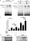
FIGURE 1.
FIGURE 1.
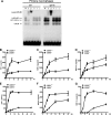
IgG immune complexes induce C/EBPβ and -δ expression and increase C/EBP DNA binding activity in macrophages. A, RAW264.7 cells were stimulated with 100 μg/ml immune complexes for the times indicated. Nuclear proteins were harvested and subjected to EMSA to measure C/EBP DNA binding activity. B, RAW264.7 cells were treated or left untreated with 100 μg/ml IgG immune complexes for 4 h. The nuclear extracts were harvested for gel supershift assay to identify which C/EBP family member DNA binding activity was regulated by IgG immune complex treatment. N, α, β, δ, ϵ, and γ, normal rabbit IgG, anti-C/EBPα antibody, anti-C/EBPβ antibody, anti-C/EBPδ antibody, anti-C/EBPϵ antibody, and anti-C/EBPγ antibody, respectively. The arrows indicate C/EBP complex and supershift species. C, RAW264.7 cells were transiently transfected with a total of 0.5 μg of the indicated DNA. 24 h after transfection, the cells were treated with 100 μg/ml IgG immune complex for 5 h. Cell lysates were used to perform a luciferase activity assay. Luminometer values were normalized for expression from a co-transfected thymidine kinase reporter gene. The data were expressed as means of three experiments ± S.E. (error bars). RAW264.7 cells were stimulated with 100 μg/ml IgG immune complexes for different time periods. Then total cellular RNA was isolated for RT-PCR with primers for C/EBPβ, C/EBPδ, and GAPDH, respectively (D). The level of GAPDH is shown at the bottom as a loading control. E, the total proteins were extracted to conduct Western blot using rabbit anti-C/EBPβ antibody, rabbit anti-C/EBPδ antibody, and rabbit anti-GAPDH antibody, respectively. The level of GAPDH is shown at the bottom as a loading control.
FIGURE 2.
FIGURE 2.
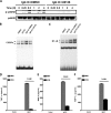
C/EBPβ and -δ silencing down-regulates IgG immune complex-induced TNF-α, MIP-2, and MIP-1α production from macrophages. A and F, RAW264.7 cells were transiently transfected with control siRNA, C/EBPβ siRNA (A), or C/EBPδ siRNA (F), respectively. 12 h later, RNAs were extracted and subjected to RT-PCR to examine the expression of C/EBPβ, C/EBPδ, and GAPDH, respectively. The level of GAPDH is shown at the bottom as a loading control. B and G, RAW264.7 cells were transiently transfected with control siRNA, C/EBPβ siRNA (B), or C/EBPδ siRNA (G). 12 h later, cells were incubated with or without 100 μg/ml IgG IC for 4 h. Total RNA was extracted, and semiquantitative RT-PCR was performed to examine expression of TNF-α, MIP-2, and MIP-1α, respectively. C–E and H–J, RAW264.7 cells were transiently transfected with control siRNA, C/EBPβ siRNA (C–E), or C/EBPδ (H–J), respectively. 12 h after transfection, the cells were stimulated with or without 100 μg/ml IgG immune complex for 5 h. The supernatants were harvested, and ELISA was performed to determine the production of TNF-α (C–E and H–J). Data are presented as mean ± S.E. (error bars) (n = 7–12).
FIGURE 3.
FIGURE 3.
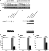
C/EBPβ and -δ are critical regulators of cytokine and chemokine production in IgG IC-stimulated macrophages. A, primary peritoneal macrophages obtained from wild type were treated or left untreated with 100 μg/ml IgG immune complexes for 4 h. The nuclear extracts were harvested for a gel supershift assay to identify which C/EBP family member DNA binding activity was regulated by IgG immune complex treatment. N, α, β, δ, ϵ, and γ, normal rabbit IgG, anti-C/EBPα antibody, anti-C/EBPβ antibody, anti-C/EBPδ antibody, anti-C/EBPϵ antibody, and anti-C/EBPγ antibody, respectively. The arrows indicate C/EBP binding bands and supershift bands. B–D and E–G, primary peritoneal macrophages obtained from corresponding wild type, C/EBPβ knock-out (B–D), and C/EBPδ knock-out (E–G) mice were treated with 100 μg/ml IgG immune complex for different time periods, and supernatants were subjected to ELISA analysis for TNF-α, MIP-2, and MIP-1α production. Data are presented as mean ± S.E. (error bars) (n = 6).
FIGURE 4.
FIGURE 4.
FcγRs play important roles in C/EBP-mediated cytokine and chemokine production in macrophages. A–C, primary peritoneal macrophages obtained from corresponding wild type and FcR γ-chain-deficient mice were treated with 100 μg/ml IgG IC for 5 h, and supernatants were subjected to ELISA analysis for TNF-α (A), MIP-2 (B), and MIP-1α (C) production. Data are presented as mean ± S.E. (error bars) (n = 8). D, primary peritoneal macrophages obtained from corresponding wild type and FcR γ-chain-deficient mice were treated or left untreated with 100 μg/ml IgG immune complexes for 4 h. The nuclear proteins were harvested and subjected to EMSA to measure C/EBP DNA binding activity.
FIGURE 5.
FIGURE 5.
ERK1/2 is involved in IgG IC-induced C/EBPβ and -δ activation and subsequent cytokine/chemokine production in macrophages. A, RAW264.7 cells were treated with 100 μg/ml IgG immune complexes in the presence or absence of ERK1/2 inhibitor, U0126 (10 μm), for the indicated time periods. Total proteins were subjected to Western blot by using rabbit anti-phospho-p44/42 (p-p44/42) antibody and rabbit anti-p44/42 antibody, respectively. B and C, RAW264.7 cells were treated with 100 μg/ml IgG immune complexes in the presence or absence of ERK1/2 inhibitor, U0126 (10 μm), for 4 h. The nuclear proteins were subjected to EMSA for C/EBP DNA binding (B) and NF-κB DNA binding (C). D–F, RAW264.7 cells were treated with 100 μg/ml IgG immune complexes in the presence or absence of ERK1/2 inhibitor, U0126 (10 μm), for 5 h, and supernatants were subjected to ELISA analysis for TNF-α (D), MIP-2 (E), and MIP-1α (F) production. Data are presented as mean ± S.E. (error bars) (n = 10).
FIGURE 6.
FIGURE 6.
p38 MAPK is involved in IgG IC-induced C/EBPβ and -δ activation and subsequent cytokine/chemokine production in macrophages. A, RAW264.7 cells were treated with 100 μg/ml IgG immune complexes in the presence or absence of p38 MAPK inhibitor VII (10 μm) for the indicated time periods. Total proteins were subjected to Western blot by using rabbit anti-phospho-p38 (p-p38) antibody and rabbit anti-p38 antibody, respectively. B and C, RAW264.7 cells were treated with 100 μg/ml IgG immune complexes in the presence or absence of p38 MAPK inhibitor VII (10 μm) for 4 h. The nuclear proteins were subjected to EMSA for C/EBP DNA binding (B) and NF-κB DNA binding (C). D–F, RAW264.7 cells were treated with 100 μg/ml IgG immune complexes in the presence or absence of p38 MAPK inhibitor VII (10 μm) for 5 h, and supernatants were subjected to ELISA analysis for TNF-α (D), MIP-2 (E), and MIP-1α (F) production. Data are presented as mean ± S.E. (error bars) (n = 10).
FIGURE 7.
FIGURE 7.
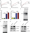
C5a signaling enhances IgG immune complex-induced C/EBP activation and cytokine and chemokine production in macrophages. A–C, mouse peritoneal macrophages were treated with 10 nm recombinant human C5a, 100 μg/ml IgG immune complexes, or a combination of both stimuli for different time points, and supernatants were subjected to ELISA analysis for TNF-α (A), MIP-2 (B), and MIP-1α (C) production. Data are presented as mean ± S.E. (n = 7). D and E, RAW264.7 cells were transiently transfected with a total of 0.5 μg of DNA. 24 h after transfection, the cells were treated with 10 nm recombinant human C5a, 100 μg/ml IgG immune complexes, or a combination of both stimuli for 4 h. Cell lysates were used to perform the luciferase activity assay. Luminometer values were normalized for expression from a co-transfected thymidine kinase reporter gene. The data were expressed as means of three experiments ± S.E. (error bars). F and G, peritoneal macrophages obtained from wild type and C5aR-deficient mice were treated with or without 100 μg/ml IgG IC for 4 h, and nuclear proteins were subjected to EMSA for C/EBP (F) or NF-κB (G) DNA binding activity. H and I, RAW264.7 cells were treated with 100 μg/ml IgG immune complex or IgG immune complex plus 10 nm rhC5a for 4 h. Total proteins were extracted and subjected to Western blot by using rabbit anti-phospho-p44/42 (p-p44/42) antibody, rabbit anti-p44/42 antibody, and rabbit anti-GAPDH (H) and using rabbit anti-phospho-p38 (p-p38) antibody, rabbit anti-p38 antibody, and rabbit anti-GAPDH (I).

Similar articles

Cited by

References

    1. Nimmerjahn F., Ravetch J. V. (2008) Fcγ receptors as regulators of immune responses. Nat. Rev. Immunol. 8, 34–47 - PubMed
    1. Halstead S. B., Mahalingam S., Marovich M. A., Ubol S., Mosser D. M. (2010) Intrinsic antibody-dependent enhancement of microbial infection in macrophages. Disease regulation by immune complexes. Lancet Infect. Dis. 10, 712–722 - PMC - PubMed
    1. Fernández N., Renedo M., García-Rodríguez C., Sánchez Crespo M. (2002) Activation of monocytic cells through Fc γ receptors induces the expression of macrophage-inflammatory protein (MIP)-1 α, MIP-1 β, and RANTES. J. Immunol. 169, 3321–3328 - PubMed
    1. Gao H., Neff T., Ward P. A. (2006) Regulation of lung inflammation in the model of IgG immune-complex injury. Annu. Rev. Pathol. 1, 215–242 - PubMed
    1. Bayón Y., Alonso A., Sánchez Crespo M. (1997) Stimulation of Fc γ receptors in rat peritoneal macrophages induces the expression of nitric oxide synthase and chemokines by mechanisms showing different sensitivities to antioxidants and nitric oxide donors. J. Immunol. 159, 887–894 - PubMed

Publication types

MeSH terms

Substances

LinkOut - more resources